 Initializing download.
-
 By César Adrián Gómez Valdivia, MD
Fundación Hospital Nuestra Señora de la Luz IAP
Co-author(s): @eyemissu2 - Uploaded on Aug 12, 2025.
- Last modified by Joshua Friedman on Aug 13, 2025.
- Rating
- Appears in
- Miscellaneous
- Condition/keywords
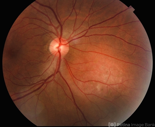
- amelanotic melanoma
- Photographer
- @eyemissu2
- Imaging device
-
Fundus camera
TOPCON TRC-50DX - Description
- This case highlights an amelanotic melanoma, an atypical presentation of a choroidal melanoma lacking the characteristic pigmentation. These lesions can easily be mistaken for choroidal hemangiomas, metastases, or inflammatory masses. Clinically, the lesion appears as a dome-shaped, yellowish subretinal mass, often associated with subretinal fluid, lipofuscin deposition, or retinal detachment. The absence of pigment can delay diagnosis, making multimodal imaging essential. Diagnostic tools: • B-scan ultrasound: low to medium internal reflectivity • OCT: overlying subretinal fluid and RPE elevation • FAF: orange pigment and RPE disruption • ICG/FA: variable, often hypofluorescent core Important: Prompt referral to ocular oncology is critical for management and prognosis.


