 Initializing download.
-
 By César Adrián Gómez Valdivia, MD
Fundación Hospital Nuestra Señora de la Luz IAP
Co-author(s): @eyemissu2 - Uploaded on Aug 12, 2025.
- Last modified by Joshua Friedman on Aug 13, 2025.
- Rating
- Appears in
- Miscellaneous
- Condition/keywords
- amelanotic melanoma
- Photographer
- @eyemissu2
- Imaging device
-
Fundus camera
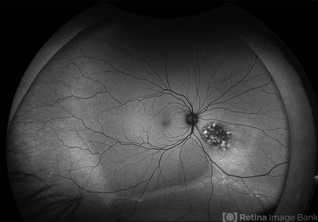
California ICG OPTOS - Description
- This FAF image reveals a hypoautofluorescent mass with areas of dense hyperautofluorescent stippling—a classic pattern suggestive of an amelanotic choroidal melanoma. Amelanotic melanoma is a rare variant of uveal melanoma, accounting for only a minority of cases. Unlike pigmented melanomas, these lesions lack melanin, making them more challenging to detect on conventional color fundus imaging. FAF Characteristics: • Central hypoautofluorescence: due to loss or compression of the RPE • Peripheral hyperautofluorescent speckling: consistent with lipofuscin accumulation or RPE disruption • Often associated with subretinal fluid or orange pigment seen clinically Location: Juxtapapillary, with potential optic nerve involvement—a factor that complicates both diagnosis and


