-
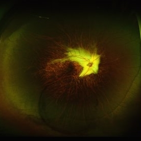
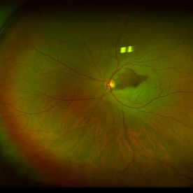
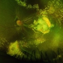
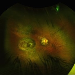
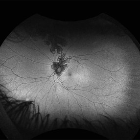
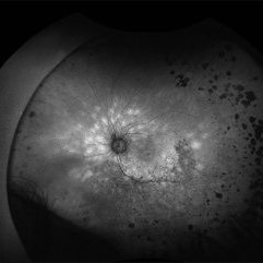
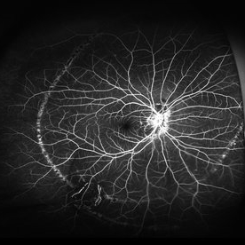 Schlaegel Line
Mar 14 2024 by César Adrián Gómez Valdivia, MD
Hyperfluorescent, concentric, chorioretinal striae on fluoroangiography.
Photographer: Erika Paulina Ornelas Cazares
Imaging device: California ICG OPTOS
Condition/keywords: line, PHOS
-
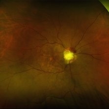
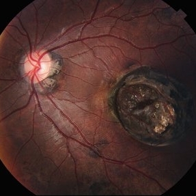
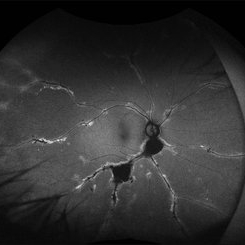
 Optic Nerve Melanocytoma
Mar 14 2024 by César Adrián Gómez Valdivia, MD
Benign neoplasm with seldom malignant transformation.
Photographer: Erika Paulina Ornelas Cazares
Imaging device: TOPCON TRC-50DX
Condition/keywords: disc, Melanocytoma, Nerve, Optic, optic disc melanocytoma
-
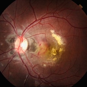
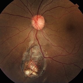
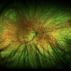
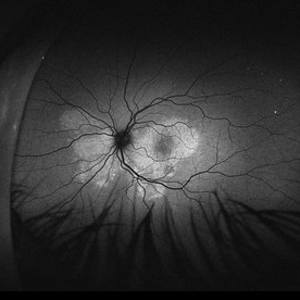
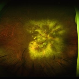
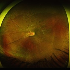
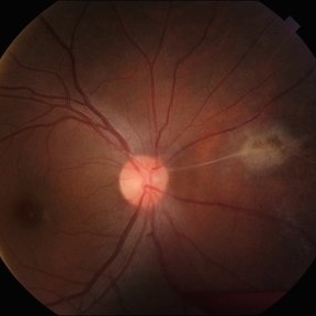
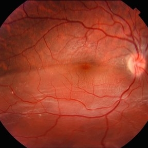
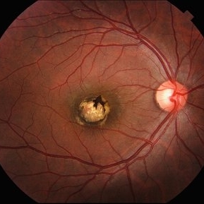
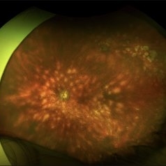
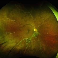
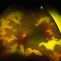
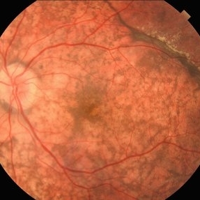
 Degenerative Myopia
Apr 21 2024 by César Adrián Gómez Valdivia, MD
Degenerative Myopia
Photographer: Erika Paulina Ornelas Cazares
Imaging device: Topcon TRC-50 DX
Condition/keywords: degenerative myopia, myopia
-
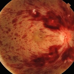
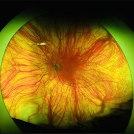
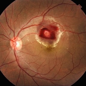
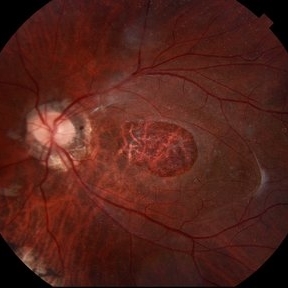
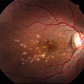
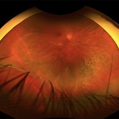
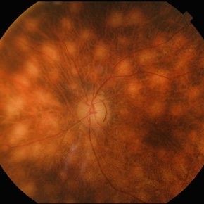
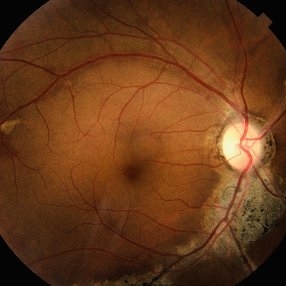
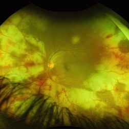
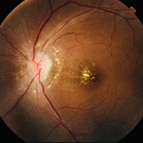
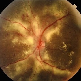 Hypertensive Retinopathy
Apr 21 2024 by César Adrián Gómez Valdivia, MD
Hypertensive Retinopathy
Photographer: Erika Paulina Ornelas Cazares
Imaging device: TOPCON TRC-50DX
Condition/keywords: hypertensive retinopathy
-
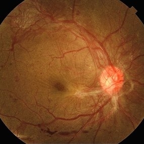
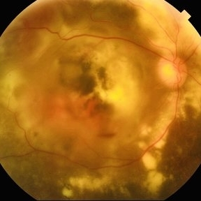
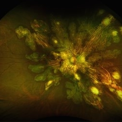
 Serpiginous Choroidopathy
Apr 21 2024 by César Adrián Gómez Valdivia, MD
Gray-yellowish subretinal infiltrates that usually spread centrifugally from the peripapillary region in a serpiginous (snake-like) manner. Active lesions show a leading edge and resolve with subsequent RPE and choriocapillary atrophy.
Photographer: @eyemissu2
Imaging device: TOPCON TRC-50DX
Condition/keywords: macula serpiginous choroidopathy
-
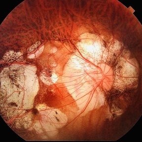
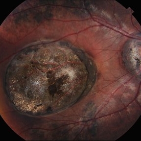
 Retinal Colomoba
Jul 21 2024 by César Adrián Gómez Valdivia, MD
Retinal Coloboma found in a female 41 year old patient. Iris, Lens, Ciliary Body, Zonules, Choroid and Retina were involved.
Photographer: Erika Paulina Ornelas Cazares
Imaging device: TOPCON TRC-50DX
Condition/keywords: coloboma
-
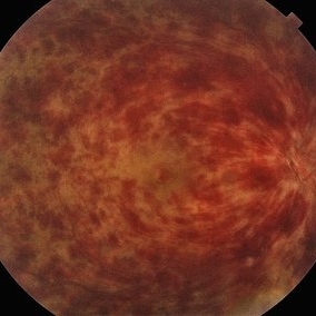
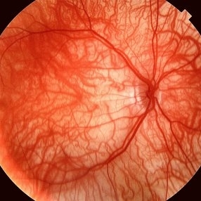
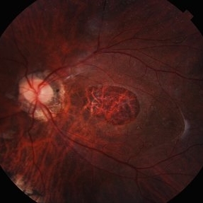
 Central Retinal Vein Occlusion
Jul 21 2024 by César Adrián Gómez Valdivia, MD
Central Retinal Vein Occlusion found in a 72 year old patient with history of uncontrolled Hypertension. Non-Ischemic Variant.
Photographer: Erika Paulina Ornelas Cazares
Imaging device: TOPCON TRC-50DX
Condition/keywords: central retinal vein occlusion (CRVO)
-
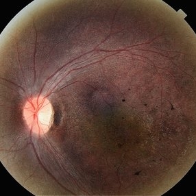
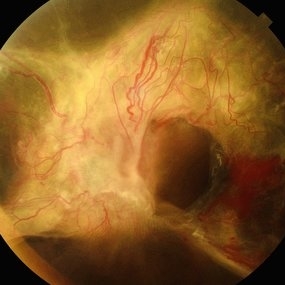
 Extensive Neovascularization
Jul 21 2024 by César Adrián Gómez Valdivia, MD
Macular Traction and Extensive Neovascularization found in a 66 year-old patient with history of uncontrolled Type 2 Diabetes Mellitus.
Photographer: Erika Paulina Ornelas Cazares
Imaging device: TOPCON TRC-50DX
Condition/keywords: diabetic retinopathy, neovascularization (NV)
-
 Ischemic Central Retinal Vein Occlusion
Aug 6 2024 by César Adrián Gómez Valdivia, MD
Fundus photograph of an 80 year old man with and acute central retinal vein occlusion, ischemic variant.
Photographer: @eyemissu2
Condition/keywords: central retinal vein occlusion (CRVO), ischemic CRVO
-
 Optic Disc Coloboma
Aug 10 2024 by César Adrián Gómez Valdivia, MD
Optic Disc Coloboma found in an 8YO patient. Findings were bilateral. Unlike the morning glory disc, the ODC has no central glial tuft and the disc vasculature is usually normal.
Photographer: @eyemissyou2
Imaging device: Topcon
Condition/keywords: Coloboma, coloboma of optic disc, optic disc
-
 Foveal Hypoplasia / Ocular Albinism
Aug 25 2024 by César Adrián Gómez Valdivia, MD
Fundus photograph of a 6-year-old female patient with foveal hypoplasia, ocular albinism and pendular nystagmus. Findings were bilateral. Retinal and choroidal vasculature are exquisitely beautiful.
Photographer: @eyemissu2
Imaging device: TOPCON TRC-50DX
Condition/keywords: Albinism, foveal hypoplasia, ocular albinism, vascula
-
 Foveal Hypoplasia / Ocular Albinism
Aug 25 2024 by César Adrián Gómez Valdivia, MD
Fundus photograph of a 6-year-old female patient with foveal hypoplasia, ocular albinism and pendular nystagmus. Findings were bilateral. Retinal and Choroidal vasculature are exquisitely beautiful.
Photographer: @eyemissu2
Imaging device: TOPCON TRC-50DX
Condition/keywords: albinism, foveal hypoplasia, ocular albinism
-
 Foveal Hypoplasia / Ocular Albinism
Aug 29 2024 by César Adrián Gómez Valdivia, MD
Fundus photograph of a 64-year-old female patient with foveal hypoplasia, ocular albinism and pendular nystagmus. Findings were bilateral. Retinal and choroidal vasculature are exquisitely beautiful.
Photographer: @eyemissu2
Imaging device: California ICG OPTOS
Condition/keywords: foveal hypoplasia, ocular albinism
-
 Foveal Hypoplasia / Ocular Albinism
Aug 29 2024 by César Adrián Gómez Valdivia, MD
Fundus photograph of a 6-year-old female patient with foveal hypoplasia, ocular albinism and pendular nystagmus. Findings were bilateral. Retinal and choroidal vasculature are exquisitely beautiful.
Photographer: @eyemissu2
Imaging device: TOPCON TRC-50DX
Condition/keywords: foveal hypoplasia, ocular albinism
-
 Stargardt Disease
Aug 29 2024 by César Adrián Gómez Valdivia, MD
Fundus photograph of a 10 year-old male patient with Stargardt Disease. Findings were Bilateral.
Photographer: @eyemissu2
Imaging device: TOPCON TRC-50DX
Condition/keywords: Stargardt disease
-
 Vasoproliferative Tumor
Aug 29 2024 by César Adrián Gómez Valdivia, MD
Inferior retinal vasoproliferative tumor found in a 66 year-old female patient. Asymptomatic.
Photographer: @eyemissu2
Imaging device: California ICG OPTOS
Condition/keywords: Vasoproliferative Tumor
-
 Straatsma Syndrome
Aug 29 2024 by César Adrián Gómez Valdivia, MD
Fundus photograph of a 11 year-old female patient with unilateral myelinated retinal nerve fibers, axial myopia, amblyopia and strabismus.
Photographer: @eyemissu2
Imaging device: California ICG OPTOS
Condition/keywords: Straatsma Syndrome
-
 Sunset Glow Fundus
Oct 14 2024 by César Adrián Gómez Valdivia, MD
“Sunset Glow Fundus” found in a 65 year-old male patient diagnosed with Voght-Koyanagi-Harada disease, convalescent stage. Choroidal depigmentation occurs several months after the uveitic stage, leading to a pale disc with a bright red-orange choroid.
Photographer: @eyemissu2
Imaging device: OPTOS
Condition/keywords: sunset, Sunset Glow Fundus, VKH
-
 Sunset Glow Fundus
Oct 14 2024 by César Adrián Gómez Valdivia, MD
"Sunset Glow Fundus” found in a 65 year-old male patient diagnosed with Voght-Koyanagi-Harada disease, convalescent stage. Choroidal depigmentation occurs several months after the uveitic stage, leading to a pale disc with a bright red-orange choroid.
Photographer: @eyemissu2
Imaging device: California ICG OPTOS
Condition/keywords: sunset, Sunset Glow Fundus, vkh
-
 Sunset Glow Fundus
Oct 14 2024 by César Adrián Gómez Valdivia, MD
"Sunset Glow Fundus” found in a 65 year-old male patient diagnosed with Voght-Koyanagi-Harada disease, convalescent stage. Choroidal depigmentation occurs several months after the uveitic stage, leading to a pale disc with a bright red-orange choroid.
Photographer: @eyemissu2
Imaging device: TOPCON TRC-50DX
Condition/keywords: sunset, Sunset Glow Fundus
-
 Sunset Glow Fundus
Oct 14 2024 by César Adrián Gómez Valdivia, MD
"Sunset Glow Fundus” found in a 65 year-old male patient diagnosed with Voght-Koyanagi-Harada disease, convalescent stage. Choroidal depigmentation occurs several months after the uveitic stage, leading to a pale disc with a bright red-orange choroid.
Photographer: @eyemissu2
Imaging device: TOPCON TRC-50DX
Condition/keywords: Sunset, Sunset Glow Fundus
-
 Serous Retinal Detachment
Oct 14 2024 by César Adrián Gómez Valdivia, MD
Serous Retinal Detachment found in a 19 year-old female patient with suspected Vogt-Koyanagi-Harada disease. Findings were bilateral. Patient was admitted for Methylprednisolone and Cyclophosphamide treatment.
Photographer: @eyemissu2
Imaging device: California ICG OPTOS
Condition/keywords: serous retinal detachment, Vogt-Koyanagi-Harada
-
 Serous Retinal Detachment
Oct 14 2024 by César Adrián Gómez Valdivia, MD
Serous Retinal Detachment found in a 19 year-old female patient with suspected Vogt-Koyanagi-Harada disease. Findings were bilateral. Patient was admitted for Methylprednisolone and Cyclophosphamide treatment.
Photographer: @eyemissu2
Imaging device: California ICG OPTOS
Condition/keywords: serous retinal detachment, vkh, Vogt-Koyanagi-Harada
-
 Acute Syphilitic Posterior Placoid Chorioretinitis
Oct 16 2024 by César Adrián Gómez Valdivia, MD
Fundus autofluorescence image of an acute syphilitic posterior placoid chorioretinitis found in a HIV positive 28 YO male patient with suspected neurosyphilis. A beautiful butterfly autofluorescence pattern can be appreciated.
Photographer: @eyemissu2
Imaging device: California ICG OPTOS
Condition/keywords: acute syphilitic posterior placoid chorioretinitis, chorioretinitis, syphilis
-
 Serpiginous Choroidopathy
Oct 19 2024 by César Adrián Gómez Valdivia, MD
Fundus photograph of a 29-year-old woman with Serpiginous Choroidopathy. Finings were bilateral.
Photographer: @eyemissu2
Imaging device: California ICG OPTOS
Condition/keywords: macula serpiginous choroidopathy, serpiginous choroiditis, serpiginous like choroiditis
-
 Acute Syphilitic Posterior Placoid Chorioretinitis
Oct 20 2024 by César Adrián Gómez Valdivia, MD
Fundus autofluorescence image of an acute syphilitic posterior placoid chorioretinitis found in a HIV positive 28 YO male patient with suspected neurosyphilis. A beautiful butterfly autofluorescence pattern can be appreciated.
Photographer: @eyemissu2
Imaging device: California ICG OPTOS
Condition/keywords: acute syphilitic posterior placoid chorioretinitis
-
 Choroidal Fracture
Oct 27 2024 by César Adrián Gómez Valdivia, MD
Fundus photograph of a traumatic choroidal fracture & extra-macular sub-retinal hemorrhage.
Photographer: @eyemissu2
Imaging device: TOPCON TRC-50DX
Condition/keywords: Choroidal Fracture
-
 Ruptured Retinal Arterial Macro-Aneurysm
Oct 27 2024 by César Adrián Gómez Valdivia, MD
Ruptured retinal arterial macro-aneurysm found in a 56 YO female patient with history of untreated hypertension. Round or fusiform dilation of a retinal arteriole is usually seen within a third degree branch of one of the four main arcade arteries. Most common location for a symptomatic macroaneurysm is from a branch of the superotemporal arcade.
Photographer: @eyemissu2
Imaging device: TOPCON TRC-50DX
Condition/keywords: ruptured macroaneurysm
-
 Hypertensive Retinopathy
Oct 27 2024 by César Adrián Gómez Valdivia, MD
Fundus photograph of a 62 year-old woman with history of untreated hypertension and chronic kidney disease. Findings were bilateral.
Photographer: @eyemissu2
Imaging device: TOPCON TRC-50DX
Condition/keywords: hypertensive retinopathy
-
 Ciliary Artery Sparing Central Retinal Artery Occlusion (CRAO)
Oct 27 2024 by César Adrián Gómez Valdivia, MD
Ciliary artery sparing central retinal artery occlusion (CRAO) is a condition where the central retinal artery is blocked, but the cilioretinal artery is patent, allowing for the preservation of central vision.
Photographer: @eyemissu2
Imaging device: California ICG OPTOS
Condition/keywords: Ciliary artery sparing central retinal artery occlusion (CRAO), ciliary artery sparring, oclussion
-
 Giant Persistent Macular Hole
Dec 6 2024 by César Adrián Gómez Valdivia, MD
Giant Persistent Macular Hole found in a 48 year-old male patient one year after vitrectomy.
Photographer: @eyemissu2
Imaging device: TOPCON TRC-50DX
Condition/keywords: macular, macular hole
-
 Macular Dragging
Dec 12 2024 by César Adrián Gómez Valdivia, MD
Macular dragging found in a 14 year-old female patient with ROP history. Findings were bilateral.
Photographer: @eyemissu2
Imaging device: TOPCON TRC-50DX
Condition/keywords: macular dragging, rop, ROP MACULAR DRAGGING
-
 Cilioretinal Artery Occlusion
Dec 12 2024 by César Adrián Gómez Valdivia, MD
Cilioretinal artery occlusion found in a 25 year-old male patient with history of monocular, sudden, painless vision loss.
Photographer: @eyemissu2
Imaging device: TOPCON TRC-50DX
Condition/keywords: cilioretinal artery occlusion, oclussion
-
 Morning Glory Anomaly
Jan 5 2025 by César Adrián Gómez Valdivia, MD
Morning Glory Anomaly found in a 10 year-old male patient with poor visual acuity and strabismus. Findings were unilateral.
Photographer: @eyemissu2
Imaging device: TOPCON TRC-50DX
Condition/keywords: Morning Glory Anomaly, Morning Glory Syndrome
-
 Hamartoma of the Retina and Retinal Pigment Epithelium
Jan 5 2025 by César Adrián Gómez Valdivia, MD
Hamartoma of the retina and retinal pigment epithelium found in a 10 year-old male patient with type 2 neurofibromatosis history. Overlaying fibrous proliferation can be appreciated. Findings were unilateral.
Photographer: @eyemissu2
Imaging device: TOPCON TRC-50DX
Condition/keywords: hamartoma, retinal pigment epithelium (RPE) hamartoma
-
 Hypertensive Retinopathy
May 26 2025 by César Adrián Gómez Valdivia, MD
Fundus photograph of a 62 year-old woman with history of untreated hypertension and chronic kidney disease. Findings were bilateral.
Photographer: @eyemissu2
Imaging device: OPTOS
Condition/keywords: hypertensive retinopathy
-
 Optic Nerve Metastasis
May 26 2025 by César Adrián Gómez Valdivia, MD
Fundus photograph of a 62 year-old woman with breast cancer history presented to the ER with decreased visual acuity. Optic nerve metastasis were found. Findings were bilateral.
Photographer: @eyemissu2
Imaging device: California ICG OPTOS
Condition/keywords: nerve, Optic, retinal metastasis
-
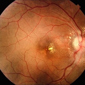 Infectious Neuroretinitis
May 26 2025 by César Adrián Gómez Valdivia, MD
Neuroretinitis found in a 38 year-oldmale patient with IV drugs abuse history. Findings were bilateral. The lipid-rich component of the exudate is able to penetrate into the outer plexiform layer, creating what is clinically seen as a macular star pattern.
Photographer: @eyemissu2
Imaging device: TOPCON TRC-50DX
Condition/keywords: neuroretinitis
-
 ROP / Disk Dragging
May 27 2025 by César Adrián Gómez Valdivia, MD
Macular dragging found in a 14 year-old female patient with ROP history. Findings were bilateral.
Photographer: @eyemissu2
Imaging device: California ICG OPTOS
Condition/keywords: Disk Dragging
-
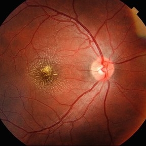 Macular Star
May 27 2025 by César Adrián Gómez Valdivia, MD
Macular Star found in a 31 year-old male patient with suspected Cat Scratch Disease. Typical intraocular presentations include neuroretinitis with optic nerve edema, macular star formation, and discrete white retinal or choroidal lesions. Findings were unilateral.
Photographer: @eyemissu2
Imaging device: TOPCON TRC-50DX
Condition/keywords: macular star
-
 Multiple evanescent White Dot Syndrome (MEWDS)
May 27 2025 by César Adrián Gómez Valdivia, MD
Fundus photograph of a 21 year-old female patient with suspected Multiple Evanescent White Dot Syndrome (MEWDS). The White Dot Syndromes produce yellow-white retinal lesions classically located at the retinal pigment epithelium or outer retina and are found primarily in young adults. Symptoms of MEWDS include unilateral blurred vision, visual field loss, photopsias, and floaters.
Photographer: @eyemissu2
Imaging device: TOPCON TRC-50DX
Condition/keywords: multiple evanescent white dot syndrome (MEWDS)
-
 Coats Disease
May 27 2025 by César Adrián Gómez Valdivia, MD
Fundus photograph of an 8 year-old male patient with Coats disease. Vascular leakage causes hard exudates which may be peripheral (near the vascular abnormalities) or midperipheral and central (at the macula). Findings were bilateral.
Photographer: @eyemissu2
Imaging device: California ICG OPTOS
Condition/keywords: Coats disease
-
 Coats Disease
May 27 2025 by César Adrián Gómez Valdivia, MD
Fluorescein Angiography on an 8 year-old male patient with Coats disease. Vascular leakage causes hard exudates which may be peripheral (near the vascular abnormalities) or midperipheral and central (at the macula. Findings were bilateral.
Photographer: @eyemissu2
Imaging device: California ICG OPTOS
Condition/keywords: Coats disease
-
 Franceschetti's Sign
Jun 5 2025 by César Adrián Gómez Valdivia, MD
Franceschetti's sign found in a 22 year-old female patient diagnosed with ocular toxoplasmosis. These bands typically link an old scar to the optic disc, indicative of previous inflammation. Findings were unilateral.
Photographer: @eyemissu2
Imaging device: TOPCON TRC-50DX
Condition/keywords: chorio, Franceschetti's Sign, toxoplasmosis
-
 Wagon-Wheel Lesion
Jun 5 2025 by César Adrián Gómez Valdivia, MD
Wagon-wheel lesion found in a 12 year-old male patient diagnosed with congenital toxoplasmosis. Findings were bilateral.
Photographer: @eyemissu2
Imaging device: TOPCON TRC-50DX
Condition/keywords: toxoplasmosis, Wagon-wheel lesion
-
 Wagon-Wheel Lesion
Jun 5 2025 by César Adrián Gómez Valdivia, MD
Wagon-wheel lesion found in a 12 year-old male patient diagnosed with congenital toxoplasmosis. Findings were bilateral.
Photographer: @eyemissu2
Imaging device: California ICG OPTOS
Condition/keywords: toxoplasmosis, Wagon-wheel lesion
-
 Wagon-Wheel Lesion
Jun 5 2025 by César Adrián Gómez Valdivia, MD
Wagon-wheel lesion found in a 12 YO male patient diagnosed with congenital toxoplasmosis. Findings were bilateral.
Photographer: @eyemissu2
Imaging device: TOPCON TRC-50DX
Condition/keywords: toxoplasmosis chorioretinitis, Wagon-wheel lesion
-
 Retinal Detachment
Jun 5 2025 by César Adrián Gómez Valdivia, MD
Fundus Photograph of a 19 year-old male patient with a RRD due to a Retinal Dialysis. Subretinal fluid and retinal folding can be appreciated.
Photographer: @eyemissu2
Imaging device: TOPCON TRC-50DX
Condition/keywords: retinal detachment
-
 Retinal Detachment
Jun 5 2025 by César Adrián Gómez Valdivia, MD
Fundus Photograph of a 19 year-old male patient with a RRD due to a Retinal Dialysis. Subretinal fluid and retinal folding can be appreciated.
Photographer: @eyemissu2
Imaging device: OPTOS
Condition/keywords: retinal detachment
-
 Macular Coloboma
Jun 5 2025 by César Adrián Gómez Valdivia, MD
Macular Coloboma found in a 28 year-old male patient, visual acuity was 20/60. Resulting due to fusion failure of the optic fissure, colobomas are commonly found in the infero-nasal quadrant. If the retina is involved, it is reduced to glial tissue with no underlying RPE or choroid. This appears as an area of whitening often with pigment deposition at the junction of the coloboma and normal retina. Findings were bilateral.
Photographer: @eyemissu2
Imaging device: TOPCON TRC-50DX
Condition/keywords: coloboma
-
 Idiopathic Multifocal Choroiditis
Jun 14 2025 by César Adrián Gómez Valdivia, MD
Fundus Autofluorescence of an 46 YO female patient diagnosed with Idiopathic Multifocal Choroiditis. Findings were bilateral.
Photographer: @eyemissu2
Imaging device: California ICG OPTOS
Condition/keywords: choroiditis
-
 Serpiginous Choroidopathy
Jun 14 2025 by César Adrián Gómez Valdivia, MD
Fundus Autofluorescence of a 29-year-old woman with Serpiginous Choroidopathy. Finings were bilateral.
Photographer: @eyemissu2
Imaging device: California ICG OPTOS
Condition/keywords: Serpiginous Choroidopathy
-
 Serpiginous Choroidopathy
Jun 14 2025 by César Adrián Gómez Valdivia, MD
Fundus Autofluorescence of a 29-year-old woman with Serpiginous Choroidopathy. Finings were bilateral.
Photographer: @eyemissu2
Imaging device: California ICG OPTOS
Condition/keywords: Serpiginous Choroidopathy
-
 CMV Retinitis
Jun 14 2025 by César Adrián Gómez Valdivia, MD
Fundus Photograph of a 28YO male patient diagnosed with Cytomegalovirus Retinitis. Findings were unilateral. Visual acuity was 20/25 on SC. CMV is the most common opportunistic ocular pathogen in advanced HIV infection, typically occurring when CD4+ T-cell counts fall below 50 cells/µL
Photographer: @eyemissu2
Imaging device: California ICG OPTOS
Condition/keywords: CMV retinitis
-
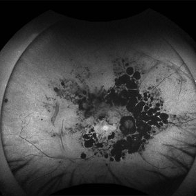
 Pigmented Paravenous Retinochoroidal Atrophy
Jun 18 2025 by César Adrián Gómez Valdivia, MD
Fundus Autofluorescence Image of a 42YO female patient diagnosed with Pigmented Paravenous Retinochoroidal Atrophy. Findings were bilateral. Image shows hypoautofluorescence in the affected areas due to overall loss of RPE cells and thus lower lipofuscin levels.
Photographer: @eyemissu2
Imaging device: California ICG OPTOS
Condition/keywords: pigmented paravenous chorioretinal atrophy (PPCRA)
-
 Birdshot Retinochoroidopathy
Jun 18 2025 by César Adrián Gómez Valdivia, MD
Fundus photograph of a 86 YO female patient diagnosed with Birdshot Retinochoroidopathy. Characteristically multifocal cream-colored or yellow-orange, oval or round lesions that emerge from around the optic nerve can be appreciated.
Photographer: @eyemissu2
Imaging device: California ICG OPTOS
Condition/keywords: Birdshot Retinochoroidopathy
-
 Birdshot Retinochoroidopathy
Jun 18 2025 by César Adrián Gómez Valdivia, MD
Fundus photograph of a 86 YO female patient diagnosed with Birdshot Retinochoroidopathy. Characteristically multifocal cream-colored or yellow-orange, oval or round lesions that emerge from around the optic nerve can be appreciated.
Photographer: @eyemissu2
Imaging device: TOPCON TRC-50DX
Condition/keywords: Birdshot Retinochoroidopathy
-
 Birdshot Retinochoroidopathy
Jun 18 2025 by César Adrián Gómez Valdivia, MD
Fundus Autofluorescence of a 86 YO female patient diagnosed with Birdshot Retinochoroidopathy.
Photographer: @eyemissu2
Imaging device: California ICG OPTOS
Condition/keywords: birdshot retinochoroidopathy
-
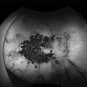
 Pigmented Paravenous Retinochoroidal Atrophy
Jun 18 2025 by César Adrián Gómez Valdivia, MD
Fundus photograph Image of a 42YO female patient diagnosed with Pigmented Paravenous Retinochoroidal Atrophy. Findings were bilateral. Image shows hypoautofluorescence in the affected areas due to overall loss of RPE cells and thus lower lipofuscin levels.
Photographer: @eyemissu2
Imaging device: California ICG OPTOS
Condition/keywords: Pigmented Paravenous Retinochoroidal Atrophy
-
 Pigmented Paravenous Retinochoroidal Atrophy
Jun 18 2025 by César Adrián Gómez Valdivia, MD
Fundus photograph of a 42YO female patient diagnosed with Pigmented Paravenous Retinochoroidal Atrophy. Findings were bilateral. Image shows hypoautofluorescence in the affected areas due to overall loss of RPE cells and thus lower lipofuscin levels.
Photographer: @eyemissu2
Imaging device: TOPCON TRC-50DX
Condition/keywords: Pigmented Paravenous Retinochoroidal Atrophy
-
 Serpiginous Choroidopathy
Jun 23 2025 by César Adrián Gómez Valdivia, MD
Fundus photograph of a 29 year-old female patient diagnosed with Serpiginous Choroidopathy. Finings were bilateral. The most common complication of SC is choroidal neovascularization affecting up to 35% of patients. Other reported complications are subretinal fibrosis, cystoid macular edema, branch vein occlusion, serous retinal detachment, optic disc neovascularization, and anterior uveitis.
Photographer: @eyemissu2
Imaging device: California ICG OPTOS
Condition/keywords: serpiginous choroiditis
-
 Serpiginous Choroidopathy
Jun 23 2025 by César Adrián Gómez Valdivia, MD
Fundus photograph of a 29 year-old female patient diagnosed with Serpiginous Choroidopathy. Finings were bilateral. The most common complication of SC is choroidal neovascularization affecting up to 35% of patients. Other reported complications are subretinal fibrosis, cystoid macular edema, branch vein occlusion, serous retinal detachment, optic disc neovascularization ,and anterior uveitis.
Photographer: @eyemissu2
Imaging device: TOPCON TRC-50DX
Condition/keywords: serpiginous choroiditis
-
 Cytomegalovirus Retinitis
Jun 23 2025 by César Adrián Gómez Valdivia, MD
Fundus Photograph of a 28 year-old male patient diagnosed with Cytomegalovirus Retinitis. Findings were unilateral. Visual acuity was 20/25 on SC. CMV is the most common opportunistic ocular pathogen in advanced HIV infection, typically occurring when CD4+ T-cell counts fall below 50 cells/µL
Photographer: @eyemissu2
Imaging device: California ICG OPTOS
Condition/keywords: Cytomegalovirus Retinitis
-
 Annular Tractional Retinal Detachment
Jul 5 2025 by César Adrián Gómez Valdivia, MD
Fundus photograph of an 66 YO female patient diagnosed with advanced proliferative diabetic retinopathy.
Photographer: @eyemissu2
Imaging device: TOPCON TRC-50DX
Condition/keywords: tractional retinal detachment
-
 Giant Persistent Macular Hole
Jul 5 2025 by César Adrián Gómez Valdivia, MD
Giant Persistent Macular Hole found in a 48YO male patient 1 year after vitrectomy.
Photographer: @eyemissu2
Imaging device: TOPCON TRC-50DX
Condition/keywords: macular hole
-
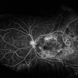
 Schlaegel Line
Jul 7 2025 by César Adrián Gómez Valdivia, MD
Fluorescein Angiography showing a Schlaegel Line in a patient with suspected PHOS
Photographer: @eyemissu2
Imaging device: California ICG OPTOS
Condition/keywords: Schlaegel Line
-
 Neuroretinitis
Jul 7 2025 by César Adrián Gómez Valdivia, MD
Neuroretinitis found in a 38 YO male patient with IV drugs abuse history. Findings were bilateral. The lipid-rich component of the exudate is able to penetrate into the outer plexiform layer, creating what is clinically seen as a macular star pattern.
Photographer: @eyemissu2
Imaging device: TOPCON TRC-50DX
Condition/keywords: neuroretinitis
-
 Scimitar Sign & Sunset Glow Fundus
Jul 7 2025 by César Adrián Gómez Valdivia, MD
Scimitar Sign & Sunset Glow Fundus found in a 38YO female patient diagnosed with Vogt-Koyanagi-Harada disease. Findings were bilateral.
Photographer: @eyemissu2
Imaging device: TOPCON TRC-50DX
Condition/keywords: Scimitar Sign
-
 Amelanotic Melanoma
Aug 12 2025 by César Adrián Gómez Valdivia, MD
This case highlights an amelanotic melanoma, an atypical presentation of a choroidal melanoma lacking the characteristic pigmentation. These lesions can easily be mistaken for choroidal hemangiomas, metastases, or inflammatory masses. Clinically, the lesion appears as a dome-shaped, yellowish subretinal mass, often associated with subretinal fluid, lipofuscin deposition, or retinal detachment. The absence of pigment can delay diagnosis, making multimodal imaging essential. Diagnostic tools: • B-scan ultrasound: low to medium internal reflectivity • OCT: overlying subretinal fluid and RPE elevation • FAF: orange pigment and RPE disruption • ICG/FA: variable, often hypofluorescent core Important: Prompt referral to ocular oncology is critical for management and prognosis.
Photographer: @eyemissu2
Imaging device: TOPCON TRC-50DX
Condition/keywords: amelanotic melanoma
-
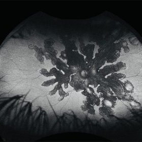
 Amelanotic Melanoma
Aug 12 2025 by César Adrián Gómez Valdivia, MD
This FAF image reveals a hypoautofluorescent mass with areas of dense hyperautofluorescent stippling—a classic pattern suggestive of an amelanotic choroidal melanoma. Amelanotic melanoma is a rare variant of uveal melanoma, accounting for only a minority of cases. Unlike pigmented melanomas, these lesions lack melanin, making them more challenging to detect on conventional color fundus imaging. FAF Characteristics: • Central hypoautofluorescence: due to loss or compression of the RPE • Peripheral hyperautofluorescent speckling: consistent with lipofuscin accumulation or RPE disruption • Often associated with subretinal fluid or orange pigment seen clinically Location: Juxtapapillary, with potential optic nerve involvement—a factor that complicates both diagnosis and
Photographer: @eyemissu2
Imaging device: California ICG OPTOS
Condition/keywords: amelanotic melanoma
A project from the American Society of Retina Specialists