Search results (0 results)
-
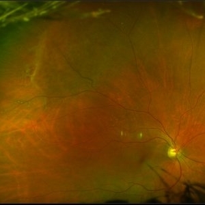
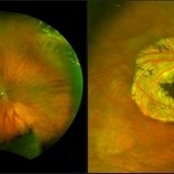 Operculated Hole and CHRPE
Jan 16 2018 by Carolyn Daley
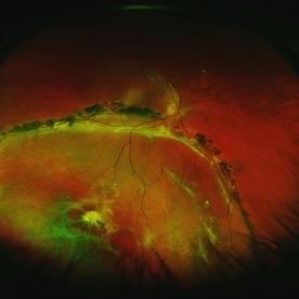
58-year-old woman with an operculated hole and CHRPE in the right eye. Patient is asymptomatic so no treatment was recommended at this time.
Photographer: Carolyn Daley
Imaging device: Optos ultra wide field image
Condition/keywords: congenital hypertrophy of the retinal pigment epithelium (CHRPE), operculated retinal hole, Optos, ultra-wide field imaging
-
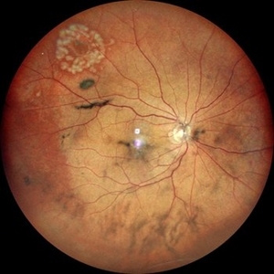
Peripheral Retinal Hole with OCT Co-localization
Sep 26 2023 by Bradley T. Smith, MD, FASRS
Peripheral asymptomatic atrophic retinal hole with OCT co localization demonstrating small cuff of sub retinal fluid. Near infrared imaging shows hyper reflectivity through hole.
Condition/keywords: atrophic hole, lattice degeneration, OCT
-
 Retinal Detachment and Lattice Degeneration
Mar 25 2025 by Korey Starkey
26 year-old patient presented at first visit with rhegmatogenous macula involving retinal detachment of the left eye. Underwent prompt surgical repair. Both eyes also present with lattice degeneration with atrophic holes.
Photographer: Korey Starkey
Condition/keywords: atrophic retinal hole, fundus photography, lattice degeneration, montage photo, Optos, OPTOS CALIFORNIA RGB, retinal detachment, retinal holes, rhegmatogenous retinal detachment, ultra-wide field imaging
-
 Retinal Detachment with Retinal Hole
Sep 30 2013 by Jason S. Calhoun
Patient in with complaints of floaters in the right eye. VA was 20/40 with no improvement. Fundus exam shows retinal detachment from 9-12 o'clock with hole at 10:30 posteriorly. Pneumatic retinopexy was performed with C3F8 Gas bubble and laser around the retinal tear in the right eye.
Photographer: Jason S. Calhoun, Department of Ophthalmology, Mayo Clinic Jacksonville, Florida
Imaging device: TOPCON TRC 50-EX
Condition/keywords: retinal hole
-
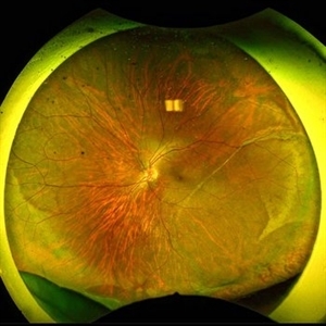
 Retinal Hole
Feb 11 2024 by Anjana Mirajkar, MS Ophthalmology
A color photo of RE of a 50 year old male showing lasered retinal hole superiorly with vitreous degeneration.
Photographer: Dr. Anjana Mirajkar -Retina Foundation, Ahmedabad
Imaging device: Mirante-Nidek
Condition/keywords: full thickness retinal hole
-
 The Bullet Ridden Retina
Feb 17 2024 by SHISHIR VERGHESE, MS, FVRS, FAICO (Retina)
Fundus image obtained of a case of lasered branch retinal vein occlusion (BRVO) with fibrovascular proliferation (FVP) where the laser marks have given way to multiple small retinal holes due to traction from the same.
Photographer: DIVYA SHAJI
Imaging device: NIDEK MIRANTE
Condition/keywords: BRVO, chronic retinal detachment
-
 Full-thickness Macular Hole
Aug 28 2012 by Sharon Fekrat, MD FACS FASRS
65 year old woman with a recurrent full thickness macular hole following previous 20 g pars plana vitrectomy in the right eye as well as an iatrogenic retinal hole in the papillomacular bundle. Both retinal defects are captured here in this Zeiss Stratus OCT image.
Photographer: Michael P. Kelly, FOPS Director, Duke Eye Labs, Duke University Eye Center, Durham, NC
Imaging device: Zeiss Cirrus
Condition/keywords: retinal break
-
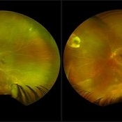
 Total retinal Detachment multiple holes
Sep 26 2022 by Denica Rodriguez
60 year old Male presented with two week old Macula off Retinal detachment with multiple tears.
Photographer: Denica Rodriguez
Imaging device: Optos California
Condition/keywords: color fundus photograph, color photo, macula-off, optos, pseudocolor, Retinal detachment, retinal holes, retinal tear, Retinal tear with detachment, superior arcade, superior field, superior retina, total retinal detachment
-
 Meridional Fold
Nov 9 2012 by Norman Byer
This is the same lesion as in the previous photograph. With the scleral indentation placed more posterior, we now can see that the fold ends over a small collection of subretinal fluid and that there is a very tiny retinal hole just below the posterior end of the retinal fold.
Condition/keywords: peripheral cystoid degeneration, retinal fold, retinal hole, scleral indentation, subretinal fluid
-
 Asymptomatic Superior Retinal Detachment
May 5 2016 by Steven J Ryder, MD
38-year-old African American female with moderate myopia (-4.50 Sph OU) and asymptomatic superior retinal detachment in the right eye. Montage fundus photography showing localized retinal detachment superiorly with single full-thickness retinal break at 12:00.
Photographer: Luis Bernhard, Miami VA Healthcare System
Imaging device: Topcon
Condition/keywords: asymptomatic, full thickness retinal hole, myopia, retinal break, retinal detachment with retinal defect
-
 Chronic Inferior Retinal Detachment
Mar 1 2017 by Philip J. Polkinghorne, MD
Color photograph of chronic retinal detachment with pigment demarcation line and atrophic holes visible. The vision was recorded at 20/20, and follow up is 3 years.
Photographer: Alex Fraser
Condition/keywords: atrophic retinal hole, demarcation line
-
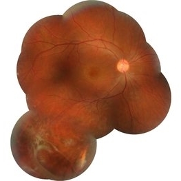 Juvenile Retinoschisis
Oct 10 2015 by Hamid Ahmadieh, MD
Merged color fundus photograph of the right eye of a 30-year-old man with juvenile retioschisis. Involvement of the retinal periphery with typical large inner layer retinal holes is visible.
Photographer: Shabnam Pooreh, Negah Eye Center, Tehran, Iran
Condition/keywords: color fundus photograph, inner layer holes, juvenile retinoschisis
-
 Lattice Lesion
Nov 9 2012 by Norman Byer
In this 47-year-old woman, this lattice lesion with a small hole in the right end has led to a subclinical retinal detachment which extends to the margin of the subtle yellowish zone almost at the upper edge of the photograph. This patient did not desire surgery, and the area of detachment has changed only a small amount in the past eight years. The risk of a clinical retinal detachment developing from lattice degeneration is less than 1 percent. In those cases where it does though, about 3 quarters are caused by a tractional tear and about one quarter are caused by an atrophic hole as in this case.
Condition/keywords: atrophic retinal hole, lattice degeneration, lattice lesion, retinal hole, yellowish zone
-
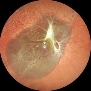
 Operculated Retinal Hole in Retinal Detachment
Oct 12 2012 by Jeffrey G. Gross, MD, FASRS
Operculated retinal hole in retinal detachment.
Condition/keywords: operculated retinal hole, retinal degeneration
-
 1 year Follow Up after Scleral Buckle Surgery in a Young Patient
May 18 2023 by Jesus Lozano, MD
25 year old man after Scleral Buckle Surgery + laser Retinopexy do to RRD macula off with ínfero temporal mid peripheral retinal holes in an area of lattice degeneration. Final VA 6/9.
Imaging device: Optos
Condition/keywords: scleral buckle
-
 Acute Retinal Detachment
Oct 12 2012 by Jeffrey G. Gross, MD, FASRS
Acute RD with operculated retinal hole.
Condition/keywords: acute retinal detachment, operculated retinal hole
-
 ARN (#1) Initial Photo
May 27 2019 by John S. King, MD
60-year-old African American female who had been treated for iridocyclitis for at least a week sent in for vitritis and a nasal fundus lesion. Complaints included redness, floaters, photophobia, and decreased vision. Husband had recent shingles. Acuity was 20/60-2 with IOP of 12, and small KP in Art's triangel, 1-2+ a/c cell, 2-3+ ant vit cell, diffuse arteriolar sheathing, multiple areas of retinal whitening in periphery and mid-periphery (see Photo #1). PCR of a/c was performed, and intravitreal GCV administered, and VACV 2g qid and ASA started.... PCR positive for HZV, pred taper was started two days after presentation as the infection had begun to stablize..... Five days from presentation the vision was 20/60, inflammation and areas of retinal whitening had improved (see Photo #2).... One week later acuity was 20/30, the a/c was quiet and KP resolved; ant vitreous cell decreased; and there was further improvement in retinal appearance without any signs of retinal holes or detachment; she is now on low dose maint VACV (see photo#3)
Photographer: Maysee Yang
Imaging device: Optos CA
Condition/keywords: acute retinal necrosis, Herpes zoster
-
 ARN (#2) Five Days Since Initial Visit
May 27 2019 by John S. King, MD
60-year-old African American female who had been treated for iridocyclitis for at least a week sent in for vitritis and a nasal fundus lesion. Complaints included redness, floaters, photophobia, and decreased vision. Husband had recent shingles. Acuity was 20/60-2 with IOP of 12, and small KP in Art's triangel, 1-2+ a/c cell, 2-3+ ant vit cell, diffuse arteriolar sheathing, multiple areas of retinal whitening in periphery and mid-periphery (see Photo #1). PCR of a/c was performed, and intravitreal GCV administered, and VACV 2g qid and ASA started.... PCR positive for HZV, pred taper was started two days after presentation as the infection had begun to stablize..... Five days from presentation the vision was 20/60, inflammation and areas of retinal whitening had improved (see Photo #2).... One week later acuity was 20/30, the a/c was quiet and KP resolved; ant vitreous cell decreased; and there was further improvement in retinal appearance without any signs of retinal holes or detachment; she is now on low dose maint VACV (see photo#3)
Photographer: Maysee Yang
Imaging device: Optos CA
Condition/keywords: acute retinal necrosis, Herpes zoster
-
 ARN (#3) This is comparison between the latest visit (left) and one week prior (which is the right photo, and same one as photo #2)
May 27 2019 by John S. King, MD
60-year-old African American female who had been treated for iridocyclitis for at least a week sent in for vitritis and a nasal fundus lesion. Complaints included redness, floaters, photophobia, and decreased vision. Husband had recent shingles. Acuity was 20/60-2 with IOP of 12, and small KP in Art's triangel, 1-2+ a/c cell, 2-3+ ant vit cell, diffuse arteriolar sheathing, multiple areas of retinal whitening in periphery and mid-periphery (see Photo #1). PCR of a/c was performed, and intravitreal GCV administered, and VACV 2g qid and ASA started.... PCR positive for HZV, pred taper was started two days after presentation as the infection had begun to stablize..... Five days from presentation the vision was 20/60, inflammation and areas of retinal whitening had improved (see Photo #2).... One week later acuity was 20/30, the a/c was quiet and KP resolved; ant vitreous cell decreased; and there was further improvement in retinal appearance without any signs of retinal holes or detachment; she is now on low dose maint VACV (see photo#3)
Photographer: Maysee Yang
Imaging device: Optos CA
Condition/keywords: acute retinal necrosis, Herpes zoster
-
 Asymptomatic Lesion
Nov 9 2012 by Norman Byer
This is the same lesion as seen in the previous slide pair. Here the scleral indentation is carried more posterior revealing a tiny, round, full thickness retinal hole. This is not a tear produced by traction even though vitreous is always attached to these flaps. You will note that the hole is round and is separated by a slight distance from the flap itself. It is probably the result of long continued atrophy and devitalization of the retina. A posterior vitreous was not detached. This lesion has not changed its appearance for more than a year of observation, but the age of the hole is actually unknown.
Condition/keywords: asymptomatic, atrophy, full thickness retinal hole, posterior scleral indentation, retinal hole, round hole
-
 Asymptomatic Superior Retinal Detachment
May 5 2016 by Steven J Ryder, MD
38-year-old African American female with moderate myopia (-4.50 Sph OU) and asymptomatic superior retinal detachment in the right eye. Zeiss Cirrus OCT capturing full-thickness retinal break at 12:00 and temporal vitreoretinal traction.
Photographer: Luis Bernhard, Miami VA Healthcare System
Imaging device: Zeiss Cirrus
Condition/keywords: asymptomatic, full thickness retinal hole, retinal break, retinal detachment with retinal defect
-
 Asymptomatic Superior Retinal Detachment
May 5 2016 by Steven J Ryder, MD
38-year-old African American female with moderate myopia (-4.50 Sph OU) and asymptomatic superior retinal detachment in the right eye. Zeiss OCT capturing vertical raster scans through border of retinal detachment.
Photographer: Luis Bernhard, Miami VA Healthcare System
Imaging device: Cirrus
Condition/keywords: asymptomatic, full thickness retinal hole, retinal detachment with retinal defect
-
 Atrophic Holes in Lattice Lesion
Nov 9 2012 by Norman Byer
In this 26-year-old woman, these two atrophic holes in a lattice lesion led to a clinical retinal detachment which was operated on successfully. In retinal detachments of this type resulting from non tractional atrophic holes, it has been found that 50% occur before the age of 30 years.
Condition/keywords: atrophic retinal hole, lattice lesion
-
 Barrage Laser
Nov 5 2024 by Dr Bilal Mir
Freshly barrage lasered fundus picture of a young myope.
Photographer: Dr BILAL AHMED MIR, Mbbs Ms ophthalmology
Condition/keywords: retinal hole
-
 Bullous Retinoschisis with Outer Retinal Holes
Jun 15 2020 by Olivia Rainey
Ultra-widefield pseudocolor fundus photograph of a 56-year-old female with bullous retinoschisis with outer retinal holes affecting her right eye. The physician noted superotemporal retinoschisis in her monoculcar functioning eye. There was no demarcation line and no inner or outer layer breaks at her first appointment in February of 2020. On 6/15/20 she had a new onset outer holes and SRF tracking inferiorly. The physician recommended observation, however if this continues to progress we have discussed indications for barrier laser.
Photographer: Olivia Rainey, OCT-C, COA
Imaging device: Optos California
Condition/keywords: bullous retinoschisis, Optos, outer layer breaks, outer layer hole, pseudocolor, subretinal fluid, superior retina, ultra-wide field imaging
 Loading…