Search results (7 results)
-
 Commotio retinae
Apr 29 2022 by Otakar Dušek, M.D. Ph.D.
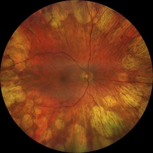
Color fundus photograph of a 24-year-old woman who was hit by a volleyball in her right eye. This caused whitening of the lower peripheral retina (Berlin's edema) i.e. commotio retinae.
Photographer: Otakar Dušek, Charles University, Prague
Imaging device: Zeiss Clarus
Condition/keywords: Berlin's edema, blunt trauma, commotio retinae
-
 Relentless Placoid Chorioretinitis
Jan 22 2021 by Renata Garcia Franco, Md
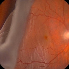
20-year-old male with reduction of vision in both eyes, scotoma and metamorphopsia. Widespread multiple chorioretinal lesions with RPE hyperplasia, which appear from posterior pole to peripheral retina.
Photographer: Fatima Hernandez, Instituto de la Retina del Bajio SC
Imaging device: Zeiss
Condition/keywords: chorioretinitis
-
 Didanosine Toxicity
Jan 27 2020 by Nimesh A. Patel, MD, FASRS
Patient with history of HIV treated with didanosine. Developed gyrate like peripheral retinal atrophy with central sparing. Vision is 20/25
Imaging device: Clarus
Condition/keywords: AIDS, didanosine, HIV
-
 Retinal Detachment with Giant Retinal Tear and Macular Hole
Jan 6 2020 by MATTEO FORLINI, MD
A 61-year-old-male patient presented with sudden diminution of vision in the right eye due to retinal detachment with giant retinal tear and macular hole. Best corrected visual acuity (BCVA) at presentation was 20/200. A 23 G vitrectomy was performed. The edges of the tear were unrolled and complete retinal re-attachment under PFCL was achieved. A 360 degree intraoperative endolaser was performed on the peripheral retina as well as around the edges of the tears. PFCL was exchanged with silicone oil 5000cs as final tamponade. At six-months follow-up retina was attached and macular hole was repaired. Best-corrected visual acuity is 20/125 at present.
Photographer: Matteo Forlini MD, San Marino Hospital, Republic of San Marino
Condition/keywords: full thickness macular hole, giant retinal tear, silicone oil
-
 Multiple Myeloma with Cytomegalovirus Retinitis
Apr 5 2018 by Kim Barrett
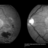
Ultra-wide field fluorescein angiogram of a 77-year-old male with multiple myeloma. Patient's angiogram presented significant peripheral retinal ischemia and cystoid macular edema. Patient tested positive for polymerase chain reaction, confirming cytomegalovirus retinitis. Patient is being treated with intravitreal ganciclovir and his current vision is 20/200.
Photographer: Kim Barrett, COA
Imaging device: Optos
Condition/keywords: cystoid macular edema (CME), fluorescein angiogram (FA), fluorescein leakage, intravitreal ganciclovir, myeloma, peripheral ischemia, positive polymerase chain reaction (PCR), ultra-wide field imaging
-
 Neovascular ARMD With Subretinal Hemorrhage, Red-Free Photos - Stereo
Nov 26 2014 by James B. Soque, CRA, OCT-C, COA, FOPS
Stereo FC, RF and FA of a 77-year-old white female with visual acuity CC 20/200-3, with left eye neovascular ARMD, drusen, and subretinal hemorrhage with hard exudates temporally. Peripheral retina reveals cobblestone degeneration.
Photographer: James Soque, CRA, COA, Island Retina, Shirley, NY
Imaging device: Topcon TRC 50 EX, with MERGE software and OIS 5 MP digital Camera
Condition/keywords: neovascular age-related macular degeneration (AMD), red-free, stereo pair
-
 ARMD With Geographic Atrophy, Peripheral Degeneration
Dec 6 2013 by James B. Soque, CRA, OCT-C, COA, FOPS
92-year-old white female with exudative macular degeneration, geographic atrophy, and peripheral retinal degeneration.
Photographer: James Soque, CRA COA, Island Retina, Shirley, New York
Imaging device: Topcon TRC 50DX with OIS 10.6.45
Condition/keywords: fundus photograph, geographic atrophy
 Loading…