Search results (23 results)
-
 Von Hippel-Lindau Syndrome
Jan 7 2025 by Jordyn Beckman
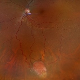
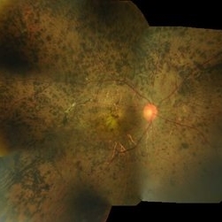
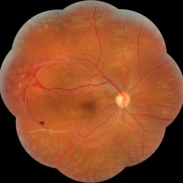
Fundus photograph of an 37 year old female presents with reddish vascular lesion with feeder vessels for possible Von Hippel-Lindau Syndrome.
Photographer: Jordyn Beckman
Imaging device: California Optos
Condition/keywords: color fundus photograph, feeder vessel, genetic disorder, pre-cryotherapy
-
 Retinal detachment
Apr 12 2023 by Ahmed Abbas Hashmi, OD
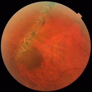
Color fundus photograph of the left eye of a 30-year-old man with asymptomatic inferior retinal detachment with pigmented demarcation line. Macula and Disc healthy.
Photographer: Ahmed Abbas Hashmi
Imaging device: Topcon TRC-NW8F
Condition/keywords: Pigmentary demarcation line, Retinal Detachment
-
 Subretinal Hemorrhage
Feb 28 2023 by Akansha Sharma
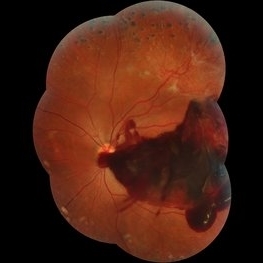
Color fundus photograph of an 84-year old male with subretinal hemorrhage associated with areas of scarring.
Photographer: Dr. Urmil Shah, Dr. Denish Patel, Dr. Akansha Sharma, Bharati Eye Hospital, Ahmedabad, Gujarat
Condition/keywords: choroidal neovascularization (CNV), subretinal hemorrhage
-
 Rhegmatogenous Retinal Detachment
Feb 26 2023 by Aditya S Kelkar, MS, FRCS, FASRS,FRCOphth
Color fundus photograph of left eye showing rhegmatogenous retinal detachment.
Photographer: Dr. Sahil Wagh, National Institute of Ophthalmology, Pune, India.
Imaging device: Zeiss Clarus 500
Condition/keywords: Retinal Detachment, rhegmatogenous retinal detachment
-
 Choroidal Melanoma with Exudative Retinal Detachment
Mar 2 2023 by Aditya S Kelkar, MS, FRCS, FASRS,FRCOphth
Color fundus photograph of the left eye of a 45 year old male showing choroidal melanoma with exudative retinal detachment.
Photographer: Dr. Pranali Surawase, National Institute of Ophthalmology, Pune, India.
Imaging device: Zeiss Clarus 500
Condition/keywords: choroidal mass, exudative retinal detachment, Retinal detachment
-
 Retinoblastoma
Nov 6 2022 by Akansha Sharma
Wide-field color fundus photograph of a 2-month old female with retinoblastoma.
Photographer: Dr. Akansha Sharma-Retina Foundation, Ahmedabad
Condition/keywords: RB gene mutation, retinoblastoma
-
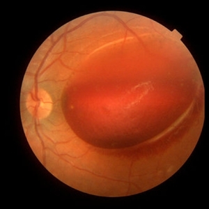
 Total retinal Detachment multiple holes
Sep 26 2022 by Denica Rodriguez
60 year old Male presented with two week old Macula off Retinal detachment with multiple tears.
Photographer: Denica Rodriguez
Imaging device: Optos California
Condition/keywords: color fundus photograph, color photo, macula-off, optos, pseudocolor, Retinal detachment, retinal holes, retinal tear, Retinal tear with detachment, superior arcade, superior field, superior retina, total retinal detachment
-
 Commotio retinae
Apr 29 2022 by Otakar Dušek, M.D. Ph.D.
Color fundus photograph of a 24-year-old woman who was hit by a volleyball in her right eye. This caused whitening of the lower peripheral retina (Berlin's edema) i.e. commotio retinae.
Photographer: Otakar Dušek, Charles University, Prague
Imaging device: Zeiss Clarus
Condition/keywords: Berlin's edema, blunt trauma, commotio retinae
-
 Traumatic Retinal Tear
Dec 5 2021 by Aditya S Kelkar, MS, FRCS, FASRS,FRCOphth
Color fundus photograph of a 34-year old man's left eye, 2 hours after a tennis ball injury, showing commotio retinae with Berlin's edema and cherry red spot in the fovea along with linear retinal tears in the temporal equatorial zone.
Photographer: Dr Sukanya Mondal. National Institute of Ophthalmology, Pune. India.
Imaging device: Zeiss Clarus 500
Condition/keywords: Berlin's edema, cherry red spot, commotio retinae, retinal tear
-
 Morning Glory Disc Anomaly
Nov 11 2020 by Yoshihiro Yonekawa, MD, FASRS
Color fundus photograph of a young boy with morning glory disc anomaly. Notice the concavity surrounding the enlarged disc, radial vasculature, and nasally dragged macula. MRI was negative for moyamoya disease, a known association.
Photographer: Alicia Thresher, Mid Atlantic Retina
Imaging device: Topcon
Condition/keywords: disc coloboma, Morning Glory Syndrome, pediatric retina
-
 Vascular Sheathing
Dec 19 2019 by Lauren Schuler
Ultra-wide field pseudocolor fundus photograph of a 68-year-old female with vascular sheathing affecting her right eye. This was noted on initial exam on 1/15/16, at patient's first appointment. This remains unchanged and patient is asymptomatic at this time.
Photographer: Lauren Schuler
Imaging device: Optos California
Condition/keywords: fundus photograph, ghost vessels, pseudocolor, vascular sheathing of retina
-
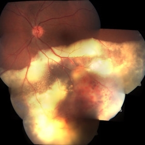
 Retinal Detachment with PVR (s/ SPR, PPV, MPV, 360 Retinectomy, PFO, PI, FAx, SO)
Aug 22 2019 by Merrick Avila
Ultra-wide field pseudocolor fundus photograph of a 64-year-old female with a treated retinal detachment with proliferative vitreoretinopathy. Patient has a history of complex retinal detachments that have been treated multiple times. On exam 8-22-19, there were large macular holes with LP vision. There was a long discussion about guarded nature of her condition and goals or trial for repair including globe sparing prevention of phthisis.
Photographer: Merrick Avila
Imaging device: Optos
Condition/keywords: diabetic retinopathy, hemorrhage, Optos, proliferative vitreoretinopathy (PVR), retinectomy, silicone oil
-
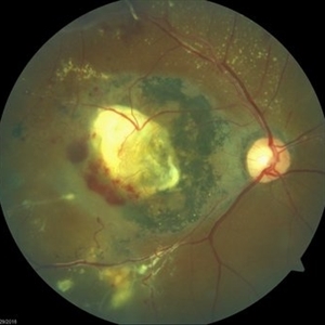 Coats' Disease Color Fundus Photograph
Apr 19 2019 by Ahmad B. Tarabishy, MD
57-year-old man referred for macular hemorrhage. Examination reveals a large macular scar along with vascular sheathing, vascular irregularity, and extensive exudation. Fluorescein angiogram revealed findings of lightbulb vascular aneurysms and capillary non-perfusion.
Imaging device: Zeiss Cirrus Photo
Condition/keywords: Coats' disease
-
 Coats' Disease
Apr 27 2018 by Brenda Fallas
3-year-old boy with unilateral Coats' Disease fundus photo.
Photographer: Brenda Fallas, Bascom Palmer Eye Institute, Miami, FL
Imaging device: Retcam III 130 degree lens
Condition/keywords: Coats' disease, color fundus photograph, retinal telangiectasia
-
 Coat's Disease
Nov 17 2016 by Amir Manor, MD
Fundus photograph of a 25-year-old man with Coat's disease.
Photographer: Galit Yair-Pur
Condition/keywords: Coats' disease, color fundus photograph
-
 Retinitis Pigmentosa
Oct 7 2015 by Avris Romario Diparaja Siahaan
Fundus photograph of a 29-year-old-man with retinitis pigmentosa in both eyes.
Photographer: Yohanes Harry Purwanto, Klinik Mata Nusantara
Imaging device: Topcon TRC 50DX IA
Condition/keywords: color fundus photograph, retinitis pigmentosa
-
 Lattice Degeneration and Choroidal Nevus
Oct 10 2015 by Hamid Ahmadieh, MD
Color fundus photograph of the right eye of a 46-year-old woman with a typical lattice degeneration and an adjacent choroidal nevus.
Photographer: Solmaz Shahmohammad, Negah Eye Center, Tehran, Iran
Condition/keywords: choroidal nevus, color fundus photograph, lattice degeneration
-
 Subhyaloid Hemorrhage
Jul 7 2015 by Hamid Ahmadieh, MD
Color fundus photograph of the left eye of a 25-year-old woman with severe subhyaloid hemorrhage due to an advanced vasoproliferative vitreoretinopathy secondary to a severe idiopathic occlusive retinal vasculitis.
Photographer: Soulmaz Shahmohammad, Negah Eye Center, Tehran, Iran
Condition/keywords: color fundus photograph, occlusive vasculitis, subhyaloid hemorrhage
-
 X-Linked Retinoschisis
Jan 31 2015 by Hamid Ahmadieh, MD
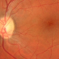
Color fundus photograph of the left eye of a 35-year-old man with x-linked retinoschisis. Please notice the foveal schisis.
Photographer: Shabnam Poureh, Negah Eye Center, Tehran, Iran
Condition/keywords: color fundus photograph, foveal schisis, x-linked retinoschisis (XLRS)
-
 Severe Neovascularization Secondary to Idiopathic Occlusive Retinal Vasculitis
Jan 17 2015 by Hamid Ahmadieh, MD
Wide- field color fundus photograph of the right eye of a 28-year-old woman with severe retinal neovascularization secondary to idiopatic occlusive retinal vasculitis.
Photographer: Solmaz Shahmohammad, Negah Eye Center, Tehran
Condition/keywords: color fundus photograph, neovascularization (NV), retinal vasculitis
-
 Subhyaloid Hemorrhage
Jun 29 2014 by Woohyok Chang, MD, PhD
Color fundus photograph of the left eye of a 65-year-old man with organizing subhyaloid hemorrhage secondary to branch retinal vein occlusion.
Photographer: Mi-Young Choi, Yeungnam University, Daegu, South Korea
Imaging device: Cannon
Condition/keywords: branch retinal vein occlusion (BRVO), subhyaloid hemorrhage
-
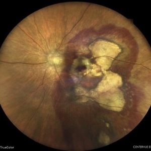
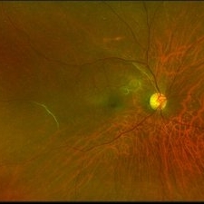
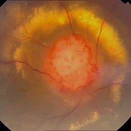 Color Photo of Optic Disc Capillary Hemangioblastoma
Mar 18 2014 by Arwa Azmeh, MD, PhD
Color fundus photograph of an 48-year-old male who complained of decreased visual acuity in his right eye over the last few months. Systemically the patient was healthy. His VA was OD Cf 3m, OS 20/20. Anterior segments were WNL in OU. IOP was WNL in OU. Fundus exam OD revealed unpigmented mass over the optic disc with retinal venous tortuosity at its edges with a ring of thick HYE surrounding it and shallow RD in this area extending to the foveal area. Several few small retinal hemorrhages were seen in the far retinal periphery which were explained to be caused by venous stasis due the optic disc tumor.
Condition/keywords: color photo, optic disc, retinal hemangioblastoma
-
 Primary Subhyaloid Hemorrhage Due to Valsalva Retinopathy
Nov 13 2013 by Hamid Ahmadieh, MD
Color fundus photograph of the left eye of a 25-year-old man with sudden drop of vision due to subhyaloid hemorrhage secondary to Valsalva retinopathy.
Photographer: Soodabeh Fooladin , Negah Eye Center, Tehran
Imaging device: TOPCON OCT
Condition/keywords: subhyaloid hemorrhage, valsalva retinopathy
 Loading…