Search results (70 results)
-
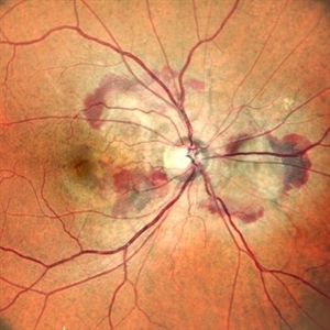
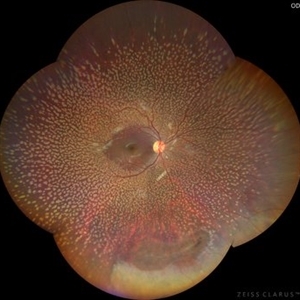
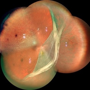
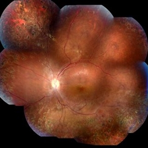
 A Large Break at the Posterior Pole With RD With PVR (S/p Old Blunt Trauma)
Jan 16 2025 by Anand Temkar
Right eye widefield fundus color photo of a 10 year old kid who noticed diminution of vision in right eye since a month. We can see the large break at the posterior pole with rolled up margins associated with retinal detachment and PVR changes.
Photographer: Dr.Anand Temkar- Retina Foundation, Ahmedabad
Imaging device: Mirante
Condition/keywords: posterior pole break, proliferative vitreoretinopathy (PVR), Retinal Detachment
-
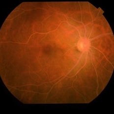
 Right Eye Color Photo With Hemorrhages in Case of CNVM With Angioid Streaks
Nov 29 2024 by Anand Temkar
A 45 year old male came with chief complaint of blurring vision in right eyes since past 4 days. His vision is 6/12 in right eye and 6/9 in left eye. His vision was 14 mmHg in right eye and 16 mmHg in left eye. He was diagnosed with Angioid Streaks in both eyes about a year ago, then he developed choroidal neovascularization in his left eye 8 months ago, for which he received AntiVEGF injections x 3. Left eye is a stable eye now. Patient presented with right eye choroidal neovascularization in a case of Angioid Streaks on recent follow up. We have advised him right eye AntiVEGF injections x 3. In this image, the right eye color photo shows bleed from CNVM in case of angioid streaks.
Photographer: Dr.Anand Temkar- Retina Foundation, Ahmedabad
Imaging device: Mirante
Condition/keywords: Angioid Streaks, choroidal neovascular membrane (CNVM)
-
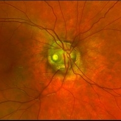
 MIDD (Maternally Inherited Diabetes and Deafness) - Right AF
Nov 30 2024 by John S. King, MD
Both right and left eyes have symmetrical ring of mottled hypo/hyper AF around the fovea and disc. The HyperAF areas correspond to RPE deposits on OCT as well as areas of blockage on FA, and drusenoid deposits seen on fundus photos. Disc drusen in right eye present as HyperAF spot 57 yo WF referred for AMD vs Pattern Dystrophy that was diagnosed 10 years ago. Reported some slow progressive vision loss in both eyes for distance and near. Denies nyctalopia or hemeralopia. Background medical history includes HTN, CVD, and DM. No family history of eye problems. Denied pentosan use. Anterior segment showed moderate cataracts (OD>OS). Posterior segment exam showed macular changes and mild NPDR. The macular appearance showed a symmetrical, paramacular ring of fleck-like drusenoid material with some faint focal areas of RPE hyperplasia. Fundus Photos, AF, OCT were performed as well as a gene test. Further questioning showed revealed that her mother and maternal grandmother had both diabetes mellitus and sensorineural hearing loss. The patient developed diabetes in her teens, and some high frequency hearing loss in her early twenties. She had not had a previous genetic test or diagnosis of MIDD. Gene testing is pending for the mitochondrial component. Invitae's retinal panel, which does not include mitochondrial disorders, only showed a variant of uncertain significance, HMCN1. I discussed this case with Dr. Freund, and it is similar to a the case report : Inoue M, Kiss S, Freund KB. MACULAR PIGMENT RINGS AS THE PRESENTING FINDING OF MITOCHONDRIAL MYOPATHY, ENCEPHALOPATHY, LACTIC ACIDOSIS, AND STROKELIKE EPISODES. Retin Cases Brief Rep. 2015 Fall;9(4):260-4. doi: 10.1097/ICB.0000000000000182. PMID: 26200388.
Photographer: Grace Melton and Carley Gunn
Imaging device: Clarus
Condition/keywords: Macular Dystrophy, Maternally Inherited Diabetes and Deafness, MIDD, Mitochondrial Disorder
-
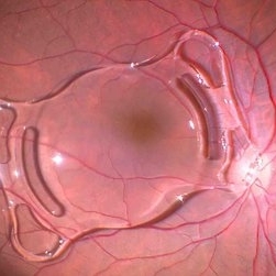 Dislocated IOL
Sep 28 2024 by Anjana Mirajkar, MS Ophthalmology
An intra operative image of right eye showing dislocated IOL sitting on the posterior pole.
Photographer: Dr. Anjana Mirajkar -Retina Foundation, Ahmedabad
Condition/keywords: dislocated IOL
-
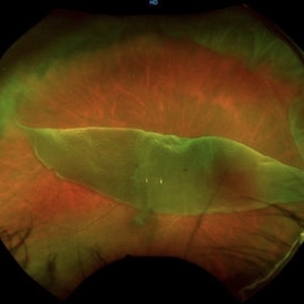
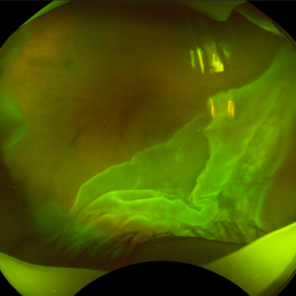
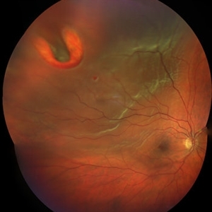
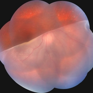 Giant Retinal Tear
Jul 15 2024 by Arthi Mohankumar , MS,MRCS ED, FICO,FAICO
Fundus montage of a 15 year old boy with Marfans syndrome who presented with defective vision in the right eye.
Photographer: Arthi Mohankumar
Condition/keywords: giant retinal tear, Retinal detachment
-
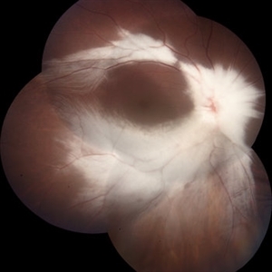
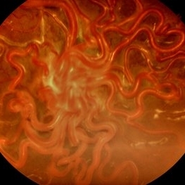 Wyburn-Mason Syndrome (Racemose Angioma)
Mar 23 2024 by Pushkar Mahale
Fundus photograph of a 10 year old child presenting with no perception of light in right eye. Fundus examination revealed dilated and tortuous retinal vessels suggestive of Racemose Hemangioma.
Photographer: Dr Pushkar Mahale
Condition/keywords: racemose hemangioma, Wyburn -Mason Syndrome
-
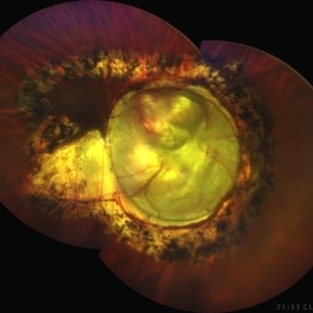
 Dislocated Crystalline Lens
Mar 19 2024 by Annaka Gooding
Ultra Wide field fundus photography of a 70 year old male who presented to clinic with a sudden increase of vision due to dropped crystalline lens secondary to severely dense cataract. Patient reported seeing a full black circle in his inferior visual field. Patient's visual acuity at time of visit was 20/100 with a +5.00 diopter lens. The physician recommended surgical intervention, and discussed surgery for PPV/PPL/IOL implantation with an ACIOL.
Photographer: Annaka Gooding, CPO
Imaging device: Optos California RGB
Condition/keywords: dislocated crystalline lens, fundus photography, inferior retina, OPTOS CALIFORNIA RGB, Right Eye, Ultra-wide field retinal imaging
-
 Dislocated Lens, Posterior OD
Jan 26 2024 by Corey Grant
OPTOS California photo presents a 71 year old male patient with a dislocated lens, posterior in the right eye. Presented on 1/26/24 with posteriorly dislocated SN60WF with a Soemmerring ring. Associated retinal hemorrhage within retinoschisis as well. This will result in a PPV/IOL exchange/SFIOL/STK for the right eye.
Photographer: Corey Grant, Ophthalmic Imager, Retina Specialist of Michigan
Imaging device: OPTOS California
Condition/keywords: color photo, IOL, OD, Optos, OPTOS CALIFORNIA, pars plana vitrectomy (PPV), retina
-
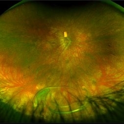
 Benign Familial Fleck Retina
Dec 21 2023 by Vishal Agrawal, MD, FRCS,FACS,FASRS
10-year male with high myopia on examination revealed diffuse flecks distributed all over fundus in both eyes sparing macula. Inferior lattice with WWOP areas were also noted in right eye.
Photographer: Dr Ayushi
Imaging device: Clarus 700
Condition/keywords: fleck retinopathy, myopia
-
 Giant Retinal Tear
Oct 24 2023 by Ivan J. Suner, MD, MBA
Fundus photograph of 49-year-old man with a giant retinal tear in the right eye.
Photographer: Norelys Alexander Jimenez, Retina Associates of Florida, Tampa, FL
Imaging device: Optos California
Condition/keywords: GIANT RETINAL TEAR
-
 Myelinated Nerve Fibre (MNF)
Jun 17 2023 by Harsh Vardhan Singh, MS
Fundus photograph of 32-year-old male having good best corrected visual acuity in both eyes with right eye having high myopia & MNF as incidental finding
Photographer: Dr Harsh Vardhan Singh, Assistant Professor, AIIMS, Guwahati
Condition/keywords: medullated nerve fibers, MNF, myelinated nerve fiber layer, myelinated nerve fibers, Nerve fiber layer arrangements, NFL
-
 Solitary large Congenital Hypertrophy of Retinal Pigment Epithelium (CHRPE)
Jul 1 2023 by Aditya S Kelkar, MS, FRCS, FASRS,FRCOphth
Right eye fundus photograph of a 42 year old asymptomatic male demonstrating a superotemporal solitary large Congenital Hypertrophy of Retinal Pigment Epithelium (CHRPE) lesion.
Photographer: Optom Komal Jangam
Imaging device: OPTOS DAYTONA
Condition/keywords: CHRPE
-
 Dislocated Lens
Apr 26 2023 by Chloe Hanifan
Ultra wide field fundus photograph of a 41-year-old male with a dislocated lens affecting his right eye. IOL noted inferior vitreous base and vitrectomy surgery for removal of IOL was recommended. Patient has history of retinitis pigmentosa as well. Patient's vision at the time of presentation was counting fingers at 2 feet.
Photographer: Chloe Hanifan
Imaging device: Optos California
Condition/keywords: dislocated lens, fundus photography, Optos, pseudocolor, retinitis pigmentosa, ULTRA WIDE FIELD
-
 Kissing Serous Choroidal Detachment
Mar 8 2023 by Annaka Gooding
Ultra-widefield fundus photograph of a 73 year old male with a Kissing serous choroidal detachment affecting his right eye. Patient presented at the office following a XEN implant and his vision was sc20/100 PH20/50+1. The physician recommended to start Prednisone treatment.
Photographer: Annaka Gooding
Imaging device: Optos California
Condition/keywords: fundus photography, Kissing Serous Choroidal Detachment, Optos, Right Eye, ultra-wide field imaging
-
 Giant Retinal Tear
Apr 21 2022 by Vaidehi Sathaye
Fundus photograph of a 16 year old male with a Rhegmatogenous Retinal Detachment secondary to a Giant Retinal Tear in the right eye.
Photographer: Dr. Vaidehi Sathaye, Retina Foundation
Condition/keywords: giant retinal tear
-
 Commotio retinae
Apr 29 2022 by Otakar Dušek, M.D. Ph.D.
Color fundus photograph of a 24-year-old woman who was hit by a volleyball in her right eye. This caused whitening of the lower peripheral retina (Berlin's edema) i.e. commotio retinae.
Photographer: Otakar Dušek, Charles University, Prague
Imaging device: Zeiss Clarus
Condition/keywords: Berlin's edema, blunt trauma, commotio retinae
-
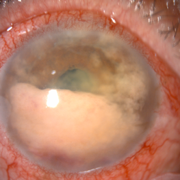 Secondary intraocular lymphoma
Apr 11 2022 by Aniruddha K Agarwal, MD
A 65-year-old male underlying nasopharyngeal non-Hodgkin’s lymphoma presented with pseudo-hypopyon and infiltration of iris from tumor on his right eye. Aqueous tap showed atypical lymphocytes.
Photographer: Kessara Pathanapitoon MD, PhD Department of Ophthalmology, Faculty of Medicine Chiang Mai University, Chiang Mai, Thailand
Condition/keywords: lymphoma, masquerade syndrome, secondary iridocyclitis (noninfectious)
-
 Thioridazine-toxicity
Apr 30 2022 by Niloofar Piri, MD
61 yo male with PMH of longstanding schizophrenia since 20s with secondary intellectual disability presented with decreased vision following a recent stroke. He was found to have bilateral chorio-retinal atrophy involving posterior pole with scalloped edges and coin shaped atrophic area at margins extending into mid-periphery, diagnosis most concerning for intermediate stage thioridazine toxicity given the history. Mother could find handwritten prescriptions from 1990s when he was on Thioridazine 800 mg daily for unknown period of time. Patient had better vision in the left eye which was affected by recent stroke and prompted him to seek medical care. Fundus photograph of the right eye is demonstrated here.
Photographer: Jacob Grodsky, MD
Condition/keywords: drug toxicity, thioridazine toxicity, toxic retinopathy
-
 Chorioretinal coloboma involving disc and macula
Mar 21 2022 by T. P . VIGNESH, MBBS,MS
Fundus photo of Right eye of a 55 year male patient revealing a fovea sparing well barraged chorioretinal coloboma involving the disc and the macula .
Photographer: Bharathi Singaravel
Imaging device: Zeiss Clarus
Condition/keywords: chorioretinal coloboma, coloboma of optic disc
-
 Total RRD with GRT with Choroidal Detachment
Apr 11 2021 by Dinesh Rungta, MBBS, DNB
Optos image of a 55-year-old male with history of sudden decrease of vision in right eye associated with flashes showing total RRD with GRT and CD.
Photographer: Dr Dinesh Rungta, Giridhar Eye Institute, Kerala, India.
Imaging device: Optos UWF Daytona Plus
Condition/keywords: detachment, giant retinal tear, re-attached retinal detachment (RRD)
-
 Retinoschisis
Mar 28 2021 by JEFFERSON R SOUSA, Tecg.º (Biomedical Systems Technology)
A 14-year-old male patient was admitted for visual evaluation. Visual acuity s/c in the right eye and 20/80 in the left eye. According to family members, he reported low vision since childhood. He had already undergone treatment with photocoagulation in another service to which he had a diagnostic hypothesis of Coats' disease. Laboratory tests were requested (HIV, TOXO, TOXOCARIASIS, ECA, VDRL, PPD). In the evaluation it was observed important exudation in the posterior pole, some vascular irregularities in the right eye. In the left eye, there is retinoschisis affecting the entire posterior pole and the region nasal to the optic disc, macula with a characteristic aspect of a cartwheel. Well exemplified by OCT-A (Structrure Deep: IPL - 25, OPL - 25).
Photographer: JEFFERSON R SOUSA - Study Center and Ophthalmological Research Dr. Andre M V Gomes, Institute Dr. Suel Abujamra São Paulo-Brazil
Imaging device: Topcon TRC-50 DX, Imaginet 4.0, angle de 50 graus. Flash 50w-s
Condition/keywords: Coats' disease, retinoschisis
-
 Lipemia Retinalis
Feb 10 2021 by KRISHNENDU NANDI, MS
Right eye fundus picture of 35-year-old young male came for routine check up, showed lipemia retinalis. Blood triglyceride level was 535 mg/dL.
Photographer: Krishnendu Nandi, Netralayam Eye Care Centre, Kolkata, India
Condition/keywords: lipemia retinalis
-
 Optic Nerve Pit Right Eye
Feb 15 2021 by Kim Barrett
A 14-year-old male presented with vision loss and VF defect. Patient was treated for presumed amblyopia with patching since age 4. He has had neurologic care for post traumatic skull fracture and brain bleed in 2012. IOP's WNL. OD is without retinoschisis or subretinal fluid. Patient is at risk of serous detachment. Current VA OD 20/200+1 PH 20/80.
Photographer: Kim Barrett C.O.A. Retina Specialist of Michigan, Grand Rapids, MI
Imaging device: Optos California
Condition/keywords: amblyopia, hemifield, Humphrey visual field, nerve, optic nerve pit, visual field defect
-
 Rhegmatogenous Retinal Detachment
Mar 3 2021 by Patrik Rajs
A 51-year-old female patient presented with inferior nasal scotoma and 5/10 vision in the right eye due to a retinal detachment with a giant retinal horseshoe tear.
Photographer: Patrik Rajs, EYE CLINIC of Jan Evangelista Purkyne University and Masaryk Hospital, Czech Republic, Ústí nad Labem
Imaging device: Clarus 700
Condition/keywords: giant retinal tear
-
 Ultra-Widefield Fundus Image of Coats' Disease with Exudative Retinal Detachment
Dec 22 2020 by Kushal S Delhiwala, MBBS, MS, FMRF,FICO, FAICO
Ultra-widefield fundus image of right eye showing Coats' disease with exudative retinal detachment (stage 3) in a 4 year old male complaining of squinting right eye. Telangiectatic vessels prominent superotemporal periphery.
Photographer: Kushal Delhiwala, Netralaya superspeciality eye hospital, Ahmedabad, Gujarat,India
Imaging device: Optos Daytona
Condition/keywords: bullous retinal detachment, Coats' disease, exudative detachment, ultra-wide field imaging
 Loading…