Search results (81 results)
-
 A Motor Vehicle Accident Causing Valsalva Retinopathy OD, While Racing A Side By Side 4 Wheel Off-Road Vehicle
May 5 2020 by John S. King, MD
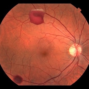
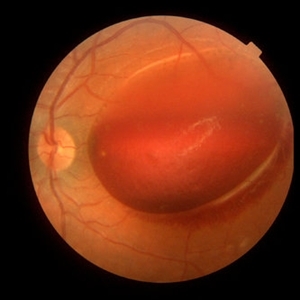
A 43-year-old white male who was injured while racing his side by side 4 wheel off-road vehicle (this is a video he showed me on his phone). He presented about three weeks after the injury. He was being seen by his local eye doctor who wanted an evaluation for the retinal heme and scotoma. His main complaint was a central/parcentral scotoma described as a greyish area in vision. Va 20/50 OD, nomotensive, no APD (by technician), anterior segment u/r; see {https://imagebank.asrs.org/file/53828/sxs-crash-during-a-race-causing-valsalva-retinopathy-od} for the fundus exam - of note there are superficial/preretinal heme, with layering of the heme superiorly; in the parafoveal region nasally there is some mottling of the RPE that may indicate an area of prior commotio retinae (also possible to have TON), which may account for his scotoma. Really bad accident, and amazingly, he had no LOC or injuries other than the right retina. Helmet and racing harness seat belt were used.
Condition/keywords: motor vehicle accident, trauma, valsalva retinopathy
-
 A Motor Vehicle Accident Causing Valsalva Retinopathy OD, While Racing A Side By Side 4 Wheel Off-Road Vehicle
Apr 29 2020 by John S. King, MD
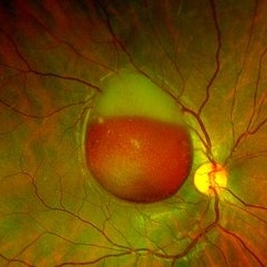
43-year-old white male who was injured while racing a side by side 4-wheel off-road vehicle (see Video: https://imagebank.asrs.org/file/53854/sxs-crash-during-a-race-causing-valsalva-retinopathy-od). He presented about three weeks after the injury. He was being seen by his local eye doctor who wanted an evaluation for the retinal heme and scotoma. His main complaint was a central/parcentral scotoma described as a greyish area in vision. Va 20/50 OD, nomotensive, no APD (by technician), anterior segment u/r; see picture for the fundus exam - of note there are superficial/preretinal heme, with layering of the heme superiorly, and small superficial heme at nasal edge of the optic disc; in the parafoveal region nasally there is some mottling of the RPE that may indicate an area of prior commotio retinae (also possible to have TON), which may account for his scotoma. Really bad accident (video), and amazingly, he had no LOC or injuries other than the right retina. Helmet and racing harness seat belt were used.
Photographer: Asli Ahmed
Imaging device: Topcon 50
Condition/keywords: valsalva retinopathy
-
 Dehemoglobinized sub-internal limiting membrane hemorrhage
Jul 29 2022 by JORGE SOBERANES
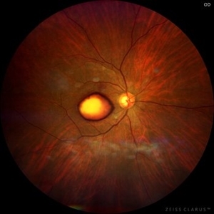
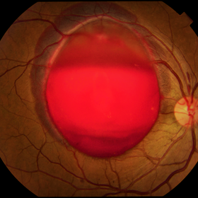
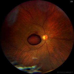
Fundus photograph of a 70-year-old man with Valsalva retinopathy manifested as premacular hemorrhage (sub-ILM) in dehemoglobinized process.
Photographer: Jorge I. Soberanes, Asociación para Evitar la Ceguera en México.
Imaging device: Zeiss Clarus 700
Condition/keywords: dehemoglobinized hemorrhage, sub-inner limiting membrane hemorrhage, valsalva retinopathy
-
 Fishing Fundus
Jul 16 2025 by Moazzam Parvez
Fundus photograph of a 31 year old woman with sub ILM hemorrhage following her yoga sessions which involves breath holding exercises .
Photographer: Moazzam Parvez , Netralayam , Kolkata
Imaging device: Topcon Maestro 2
Condition/keywords: SUB ILM hemorrhage, valsalva retinopathy
-
 OCT of a sub-internal limiting membrane hemorrhage in Valsalva retinopathy
Jul 29 2022 by JORGE SOBERANES
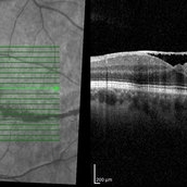
Optical coherence tomography of a 70-year-old man with a sub-internal limiting membrane due to Valsalva retinopathy
Photographer: Jorge I. Soberanes, Asociación para Evitar la Ceguera en México.
Imaging device: PLEX Elite 9000, Zeiss
Condition/keywords: OCT, sub-inner limiting membrane hemorrhage, swept source, valsalva retinopathy
-
 Optical Coherence Tomography of Valsalva Retinopathy at 2 Months
Oct 28 2024 by Andrew Jin, MD
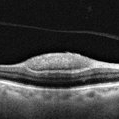
OCT of a 30 year old man with resolving valsalva retinopathy 2 months after presentation.
Condition/keywords: OCT, valsalva retinopathy
-
 Optical Coherence Tomography of Valsalva Retinopathy at Presentation
Oct 28 2024 by Andrew Jin, MD
OCT of a 30 year old man who presented with valsalva retinopathy
Condition/keywords: OCT, valsalva retinopathy
-
 Preretinal Hemorrhage
May 6 2017 by Mitzy E Torres Soriano, MD
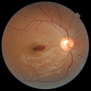
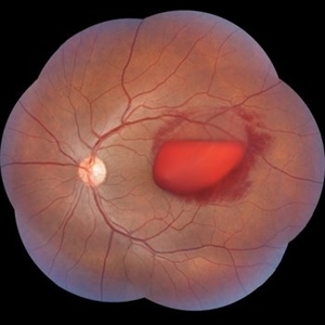
Fundus photograph of a 36-year-old-woman with a preretinal subhyaloid hemorrhage (valsalva retinopathy).
Photographer: Mitzy Torres Soriano
Condition/keywords: macular hemorrhage, premacular hemorrhage, preretinal hemorrhage, subhyaloid hemorrhage, valsalva retinopathy
-
 Primary Subhyaloid Hemorrhage due to Valsalva Retinopathy
Nov 13 2013 by Hamid Ahmadieh, MD
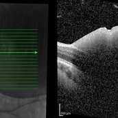
OCT images of the left eye of a 25-year-old man with sudden drop of vision due to subhyaloid hemorrhage secondary to Valsalva retinopathy.
Photographer: Soodabeh Fooladin , Negah Eye Center, Tehran
Imaging device: TOPCON OCT
Condition/keywords: optical coherence tomography (OCT), subhyaloid hemorrhage, valsalva retinopathy
-
 Primary Subhyaloid Hemorrhage Due to Valsalva Retinopathy
Nov 13 2013 by Hamid Ahmadieh, MD
Color fundus photograph of the left eye of a 25-year-old man with sudden drop of vision due to subhyaloid hemorrhage secondary to Valsalva retinopathy.
Photographer: Soodabeh Fooladin , Negah Eye Center, Tehran
Imaging device: TOPCON OCT
Condition/keywords: subhyaloid hemorrhage, valsalva retinopathy
-
---thumb.jpg/image-square;max$300,300.ImageHandler) Primary Subhyaloid Hemorrhage Due to Valsalva Retinopathy
Primary Subhyaloid Hemorrhage Due to Valsalva Retinopathy
Nov 13 2013 by Hamid Ahmadieh, MD
Infrared image of the left eye of a 25-year-old man with primary subhyaloid hemorrhage due to Valsalva retinopathy.
Photographer: Nayereh Hadipour, Negah Eye Center, Tehran
Imaging device: Heidelberg Spectralis
Condition/keywords: infrared image, subhyaloid hemorrhage, valsalva retinopathy
-
---thumb.jpg/image-square;max$300,300.ImageHandler) Primary Subhyaloid Hemorrhage Due to Valsalva Retinopathy
Primary Subhyaloid Hemorrhage Due to Valsalva Retinopathy
Nov 13 2013 by Hamid Ahmadieh, MD
FAF image of the left eye of a 25-year-old man with primary subhyaloid hemorrhage due to Valsalva retinopathy.
Photographer: Nayereh Hadipour, Negah Eye Center, Tehran
Condition/keywords: fundus autofluorescence (FAF), subhyaloid hemorrhage, valsalva retinopathy
-
---thumb.jpg/image-square;max$300,300.ImageHandler) Primary Subhyaloid Hemorrhage Due to Valsalva Retinopathy
Primary Subhyaloid Hemorrhage Due to Valsalva Retinopathy
Nov 13 2013 by Hamid Ahmadieh, MD
Arterial phase angiogram of the left eye of a 25-year-old man with primary subhyaloid hemorrhage due to Valsalva retinopathy.
Photographer: Nayereh Hadipour, Negah Eye Center, Tehran
Condition/keywords: subhyaloid hemorrhage, valsalva retinopathy
-
---thumb.jpg/image-square;max$300,300.ImageHandler) Primary Subhyaloid Hemorrhage Due to Valsalva Retinopathy
Primary Subhyaloid Hemorrhage Due to Valsalva Retinopathy
Nov 13 2013 by Hamid Ahmadieh, MD
Early venous phase angiogram of the left eye of a 25-year-old man with primary subhyaloid hemorrhage due to Valsalva retinopathy.
Photographer: Nayereh Hadipour, Negah Eye Center, Tehran
Condition/keywords: subhyaloid hemorrhage, valsalva retinopathy
-
---thumb.jpg/image-square;max$300,300.ImageHandler) Primary Subhyaloid Hemorrhage Due to Valsalva Retinopathy
Primary Subhyaloid Hemorrhage Due to Valsalva Retinopathy
Nov 13 2013 by Hamid Ahmadieh, MD
Wide-field angiogram of the left eye of a 25-year-old man with primary subhyaloid hemorrhage due to Valsalva retinopathy.
Photographer: Nayereh Hadipour, Negah Eye Center, Tehran
Condition/keywords: subhyaloid hemorrhage, valsalva retinopathy
-
---thumb.jpg/image-square;max$300,300.ImageHandler) Primary Subhyaloid Hemorrhage Due to Valsalva Retinopathy
Primary Subhyaloid Hemorrhage Due to Valsalva Retinopathy
Nov 13 2013 by Hamid Ahmadieh, MD
Midvenous phase angiogram of the left eye of a 25-year-old man with primary subhyaloid hemorrhage due to Valsalva retinopathy.
Photographer: Nayereh Hadipour, Negah Eye Center, Tehran
Condition/keywords: subhyaloid hemorrhage, valsalva retinopathy
-
 RAMA, Valsalva Retinopathy
Jul 1 2014 by John S. King, MD
Elderly female sent for possible exudative AMD. Chart review showed a prior inferotemporal mac lesion (likely arterial macroanuerysm). P/C acute painless loss of vision after severe coughing the night prior.
Photographer: Wayne A Ladlee Jr
Condition/keywords: ruptured macroaneurysm, valsalva retinopathy
-
 RAMA, Valsalva Retinopathy
Jul 1 2014 by John S. King, MD
Same patient.
Photographer: Wayne A Ladlee Jr
Imaging device: Cirrus
Condition/keywords: retinal macroaneurysm, valsalva retinopathy
-
 Retinopathy Valsalva
Jan 26 2017 by JEFFERSON R SOUSA, Tecg.º (Biomedical Systems Technology)
Male patient, 23-year-old, with low visual acuity in the right eye. In the ocular examination of the retinography, intense sub hyaloidal hemorrhage.
Photographer: JEFFERSON R SOUSA
Imaging device: Retinografo Topcon TRC-50 DX, Imaginet, campo de 35 graus. Flash 36
Condition/keywords: valsalva retinopathy
-
 Strained Retina
Sep 27 2025 by Malvika Singh
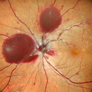
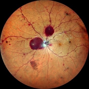
Fundus photograph of a 44 year old male showing hemorrhages at different layers.
Photographer: Dr Malvika Singh, Retina Foundation, Ahmedabad, India
Imaging device: Mirante SLO/OCT
Condition/keywords: valsalva retinopathy
-
 Strained Retina
Sep 27 2025 by Malvika Singh
Fundus photograph of a 44 year old male showing hemorrhages at different layers.
Photographer: Dr Malvika Singh, Retina Foundation, Ahmedabad, India
Imaging device: Mirante SLO/OCT
Condition/keywords: valsalva retinopathy
-
 Sub-ILM Hemorrhage (Dehemoglobinized + Red) - Valsalva Retinopathy
Jun 12 2021 by RUSHIK PATEL
Fundus Image of 21-year-old boy with sub-ILM hemorrhage (dehemoglobinized + red ) following valsalva maneuver 10 days back.
Photographer: Rushik Patel, Netralaya Super Speciality Eye Hospital, Ahmedabad, Gujarat
Imaging device: Optos
Condition/keywords: sub-inner limiting membrane hemorrhage, valsalva retinopathy
-
 Sub-internal limiting membrane hemorrhage in Valsalva retinopathy
Jul 29 2022 by JORGE SOBERANES
A fundus photography of a 70-year-old man with premacular hemorrhage (Sub-internal limiting membrane) due to Valsalva retinopathy
Photographer: Jorge I. Soberanes, Asociación para Evitar la Ceguera en México.
Imaging device: Zeiss Clarus 700
Condition/keywords: premacular hemorrhage, sub-inner limiting membrane hemorrhage, valsalva retinopathy
-
 Subhyaloid Haemorrhage - Valsalva Retinopathy
Jul 30 2023 by Maneesh M Bapaye, MD, MBA
A 25 years old paitient presented with sudden loss of vision following sudden rise intra abdominal pressure, Montage photo
Photographer: Dr.Maneesh Bapaye
Imaging device: Zeiss Fundus Camera
Condition/keywords: subhyaloid hemorrhage, valsalva retinopathy
-
 Subhyaloid Hemorrhage
Jul 31 2024 by Arthi Mohankumar , MS,MRCS ED, FICO,FAICO
A 35 year old male presented with complaints of seeing a black spot in left eye for past one day after working out in the gym the previous day. He has history of uncontrolled diabetes and hypertension. Fundus exam of the left eye revealed a sub hyaloid hemorrhage nasal to the disc with minimal background Diabetic and hypertensive changes. His baseline CBG was 200 mg/dl and BP was 170/100 He was suggested observation initially considering the nasal location. But patient found the scotoma very disturbing and eventually underwent yag hyaloidotomy
Photographer: Arthi Mohankumar
Condition/keywords: valsalva retinopathy
 Loading…