Search results (31 results)
-
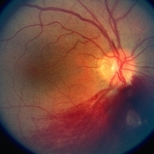
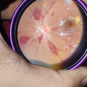
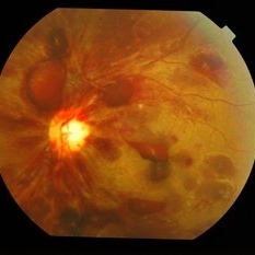
 Before & After - YAG Laser Hyaloidotomy for Subhyaloid Hemorrhage
Sep 19 2021 by Jesus Lozano, MD
23 year-old man with thrombocytopenia after chemotherapy d/t blastic plasmacytoid dendritic cell neoplasm. Developed a subhyaloid hemorrhage, and was treated with YAG Laser Hyaloidotomy.
Photographer: Yair Bet Yosef, Hadassah Medical Center. Israel
Imaging device: Optos
Condition/keywords: Dendritic cell Neoplasm, hyaloidotomy, subhyaloid hemorrhage, thrombocytopenia, vitreous hemorrhage
-
 Dengue Fever
Oct 25 2012 by Mallika Goyal, MD
Fundus photograph of the right eye of a 32-year-old gentleman with dengue fever and thrombocytopenia. Photograph shows extensive retinal and pre-retinal haemorrhages, roth spots but no dengue retinitis. Same patient as in images 1-5.
Condition/keywords: Dengue Fever, rosacea conjunctivitis, thrombocytopenia
-
 Slide 9-13
Feb 26 2019 by Lancaster Course in Ophthalmology
Superficial retinal hemorrhages. These hemorrhages have a flame-shaped appearance and are located just beneath the internal limiting membrane of the retina in a 61-year-old man with severe anemia and thrombocytopenia.
Condition/keywords: retinal hemorrhage, thrombocytopenia
-
 Thrombocytopenia
May 2 2013 by Henry J. Kaplan, MD
Superficial retinal hemorrhage in a patient with thrombocytopenia, right eye; #1.
Condition/keywords: thrombocytopenia
-
 Thrombocytopenia
May 2 2013 by Henry J. Kaplan, MD
Retinal and pre-retinal hemorrhage in thrombocytopenia, left eye; #2.
Condition/keywords: thrombocytopenia
-
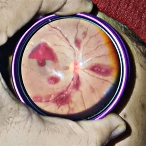 Thrombocytopenia
Sep 24 2024 by DR Rohit Gupta
Fundus photography of a 16 year old female suffering from severe thrombocytopenia. On fundus examination, multiple roth spots and subhyaloid hemorrhage were seen.
Photographer: Dr Rohit gupta
Imaging device: Samsung S21
Condition/keywords: ANEMIC RETINOPATHY, hemorrhage, leukemia, retinal hemorrhage, Roth spots, Sub hyaloid haemorrhage, thrombocytopenia
-
 Thrombocytopenia
Sep 24 2024 by DR Rohit Gupta
Fundus photography of a 16 year-old girl suffering from severe thrombocytopenia, showing flame shaped hemorrhage.
Photographer: Dr Rohit gupta
Imaging device: Samsung S21
Condition/keywords: anaemic retinopathy, flame shaped retinal hemorrhage, Haemorrhage, Roth spots, white centered retinal hemorrhage (Roth Spot), white dot syndrome
-
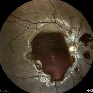
---thumb.JPG/image-square;max$300,300.ImageHandler) Thrombocytopenia
Thrombocytopenia
Dec 1 2013 by Mallika Goyal, MD
Right eye of a 16-year-old boy with thrombocytopenia and anaemia following chemotherapy for osteoscarcoma shows pre-retinal and submacular haemorrhages. The submacular component caused irreversible long term visual deterioration.
Photographer: Mallika Goyal, MD, Apollo Health City, Hyderabad, India
Condition/keywords: thrombocytopenia
-
---thumb.JPG/image-square;max$300,300.ImageHandler) Thrombocytopenia
Thrombocytopenia
Dec 1 2013 by Mallika Goyal, MD
Resolving pre-retinal and submacular heme 4 weeks after presentation.
Photographer: Mallika Goyal, MD, Apollo Health City, Hyderabad, India
Condition/keywords: thrombocytopenia
-
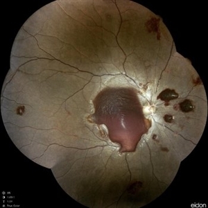
---thumb.JPG/image-square;max$300,300.ImageHandler) Thrombocytopenia
Thrombocytopenia
Dec 1 2013 by Mallika Goyal, MD
This fundus photograph of the right eye displays large geographic areas of pre-retinal hemorrhages including a central macular boat-shaped hemorrhage. This fundus image highlights pre-retinal hemorrhage that may layer horizontally.
Photographer: Mallika Goyal, MD, Apollo Hospitals, Hyderabad, India
Condition/keywords: thrombocytopenia
-
---thumb.JPG/image-square;max$300,300.ImageHandler) Thrombocytopenia
Thrombocytopenia
Dec 1 2013 by Mallika Goyal, MD
Complete resolution of pre-retinal and submacular hemorrhage 6 months after presentation. Atrophic changes at the foveal centre account for subnormal visual recovery.
Photographer: Mallika Goyal, MD, Apollo Hospitals, Hyderabad, India
Condition/keywords: thrombocytopenia
-
---thumb.JPG/image-square;max$300,300.ImageHandler) Anaemic retinopathy
Anaemic retinopathy
Oct 26 2012 by Mallika Goyal, MD
Vitreous haemorrhage in a 25-year-old gentleman with severe anaemia and thrombocytopenia.
Photographer: Mallika Goyal, MD
Condition/keywords: anaemic retinopathy, vitreous hemorrhage
-
 Anemic Retinopathy Related Retinal Hemorrhages
Nov 5 2019 by Chinmayi Vyas
Anemic retinopathy related retinal hemorrhages in a 24 years old male with Hb of 4.2gm/ dl. The manifestations of anemic retinopathy are nonspecific and may closely simulate hypertensive or diabetic retina. Retinal changes in anemia are cotton wool spots, venous tortuosity, and hemorrhages which may be present at all levels of the retina and choroid. All retinal hemorrhages can occur when Hb falls below 8 g/100 ml or if the platelet count falls below 50,000/cumm. The combination of severe anemia and thrombocytopenia is likely to produce retinal hemorrhages. The Roth’s spots or white centre hemorrhages are typically associated with bacterial endocarditis , anemia and other systemic conditions. The white center is suspected to represents focal ischemia, inflammatory or infectious infiltrate, fibrin or accumulation of neoplasticism cells.
Photographer: Dr Chinmayi Vyas
Condition/keywords: retinal hemorrhage
-
 Anemic Retinopathy Related Retinal Hemorrhages
Nov 5 2019 by Chinmayi Vyas
Anemic retinopathy related retinal hemorrhages in a 24 years old male with Hb of 4.2gm/ dl. The manifestations of anemic retinopathy are nonspecific and may closely simulate hypertensive or diabetic retina. Retinal changes in anemia are cotton wool spots, venous tortuosity, and hemorrhages which may be present at all levels of the retina and choroid. All retinal hemorrhages can occur when Hb falls below 8 g/100 ml or if the platelet count falls below 50,000/cumm. The combination of severe anemia and thrombocytopenia is likely to produce retinal hemorrhages. The Roth’s spots or white centre hemorrhages are typically associated with bacterial endocarditis , anemia and other systemic conditions. The white center is suspected to represents focal ischemia, inflammatory or infectious infiltrate, fibrin or accumulation of neoplasticism cells.
Photographer: Dr Chinmayi Vyas, Nethradhama superspeciality eye hospital , banglore, india
Imaging device: Eidon fundus imaging
Condition/keywords: anaemic retinopathy
-
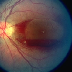
 Before & After - YAG Laser Hyaloidotomy for Subhyaloid Hemorrhage
Sep 19 2021 by Jesus Lozano, MD
23 year-old man with thrombocytopenia after chemotherapy d/t blastic plasmacytoid dendritic cell neoplasm. Developed a subhyaloid hemorrhage, and was treated with YAG Laser Hyaloidotomy.
Photographer: Yair Bet Yosef, Hadassah Medical Center. Israel
Imaging device: Optos
Condition/keywords: subhyaloid hemorrhage, vitreous hemorrhage
-
 Candida Endophthalmitis
Jan 26 2020 by Marlon García Roa, MD
Female, 30-years-old with <<< V Pregnancy Currently with 18 weeks gestation. Pathological personal history 1 month prior hospitalization for complicated acute appendicitis + pyelonephritis + severe thrombocytopenia (autoimmune treated with corticosteroids) with septic shock, appendectomy was performed, due to torpid evolution, intensive care unit with placement of central venous catheter treated with intravenous antibiotics is performed, CT scan is performed of thorax, abdomen and pelvis in search of aggregate pathology; finding multiple renal lithiasis that conditions hydronephrosis and reactivation of pyelonephritis, so he continued with antibiotic therapy and underwent endoscopic lithotomy, due to febrile persistence and with a positive blood culture result for candida Albicans, intravenous antifungals (anidulafungin) were started for 1 week, with improvement satisfactory for what was decided his discharge. During hospitalization it was required to transfuse 2 globular packages and platelet plasmapheresis as well as replacement of calcium, phosphorus and potassium. It refers to approximately 3 weeks of visual loss of the left eye associated with myodisopsia. visual acuity 20/100 Vitritis +, with floating vitreous abscess on the posterior pole, round papilla, slightly erased edges, excavation 0.3, macula without foveolar luster, conserved vein artery relationship, with vessels with multiple mineralization areas and superior peripheral lesion of ¼ diameter of papilla as ball of snow applied to retina.
Photographer: MARLON GARCIA ROA, INSTITUTO DE RETINA DEL BAJIO (INDEREB), QUERETARO, MEXICO
Condition/keywords: candida endophthalmitis
-
 Dengue Fever
Oct 25 2012 by Mallika Goyal, MD
Fundus photograph of the right eye of a 32-year-old gentleman with dengue fever and thrombocytopenia. Photograph shows extensive retinal and pre-retinal haemorrhages, roth spots but no dengue retinitis. Same patient as in images 1-5.
Condition/keywords: Dengue Fever, preretinal hemorrhage, rosacea conjunctivitis
-
 Dengue Fever
Oct 25 2012 by Mallika Goyal, MD
Fundus photograph of the left eye of a 32-year-old gentleman with dengue fever and thrombocytopenia. Photograph shows extensive retinal and pre-retinal haemorrhages, roth spots but no dengue retinitis. Same patient as in images 1-5.
Condition/keywords: Dengue Fever, preretinal hemorrhage, rosacea conjunctivitis
-
 Dengue Fever
Oct 25 2012 by Mallika Goyal, MD
Fundus photograph of the left eye of a 32-year-old gentleman with dengue fever and thrombocytopenia. Photograph shows extensive retinal and pre-retinal haemorrhages, roth spots but no dengue retinitis. Same patient as in images 1-5
Condition/keywords: Dengue Fever, preretinal hemorrhage, rosacea conjunctivitis
-
 Dengue Fever
Oct 25 2012 by Mallika Goyal, MD
Fundus photograph of the left eye of a 32-year-old gentleman with dengue fever and thrombocytopenia. Photograph shows extensive retinal and pre-retinal haemorrhages, roth spots but no dengue retinitis.
Condition/keywords: Dengue Fever, preretinal hemorrhage, rosacea conjunctivitis
-
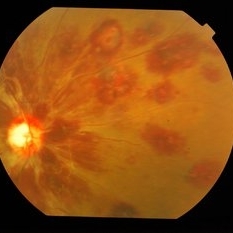
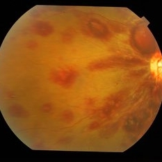
 Premacular Hemorrhage in Acute Myeloid Leukemia
Feb 10 2016 by Mallika Goyal, MD
Fundus photograph of a 45-year-old male with acute myeloid leukemia and thrombocytopenia shows premacular & retinal haemorrhage.
Photographer: Mallika Goyal, MD, Apollo Health City, Jubilee Hills, Hyderabad, India
Condition/keywords: premacular hemorrhage
-
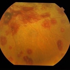
 Premacular Hemorrhage in Acute Myeloid Leukemia
Feb 10 2016 by Mallika Goyal, MD
Fundus photograph of a 45-year-old male with acute myeloid leukemia and thrombocytopenia shows premacular & retinal hemorrhage.
Photographer: Mallika Goyal, MD, Apollo Health City, Jubilee Hills, Hyderabad, India
Condition/keywords: premacular hemorrhage
-
---thumb.jpg/image-square;max$300,300.ImageHandler) Roth Spots and Retinal Hemorrhage
Roth Spots and Retinal Hemorrhage
Dec 27 2013 by David Callanan, MD
24-year-old patient, AML/ post-chemo thrombocytopenia with pre, intra, & sub-retinal.
Condition/keywords: white centered retinal hemorrhage (Roth Spot)
 Loading…