Search results (134 results)
-
 APMPPE With Serous Macular Detachment
Jun 2 2014 by Rameez N Hussain, MD
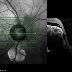
Acute posterior multifocal placoid pigment epitheliopathy (APMPPE) with serous macular detachment.
Photographer: Rameez N Hussain MD, Vitreo Retinal Services, Giridhar Eye Institute, Cochin, India
Imaging device: Zeiss FF4
Condition/keywords: acute posterior multifocal placoid pigment epitheliopathy (APMPPE), serous retinal detachment
-
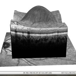 APMPPE With Serous Macular Detachment 3D SD-OCT
Jun 2 2014 by Rameez N Hussain, MD
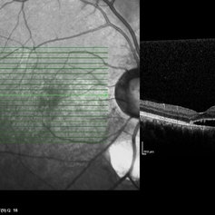
3D SD-OCT of acute posterior multifocal placoid pigment epitheliopathy (APMPPE) with serous macular detachment.
Photographer: Rameez N Hussain MD, Vitreo Retinal Services, Giridhar Eye Institute, Cochin, India
Imaging device: Heidelberg Spectralis
Condition/keywords: acute posterior multifocal placoid pigment epitheliopathy (APMPPE), serous retinal detachment
-
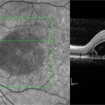 APMPPE With Serous Macular Detachment SD-OCT
Jun 2 2014 by Rameez N Hussain, MD
SD OCT image of acute posterior multifocal placoid pigment epitheliopathy (APMPPE) with serous macular detachment.
Photographer: Rameez N Hussain MD, Vitreo Retinal Services, Giridhar Eye Institute, Cochin, India
Imaging device: Heidelberg Spectralis
Condition/keywords: acute posterior multifocal placoid pigment epitheliopathy (APMPPE), serous retinal detachment
-
 B-scan Ultrasound of Choroidal Melanoma with Serous Retinal Detachment
Sep 5 2025 by Kristen Wagner
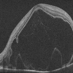
B-scan ultrasound of a choriodal melanoma with serous retinal detachment.
Photographer: Kristen Wagner, COT Tennessee Retina
Condition/keywords: B scan ultrasound, Choroidal melanoma, serous retinal detachment
-
 Central Serous Retinopathy-OCT
Jun 14 2018 by Mitzy E Torres Soriano, MD
62-year-old male patient with chronic central serous chorioretinopathy in his right eye. OCT shows serous neurosensory retinal detachment and retinal pigment epithelial detachment.
Condition/keywords: central serous chorioretinopathy (CSCR), central serous retinopathy (CSR), optical coherence tomography (OCT), pigment epithelial detachment (PED), serous retinal detachment
-
 Choroid hemangioma
Sep 7 2022 by JEFFERSON R SOUSA, Tecg.º (Biomedical Systems Technology)
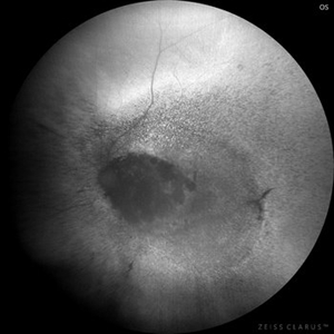
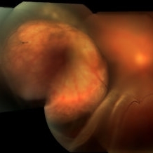
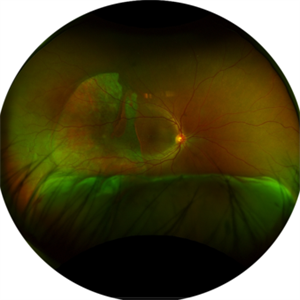
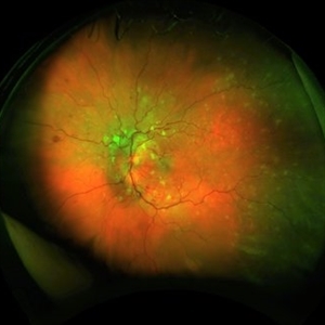
Patient 54 years old, Female, progressive loss of vision. In the multimodal evaluation of the retina showed important retinal alterations. A discreet opacity of the media impairs the quality of the images. In the Autofluorescent Background Image with a green filter, because it reaches a depth in the retinal tissue, it is able to show changes that affect the retinal pigment epithelium, it was better in this case than with the green filter. WF retinography shows an elevated, slightly reddish lesion, probable serous retinal detachment, mobilization of pigments and phantom vessels.
Photographer: JEFFERSON ROCHA DE SOUSA - Retinal Department at Instituto Dr. Suel Abujamra Sao Paulo-Brazil
Imaging device: Clarus 700 - Zeiss 135 degree images. Multimodal Evaluation
Condition/keywords: elevated retinal lesion, hemangioma, melanoma, serous retinal detachment
-
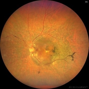
 Choroid hemangioma
Sep 7 2022 by JEFFERSON R SOUSA, Tecg.º (Biomedical Systems Technology)
Patient 54 years old, Female, progressive loss of vision. In the multimodal evaluation of the retina showed important retinal alterations. A discreet opacity of the media impairs the quality of the images. In the Autofluorescent Background Image with a green filter, because it reaches a depth in the retinal tissue, it is able to show changes that affect the retinal pigment epithelium, it was better in this case than with the green filter. WF retinography shows an elevated, slightly reddish lesion, probable serous retinal detachment, mobilization of pigments and phantom vessels.
Photographer: JEFFERSON ROCHA DE SOUSA - Retinal Department at Instituto Dr. Suel Abujamra Sao Paulo-Brazil
Imaging device: Clarus 700 - Zeiss 135 degree images. Multimodal Evaluation
Condition/keywords: elevated retinal lesion, hemangioma, melanoma, serous retinal detachment
-
 Choroidal Melanoma
Sep 7 2023 by Annaka Gooding
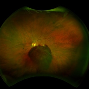
Ultra-Widefield pseudo-color and autofluorescence imaging of a 59 year old male with Choroidal Melanoma affecting his left eye. Patient reported floaters OS for months prior to examination as well as 1-2 weeks of "tunnel vision". Patient denies personal history of cancer. Patient's vision at time of examination was CF@5FT. Due to the Tumor size, the patient has developed a serous retina detachment in their inferior retina
Photographer: Annaka Gooding
Imaging device: Optos California
Condition/keywords: autofluorescence imaging, choroidal tumor, fundus photography, OPTOS CALIFORNIA, serous retinal detachment
-
 Choroidal Melanoma With a Serous Retinal Detachment
Aug 23 2018 by Nichole Lewis
63-year-old male with a large choroidal melanoma and a serous macula off retinal detachment. Vision is count fingers.
Photographer: Nichole Lewis
Condition/keywords: serous retinal detachment
-
 Choroidal Metastasis With Serous Retinal Detachment Left Eye
Sep 2 2015 by María José Marroquín Sarti
A 62-year-old woman complained of visual field loss and decreasing vision. Twenty years earlier, breast cancer was diagnosed and treated with chemotherapy and right mastectomy, four years ago, she had another treatment with chemotherapy and resection of another tumor that started to grow in the same side. Serous retinal detachment and choroidal masses were present in both eyes.
Photographer: María Jose Marroquín Sarti
Condition/keywords: choroidal metastasis, serous retinal detachment
-
 Choroidal Neovascular Membrane Evolving With Subretinal Hemorrhage
Apr 23 2021 by Andre Beckenkamp
Wide angle fundus photograph of an 82-year-old woman with dry AMD in her right eye and wet AMD in the left eye, evolving with subretinal hemorrhage and associated serous retinal detachment.
Photographer: Andre Beckenkamp
Imaging device: Optos Daytona
Condition/keywords: age-related macular degeneration (AMD), serous retinal detachment, subretinal hemorrhage, wet age-related macular degeneration (wet AMD)
-
---thumb.jpg/image-square;max$300,300.ImageHandler) Choroidal Tumor, Breast Cancer
Choroidal Tumor, Breast Cancer
Feb 13 2013 by From the Collections of Thomas M. Aaberg, MD and Thomas M. Aaberg Jr., MD
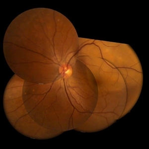
Optic nerve, pt. LB, breast cancer, serous retinal detachment.
Condition/keywords: choroidal tumor, serous retinal detachment
-
---thumb.jpg/image-square;max$300,300.ImageHandler) Choroidal Tumor, Breast Cancer
Choroidal Tumor, Breast Cancer
Feb 13 2013 by From the Collections of Thomas M. Aaberg, MD and Thomas M. Aaberg Jr., MD
Optic nerve, pt. LB, breast cancer, serous retinal detachment.
Condition/keywords: choroidal tumor, serous retinal detachment
-
 Choroidal Tumor, Breast Cancer
Feb 13 2013 by From the Collections of Thomas M. Aaberg, MD and Thomas M. Aaberg Jr., MD
Optic nerve, pt. LB, breast cancer, serous retinal detachment.
Condition/keywords: serous retinal detachment
-
 Choroidals/Serous Retinal Detachment
Mar 3 2015 by Jared Watson
20-year-old female s/p aspiration vitreal fluid for choridal effusion OS. Visible with anterior view on fundus camera.
Photographer: Jared Watson COT, University of Virginia
Condition/keywords: serous retinal detachment
-
 chronic central serous chorioretinopathy
Oct 31 2012 by Mallika Goyal, MD
Fluorescein angiogram of inferior retina of right eye with chronic CSCR shows dilation of and mild leak from retinal vessels over the inferior serous retinal detachment.
Condition/keywords: central serous chorioretinopathy (CSCR), chronic central serous chorioretinopathy (CSCR), serous retinal detachment
-
 Coats Disease
Sep 13 2013 by Maria Ana Martinez-Castellanos, MD
Peripheral fundus angiogram in a 2-years-old boy with Coat's disease.
Photographer: Maria A. Martinez-Castellanos. Asociacion para Evitar la Ceguera en Mexico
Imaging device: RetCAm II
Condition/keywords: pediatic retina, serous retinal detachment, vascular anomaly, vascular occlusions
-
 Coats' Disease
Nov 30 2018 by Darin R. Goldman, MD
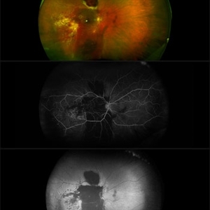
58-year-old male with a Coats’-like process in his right eye. The patient has undergone both laser photocoagulation and anti-VEGF therapy.
Photographer: Harold Rodriguez, CMA, Retina Group of Florida
Imaging device: Optomap color image, fluorescein angiogram, and fundus autofluorescence
Condition/keywords: Coats' disease, macroaneurysm, serous retinal detachment, subretinal hemorrhage
-
 Coats' Related Total Serous Retinal Detachment
Jan 19 2020 by Anfisa Ayalon, MD
Slit-lamp photograph of a 3 -year-old male with total serous retinal detachment due to Coats' disease in the right eye. S/p laser photocoagulation, cryotherapy, retinal detachment repair with scleral buckle implantation 2 years ago. Currently, the right eye has no light perception.
Photographer: Anfisa Ayalon, MD., Meir Medical Center, Kfar Saba, Israel.
Condition/keywords: blind eye, Coats' disease, retinal detachment without retinal defect, serous retinal detachment
-
 Coloboma of Disc & Choroid
Oct 6 2012 by Hamid Ahmadieh, MD
OCT image of a 25-year-old woman with serous retinal detachment secondary to coloboma of disc associated with coloboma of choroid.
Photographer: Hamid Ahmadieh, MD, Ophthalmic Research Center, Labbafinejad Medical Center, Shahid Beheshti University of Medical Sciences
Imaging device: Heidelberg Spectralis
Condition/keywords: coloboma of choroid, coloboma of optic disc, optical coherence tomography (OCT), serous retinal detachment
-
 Exudative Detachment of the Macula in Vogt-Koyanagi-Harada Syndrome
Jan 10 2018 by Peter H. Tang, MD, PhD
SD-OCT imaging of an exudative detachment of the macula in a 27-year-old male diagnosed with Vogt-Koyanagi-Harada Syndrome.
Imaging device: Zeiss Cirrus HD-OCT
Condition/keywords: exudative macula detachment, serous retinal detachment, uveitis, Vogt-Koyanagi-Harada
-
 Giant RPE Rip with Serous Retinal Detachment
Apr 30 2025 by Amber Dubey
A 50 year-old man with sudden onset diminution of vision since 3 days. Optos Image showing a fovea-sparing giant RPE rip temporally with associated serous retinal detachment inferiorly without ocular and systemic co-morbidity or antecedent history of trauma.
Photographer: Dr. Amber Dubey, Sri Sankaradeva Nethralaya, Guwahati, India
Imaging device: Optos imaging system
Condition/keywords: Optos, RPE Rip, serous retinal detachment
-
 Hypertensive Retinopathy
Aug 24 2012 by Geoffrey G. Emerson, MD, PhD, FASRS
A 35-year-old man has headaches and decreased vision. The right eye measures 20/25 and the left eye measures 3/200. The blood pressure measures 180/110.
Photographer: Geoffrey Emerson, MD, PhD, Retina Center, Minneapolis
Condition/keywords: hypertensive retinopathy, papilledema, serous retinal detachment
-
 Hypertensive retinopathy, left
Feb 23 2017 by Alla Goldberg, MD
Fundus photograph of 35-year-old man with severe hypertension (182/128).
Photographer: Sofia Rutiaga, UT Health McGovern Medical School, Cizik Eye Clinic
Condition/keywords: cotton wool spots, Elschnig's spots, hypertensive choroidopathy, hypertensive retinopathy, serous retinal detachment
 Loading…