Search results (50 results)
-
 Chronic Retinal Detachment
Oct 12 2012 by Jeffrey G. Gross, MD, FASRS
Chronic RD with multiple retinal cysts, B scan ultrasound.
Condition/keywords: B scan ultrasound, chronic retinal detachment, retinal cyst
-
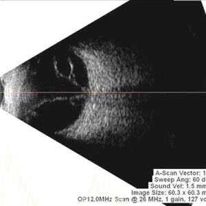
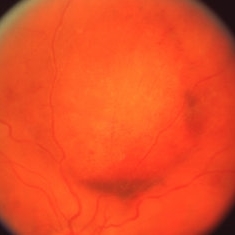
 Eye Finally Got the Ring... But the Retina Was Too Detached to Care
Nov 5 2025 by SHRADDHA RAJ SHRIVASTAVA
Left Eye B-scan ultrasound of a patient with old retinal detachment shows open funnel shaped hyperechoic membranous echoes, with high amplitude spikes on A-scan and a poor after-movement on dynamic B-scan, suggestive of retinal detachment. We can see a round echogenicity in sub-retinal location, with clear contents within, suggestive of a retinal cyst. This B-scan image is indicative of a long-standing chronic retinal detachment with secondary retinal cyst.
Photographer: Dr. Shraddha Raj Shrivastava
Condition/keywords: B scan ultrasound, chronic retinal detachment, OLD RD, open funnel RD, retinal cyst
-
 Giant Retinal Cyst
Sep 20 2025 by JORGE SOBERANES
Fundus photograph of a 45-year-old-man with a large cyst on the nasal superior side of the retina. The patient had a history of a pneumatic retinopexy two years ago and the cyst has been there since that.
Photographer: Dr. Jorge Soberanes, Asociación para Evitar la Ceguera en México (APEC), UNAM
Condition/keywords: abnormal retina, pneumatic retinopexy, retinal cyst
-
 Macrocyst in the Fovea
Feb 2 2021 by Peter J Belin, MD
36-year-old male with a white cataract and a chronic total retinal detachment for 1 year presented with a recurrent PVR detachment after primary repair 2 weeks prior. This OCT- EDI demonstrates a large retinal cyst through the fovea.
Photographer: Holly Cheshier, CRA, OCT-C, COT
Imaging device: Heidelberg Spectralis
Condition/keywords: chronic retinal detachment, proliferative vitreoretinopathy (PVR), retinal cyst, retinal macrocyst
-
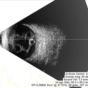
 Multiple Retinal Cysts Associated With Chronic Retinal Detachment
Sep 24 2018 by samarth mishra
Patient presented with a diminution of vision in left eye since few months. On B-scan ultrasonography multiple retinal cysts with a total retinal detachment were noted.
Photographer: Aditya Birla Sankara Nethralaya, West Bengal , Kolkata , India
Condition/keywords: B scan ultrasound, chronic retinal detachment, intraretinal cyst, retinal cyst
-
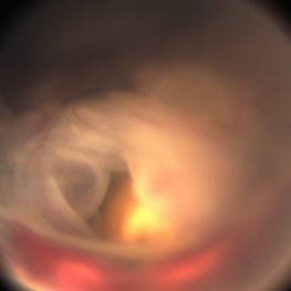
 Old RRD With Retinal Cysts and High Watermark
Apr 10 2020 by Dipak Nag, MBBS, FCPS, MSc, FRF
Intra-operative fundus picture of a 20-year-old boy showing multiple retinal cysts and high watermark in a case of old inferior retinal detachment OD.
Photographer: Dipak
Condition/keywords: high watermark, retinal cyst
-
 Retinal Cyst
Aug 14 2020 by Noy Ashkenazy, MD, MS
Fundus photograph of a 13-year-old male with a chronic retinal detachment following a penetrating ocular trauma. There is a retinal cyst and proliferative vitreoretinopathy.
Photographer: Giselle DeOliveira
Imaging device: Retcam III
Condition/keywords: chronic retinal detachment, proliferative vitreoretinopathy (PVR), retinal cyst
-
 Retinal Cyst after CRVO
Mar 27 2019 by Gary R. Cook, MD, FACS
Elderly white female s/p CRVO 3 years ago now with retinal cyst OD; V.A.= hand motions.
Condition/keywords: central retinal vein occlusion (CRVO), retinal cyst
-
---thumb.jpg/image-square;max$300,300.ImageHandler) Retinal Cyst?
Retinal Cyst?
Apr 4 2014 by H. Michael Lambert, MD
cystic structure in under retina. Etiology?
Condition/keywords: retinal cyst
-
---thumb.jpg/image-square;max$300,300.ImageHandler) Toxocariasis Cyst
Toxocariasis Cyst
Feb 13 2013 by From the Collections of Thomas M. Aaberg, MD and Thomas M. Aaberg Jr., MD
Mid-peripheral retinal cyst.
Condition/keywords: retinal cyst, toxocariasis
-
---thumb.jpg/image-square;max$300,300.ImageHandler) Toxocariasis Cyst
Toxocariasis Cyst
Feb 13 2013 by From the Collections of Thomas M. Aaberg, MD and Thomas M. Aaberg Jr., MD
Mid-peripheral retinal cyst.
Condition/keywords: retinal cyst, toxocariasis
-
 Symmetric Retinal Cysts
Aug 22 2024 by Neeket R. Patel, MD
Ultrasonography of a 55-year-old woman with chronic, symmetric retinal cysts.
Condition/keywords: Retinal Cysts
-
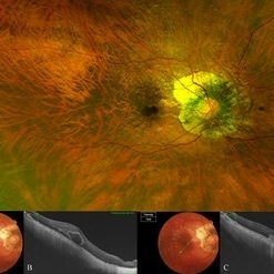 Bow-Tie Macular Hemorrhage With Cyst- Atypical Presentation of Myopic Choroidal Neovascularization
Mar 26 2021 by RUSHIK PATEL
The image of right eye of 51-year-old lady with high myopia show " Bow-Tie" macular hemorrhage (A). Optical coherence tomography (B) scan passing through hemorrhage showed intraretinal cystic lesion. During the course of intravitreal anti-VEGF injection treatment, the lesion converted into typical myopic choroidal neovascularization (C).
Photographer: Rushik Patel, Netralaya Super Speciality Eye Hospital
Condition/keywords: cyst, macular hemorrhage, myopic choroidal neovascularization (CNV)
-
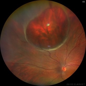
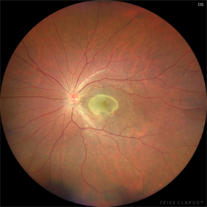
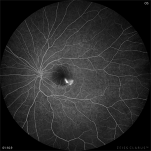
 Chronic retinal detachment changes
Apr 29 2022 by Otakar Dušek, M.D. Ph.D.
Colour fundus photo of 22-year-old woman with bulous retinal detachment number 5-9, old demarcation lines and inferotemporal periheral secondary retinal cyst.
Photographer: Otakar Dušek, Charles University, Prague
Imaging device: Zeiss Clarus
Condition/keywords: chronic retinal detachment, demarcation line, peripheral retinal cyst
-
 Chronic Retinal Detachment: Features Slide 1
Oct 22 2012 by Ronald C. Gentile, MD
Chronic retinal detachments can be associated with demarcation lines (tidemarks), subretinal bands or sheets, and retinal cysts. Fundus photo of a chronic inferior retinal detachment reveals multiple demarcation lines inferior to the center of the fovea as a result of an inferior temporal dialysis.
Photographer: The New York Eye & Ear Infirmary Department of Medical Imaging
Condition/keywords: chronic retinal detachment, demarcation line
-
 Chronic Retinal Detachment: Features Slide 2
Oct 22 2012 by Ronald C. Gentile, MD
Chronic retinal detachments can be associated with demarcation lines (tidemarks), subretinal bands or sheets, and retinal cysts. Fundus photo of a chronic retinal detachment reveals a branching subretinal band superior nasal to the macula with a portion extending to the inferior margin of the optic disc.
Photographer: The New York Eye & Ear Infirmary Department of Medical Imaging
Condition/keywords: chronic retinal detachment, subretinal bands
-
 Chronic Retinal Detachment: Features Slide 3
Oct 22 2012 by Ronald C. Gentile, MD
Chronic retinal detachments can be associated with demarcation lines (tidemarks), subretinal bands or sheets, and retinal cysts. Fundus photo of a chronic retinal detachment reveals a retinal cyst within the peripherally detached temporal retina.
Condition/keywords: chronic retinal detachment
-
 CSR with Fibrin
Jan 28 2025 by Vishal Agrawal, MD, FRCS,FACS,FASRS
A 31-year-old female was referred with a diagnosis of subretinal cysticercosis. BCVA was 20/200 OS. OCT showed a large subfoveal bacillary layer detachment (BALAD) without any scolex. FFA revealed a smoke-stack appearance. A final diagnosis of CSR with Fibrin was made and was managed conservatively. BCVA at final visit was 20/20.
Photographer: Dr Ayushi Gupta
Imaging device: Clarus 700
Condition/keywords: central serous chorioretinopathy (CSCR)
-
 CSR with Fibrin-FFA
Jan 29 2025 by Vishal Agrawal, MD, FRCS,FACS,FASRS
A 31-year-old female was referred with a diagnosis of subretinal cysticercosis. BCVA was 20/200 OS. OCT showed a large subfoveal bacillary layer detachment (BALAD) without any scolex. FFA revealed a smoke-stack appearance. A final diagnosis of CSR with Fibrin was made and was managed conservatively. BCVA at final visit was 20/20.
Photographer: Dr Ayushi Gupta
Imaging device: Clarus 700
Condition/keywords: central serous chorioretinopathy (CSCR)
-
 Cystercercosis
Sep 17 2012 by Prema Abraham, MD
Fundus photograph of 25-year-old male with subretinal cystercercosis.
Photographer: Dan Parks, Black Hills Regional Eye Institute, Rapid City South Dakota
Imaging device: Topcon 50EX
Condition/keywords: cysticercosis
-
 Cystercercosis
Sep 17 2012 by Prema Abraham, MD
Fundus photgraph of 25-year-old male with subretinal cystercercosis.
Photographer: Dan Parks, Black Hills Regional Eye Institute, Rapid City South Dakota
Imaging device: Topcon 50EX
Condition/keywords: cysticercosis
-
 Cystercercosis
Sep 17 2012 by Prema Abraham, MD
Fundus photograph of 25-year-old male with subretinal cystercercosis.
Photographer: Dan Parks, Black Hills Regional Eye Institute, Rapid City South Dakota
Imaging device: Topcon 50 EX
Condition/keywords: cysticercosis
 Loading…