Search results (66 results)
-
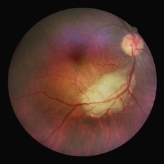
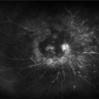
---thumb.jpg/image-square;max$300,300.ImageHandler) Behcet Disease
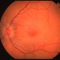
Behcet Disease
Feb 15 2013 by From the Collections of Thomas M. Aaberg, MD and Thomas M. Aaberg Jr., MD
Reprint of a fundus photograph from a patient with Behcet disease showing optic atrophy, vessel narrowing, and pigmentary changes (from Colvard et al, Arch Ophthalmol 1977;95(10):1813-7).
Condition/keywords: posterior uveitis, retinitis
-
---thumb.jpg/image-square;max$300,300.ImageHandler) Behcet Uveitis
Behcet Uveitis
Feb 15 2013 by From the Collections of Thomas M. Aaberg, MD and Thomas M. Aaberg Jr., MD
Color fundus photographs of the right eye of a patient suspected to have Behcet Uveitis. Over the course of 11 days, there is progressive optic disc edema, intraretinal whitening, hemorrhage and vessel occlusion.
Condition/keywords: Behcet's uveitis, posterior uveitis, retinitis
-
---thumb.jpg/image-square;max$300,300.ImageHandler) Behcet Uveitis
Behcet Uveitis
Feb 15 2013 by From the Collections of Thomas M. Aaberg, MD and Thomas M. Aaberg Jr., MD
Color fundus photographs of the right eye of a patient suspected to have Behcet Uveitis. Over the course of 11 days, there is progressive optic disc edema, intraretinal whitening, hemorrhage and vessel occlusion. Fluorescein angiography confirms impaired retinal perfusion secondary to vessel occlusion.
Condition/keywords: posterior uveitis, retinitis
-
 BSC CME OS
Nov 10 2012 by Pauline T Merrill, MD, FASRS
Fundus photograph left eye of a 42-year-old Caucasian male with birdshot retinochoroidopathy (HLA-A29+) and cystoid macular edema (CME)
Condition/keywords: birdshot retinochoroidopathy, cystoid macular edema (CME), posterior uveitis, uveitis
-
 Childhood Acquired Ocular Toxoplasmosis
Sep 13 2023 by Deepak Bhojwani, MS
Fundus image of a 16 year old boy diaagnosed with Ocular Toxoplasmosis since the age of 10 years showing the classic toxo chorioretinitis scar on the posterior pole. Luckily the scar is loacted juxtatemporal to fovea on OCT and so the boy has good vision of 20/30.
Photographer: DR DEEPAK BHOJWANI
Imaging device: OPTCAL COHERENCE TOMOGRAPHY
Condition/keywords: posterior uveitis, toxo chorioretinitis
-
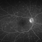
 MEWDS FA Early Phase
Jun 29 2018 by Shree K. Kurup, MD
Fluorescein angiogram (early phase) in young female with MEWDS.
Photographer: Moises Castro
Condition/keywords: multiple evanescent white dot syndrome (MEWDS), posterior uveitis
-
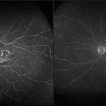
 MEWDS FA Late Phase
Jun 29 2018 by Shree K. Kurup, MD
Late phase FA young female with MEWDS.
Photographer: Moises Castro
Condition/keywords: multiple evanescent white dot syndrome (MEWDS), posterior uveitis
-
 MEWDS Mid FA
Jun 29 2018 by Shree K. Kurup, MD
Fluorescein angiogram (mid) on young female with MEWDS with late leakage in other frames
Photographer: Moises Castro
Condition/keywords: posterior uveitis
-
 MEWDS-Color
Jun 29 2018 by Shree K. Kurup, MD
Multiple evanescent white dot syndrome in young female.
Photographer: Moises Castro
Condition/keywords: choroiditis, creamy lesions, multiple evanescent white dot syndrome (MEWDS), posterior uveitis, uveitis
-
 Neovascularization in Posterior Uveitis
Jul 27 2023 by Zach Seim
An ultra-widefield fluorescein angiogram of a 72 year old male with Posterior Uveitis and Neovascularization affecting the right eye. Patient's vision at the time of the image was Dcc 20/25. Dr. Korot states that the fluorescein angiogram shows patchy leakage throughout both eyes, with peripheral nonperfusion and secondary neovascularization. The patient was asked to get an extensive serological workup in an effort to identify any systemic autoimmune or infectious etiology as the cause for their severe inflammation.
Photographer: Zach Seim
Imaging device: OPTOS California
Condition/keywords: fluorescein angiogram (FA), FLUORESCEIN ANGIOGRAPHY, fluorescein leakage, neovascularization (NV), Optos, OPTOS CALIFORNIA, posterior uveitis, right eye, ultra-wide field imaging, ultra-widefield image
-
 Ocular toxoplasmosis
Mar 5 2023 by Sergio Emilio Sifuentes Renteria, MD
Color fundus photograph of the right eye of a patient with HIV-infection and concomitant ocular toxoplasmosis.
Photographer: Sergio Emilio Sifuentes Rentería - Clínica Especializada Condesa Iztapalapa
Condition/keywords: HIV, infectious uveitis, posterior uveitis, toxoplasmosis, toxoplasmosis chorioretinitis
-
 Posterior Uveitis
Jul 5 2024 by Zach Seim
FA/ICG OS of a 39 year old female with Posterior Uveitis. VA at time of photos was Dsc 20/20-1.
Photographer: Zach Seim
Imaging device: Optos California
Condition/keywords: FA, indocyanine green (ICG) angiography, Optos, OPTOS CALIFORNIA, posterior uveitis
-
 Posterior Uveitis
Jul 5 2024 by Zach Seim
Fluorescein Angiogram of a 73 year old Male with Posterior Uveitis OS. Patient presented with VA of DCC 20/80-1 OS at this visit.
Photographer: Zach Seim
Imaging device: Optos California
Condition/keywords: fa, fluorescein angiogram (FA), Optos, OPTOS CALIFORNIA, posterior uveitis
-
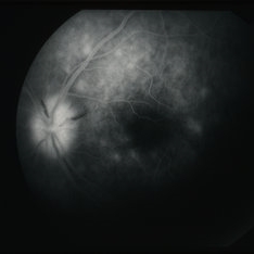
---thumb.jpg/image-square;max$300,300.ImageHandler) Posterior Uveitis
Posterior Uveitis
Feb 15 2013 by From the Collections of Thomas M. Aaberg, MD and Thomas M. Aaberg Jr., MD
Schematic diagram depicting Posterior Uveitis characterized by diffuse retinal whitening and exudative retinal detachment.
Condition/keywords: posterior uveitis, retinitis
-
---thumb.jpg/image-square;max$300,300.ImageHandler) Posterior Uveitis
Posterior Uveitis
Feb 15 2013 by From the Collections of Thomas M. Aaberg, MD and Thomas M. Aaberg Jr., MD
Reprint of schematic drawings depicting Posterior Uveitis showing acute retinal lesions and vitreous debris (left panel) that are eventually replaced by areas of well-defined retinal pigment epithelial atrophy (right panel).
Condition/keywords: posterior uveitis, retinitis
-
---thumb.jpg/image-square;max$300,300.ImageHandler) Posterior Uveitis
Posterior Uveitis
Feb 15 2013 by From the Collections of Thomas M. Aaberg, MD and Thomas M. Aaberg Jr., MD
Color photograph of the mid-peripheral retina showing scattered intraretinal hemorrhage and foci of retinal whitening consistent with posterior uveitis, such as Behcet disease.
Condition/keywords: posterior uveitis, retinitis
-
---thumb.jpg/image-square;max$300,300.ImageHandler) Posterior Uveitis
Posterior Uveitis
Feb 15 2013 by From the Collections of Thomas M. Aaberg, MD and Thomas M. Aaberg Jr., MD
Progression of disease showing expansion and confluence of areas of retinal whitening over the course of 7 days.
Condition/keywords: posterior uveitis, retinitis
-
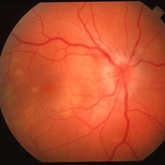 Posterior Uveitis
Apr 8 2019 by Gary R. Cook, MD, FACS
37-year-old white male with mild vitritis, optic disc hyperemia and edema, peripapillary hemorrhages and yellow-white spots in temporal macula OD; V.A. = 20/30.
Imaging device: Topcon VT-50
Condition/keywords: posterior uveitis
-
 Posterior Uveitis
Apr 8 2019 by Gary R. Cook, MD, FACS
37-year-old white male with mild vitritis, optic disc hyperemia and edema, and a couple of peripapillary NFL hemorrhages OS; V.A. = 20/20-1
Imaging device: Topcon VT-50
Condition/keywords: posterior uveitis
-
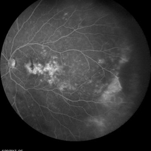
 Posterior Uveitis
Apr 8 2019 by Gary R. Cook, MD, FACS
Mid-phase (64 seconds) fluorescein angiogram image showing mild leakage and early staining of the yellow-white spots in the temporal macula of the right eye; V.A. = 20/30.
Imaging device: Topcon VT-50
Condition/keywords: FA mid phase, fluorescein angiogram (FA), posterior uveitis
-
 Posterior Uveitis
Apr 8 2019 by Gary R. Cook, MD, FACS
Late-phase (219 seconds) fluorescein angiogram image of the left eye showing late staining of the optic disc and of numerous spots deep to the retina; also blocked fluorescence from the 2 NFL hemorrhages on the optic disc; V.A. = 20/20-1
Imaging device: Topcon VT-50
Condition/keywords: FA late phase, fluorescein angiogram (FA), posterior uveitis
-
 Posterior Uveitis with Cystoid Macular Edema
Jan 18 2018 by Olivia Rainey
Ultra-wide field fluorescein angiogram of a 59-year-old female with posterior uveitis and chronic cystoid macular edema affecting her left eye. Interestingly, she has peripheral capillary nonperfusion inferotemporal, which could be driving CME.
Photographer: Olivia Rainey
Imaging device: Heidelberg Spectralis
Condition/keywords: 102 degrees, cystoid macular edema (CME), fluorescein leakage, Heidelburg Spectralis, left eye, peripheral retinal nonperfusion, posterior uveitis, ultra-wide field imaging
-
 Posterior Uveitis with Macular Edema
Jul 9 2024 by Korey Starkey
Ultra-wide field angiography of a 70 year old female with cystoid macular edema secondary to posterior uveitis. Patient's vision was Dcc20/200 at time of visit.
Photographer: Korey Starkey
Imaging device: Optos
Condition/keywords: cystoid macular edema (CME), fluorescein angiogram (FA), FLUORESCEIN ANGIOGRAPHY, hyperfluorescence, posterior uveitis, ULTRA WIDE FIELD, ultra-widefield image, vitreous debris
-
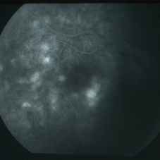
 Serpiginous Choroiditis
Sep 22 2019 by Haider Ali
35-year-old female presented with decrease in vision in her left eye for last 4 days and in right eye for last 8 days. Her right eye was previously involved in a similar episode about 5-6 months ago for which she was treated with oral steroids.
Photographer: Dr Haider Ali Chaudhry, Madinah Teaching Hospital, Faisalabad
Condition/keywords: acute posterior multifocal placoid pigment epitheliopathy (APMPPE), macula serpiginous choroidopathy, posterior uveitis, serpiginous choroiditis, uveitis, white dot lesions, white dot syndrome
 Loading…