 Initializing download.
-
 By Gary R. Cook, MD, FACS
- Uploaded on Apr 8, 2019.
- Last modified by Caroline Bozell on Aug 2, 2019.
- Rating
- Appears in
- POSTERIOR UVEITIS
- Condition/keywords
- posterior uveitis, FA late phase
- Imaging device
-
Fundus camera
Topcon VT-50 - Description
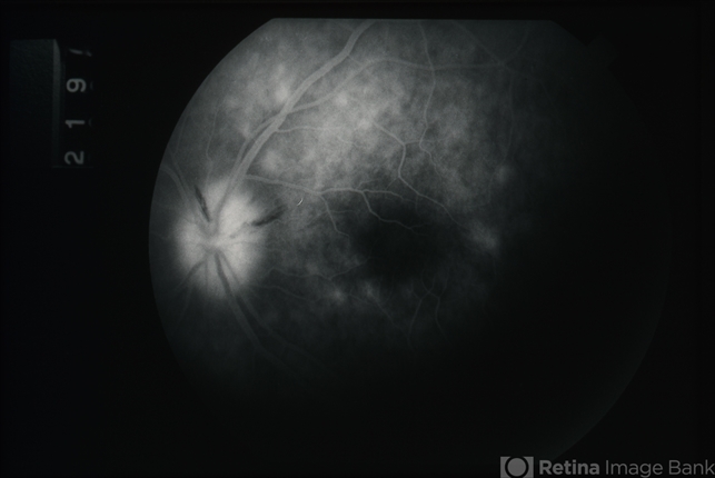
- Late-phase (219 seconds) fluorescein angiogram image of the left eye showing late staining of the optic disc and of numerous spots deep to the retina; also blocked fluorescence from the 2 NFL hemorrhages on the optic disc; V.A. = 20/20-1


---thumb.jpg/image-square;max$79,0.ImageHandler "Posterior Uveitis")
---thumb.jpg/image-square;max$79,0.ImageHandler "Posterior Uveitis")
---thumb.jpg/image-square;max$79,0.ImageHandler "Posterior Uveitis")
---thumb.jpg/image-square;max$79,0.ImageHandler "Behcet Uveitis")
---thumb.jpg/image-square;max$79,0.ImageHandler "Behcet Uveitis")
---thumb.jpg/image-square;max$79,0.ImageHandler "Behcet Disease")
---thumb.jpg/image-square;max$79,0.ImageHandler "Posterior Uveitis")