Search results (24 results)
-
 Choroidal Osteoma
Nov 21 2014 by Thomas A. Ciulla, MD, MBA, FASRS
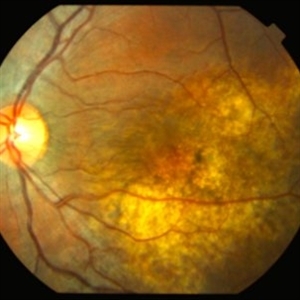
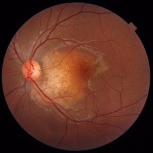
This 13-year-old girl presented with mild painless progressive blurring of central vision left eye over the past several months. Visual acuity was 20/25. In the affected left eye, retinal examination revealed a relatively flat, lightly pigmented lesion, with well-defined and scalloped edges. Clumps of associated pigment were noted.
Photographer: Thomas Steele
Condition/keywords: choroidal neovascular membrane (CNVM), choroidal neovascularization (CNV), choroidal osteoma, macular choroidal osteoma
-
 Choroidal Osteoma
Nov 21 2014 by Thomas A. Ciulla, MD, MBA, FASRS
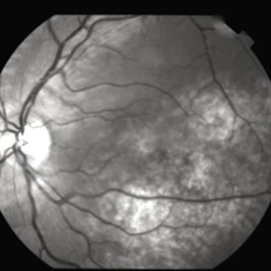
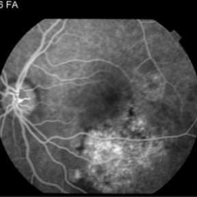
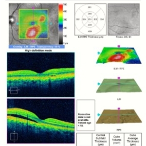
This 13-year-old girl presented with mild painless progressive blurring of central vision left eye over the past several months. Visual acuity was 20/25. In the affected left eye, retinal examination revealed a relatively flat, lightly pigmented lesion, with well-defined and scalloped edges. Clumps of associated pigment were noted. This OCT image shows subretiinal fluid just inferior to the fovea. Choroidal osteoma can be associated with the development of subretinal neovascularization (particularly at the edges of the osteoma).
Photographer: Thomas Steele
Condition/keywords: choroidal neovascular membrane (CNVM), choroidal neovascularization (CNV), choroidal osteoma, macular choroidal osteoma
-
 Choroidal Osteoma
Nov 21 2014 by Thomas A. Ciulla, MD, MBA, FASRS
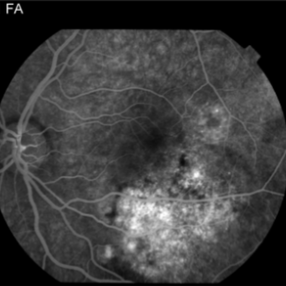
This OCT image shows subretiinal fluid just inferior to the fovea. Choroidal osteoma can be associated with the development of subretinal neovascularization (particularly at the edges of the osteoma).
Condition/keywords: choroidal neovascular membrane (CNVM), choroidal neovascularization (CNV), choroidal osteoma, macular choroidal osteoma
-
 Choroidal Osteoma
Aug 23 2012 by Gerardo Garcia-Aguirre, MD
Fundus photograph of the left eye of a 32 year-old male patient showing a choroidal osteoma. The right eye also has a choroidal osteoma with choroidal neovascularization.
Photographer: Ricardo Montoya, Asociación para Evitar la Ceguera en México
Condition/keywords: macular choroidal osteoma
-
 Choroidal Osteoma
Nov 10 2022 by Tandava Krishnan
B scan imaging of the eye with choroidal osteoma showing high reflective choroidal lesion with Posterior shadowing suggestive of a bone like lesion
Condition/keywords: choroidal osteoma, choroidal tumor, choroidal tumour, macular choroidal osteoma
-
 Choroidal osteoma
Nov 10 2022 by Tandava Krishnan
Right eye fundus picture of a patient with Creamy yellow choroidal lesion suggestive of Choroidal osteoma
Condition/keywords: choroidal osteoma, choroidal tumor, choroidal tumour, macular choroidal osteoma
-
 Choroidal Osteoma
Apr 17 2025 by Gustavo Uriel Fonseca Aguirre
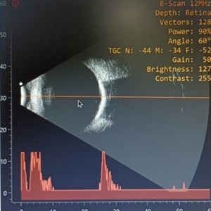
Top (B-mode): The longitudinal scan reveals a hyperechoic, flat, and well-demarcated macular lesion with posterior acoustic shadowing, pathognomonic for choroidal osteoma. Bottom (A-mode): Standardized tracing shows a tall initial spike (100% reflectivity) at the tumor surface with rapid decay to acoustic silence, confirming sound absorption by calcified tissue. This pattern remains unchanged at variable gain settings.
Photographer: Gustavo U. Fonseca Aguirre, Hospital Conde de Valenciana, Ciudad de México
Condition/keywords: choroidal osteoma, macular choroidal osteoma
-
 Choroidal Osteoma
Apr 17 2025 by Gustavo Uriel Fonseca Aguirre
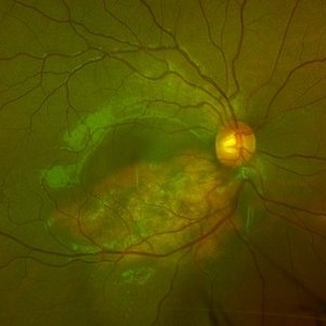
Scanning laser ophthalmoscopy reveals a well-circumscribed, yellowish-white choroidal osteoma overlying the macular region and extending into the inferior temporal vascular arcade. Retinal vessels course normally over the tumor surface, with no evidence of subretinal fluid or hemorrhage. The surrounding retina shows preserved architecture without secondary degenerative changes.
Photographer: Gustavo U. Fonseca Aguirre, Hospital Conde de Valenciana, Ciudad de México
Condition/keywords: choroidal osteoma, macular choroidal osteoma
-
 Choroidal Osteoma
Jun 2 2018 by awaneesh m upadhyay, MBBS, DNB
23-year-old patient's fundus photograph having complaints of defective vision, metamorphosia over 6 months shows yellow orange elevated well defined submacular lesion with normal overlying retinal vessels and normal disc . Vision left eye is 20/80.
Photographer: Hiteshwar Saikia
Condition/keywords: macular choroidal osteoma
-
 Choroidal Osteoma
Jan 3 2022 by Thirumalesh Mochi Basavaraj, MD
Fundus photograph of a young female in her second decade with a choroidal mass lesion with calcification suggestive of choroidal osteomalacia.
Photographer: Putta Swamy, Narayana Nethralaya
Imaging device: Topcon DRI Triton
Condition/keywords: macular choroidal osteoma
-
 Choroidal Osteoma 1
Oct 5 2012 by Ronald C. Gentile, MD
A cream colored choroidal osteoma involving the temporal macula in a young women
Photographer: The New York Eye & Ear Infirmary Department of Medical Imaging
Condition/keywords: choroidal tumor, macular choroidal osteoma
-
 Choroidal Osteoma 2
Oct 5 2012 by Ronald C. Gentile, MD
Magnified view of the macular choroidal osteoma with visible internal vascularity.
Photographer: The New York Eye & Ear Infirmary Department of Medical Imaging
Condition/keywords: choroidal tumor, macular choroidal osteoma
-
 Choroidal Osteoma 3
Oct 5 2012 by Ronald C. Gentile, MD
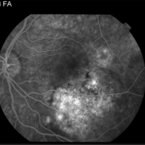
Fluorescein angiography of the macular choroidal osteoma in the early arterial-venous phase highlighting its internal vascularity.
Photographer: The New York Eye & Ear Infirmary Department of Medical Imaging
Condition/keywords: macular choroidal osteoma
-
 Choroidal Osteoma 4
Oct 5 2012 by Ronald C. Gentile, MD
Fluorescein angiography of the macular choroidal osteoma in the late phase of the angiogram with late staining of the tumor highlighting its negative staining internal vascularity.
Photographer: The New York Eye & Ear Infirmary Department of Medical Imaging
Condition/keywords: macular choroidal osteoma
-
 Choroidal Osteoma 5
Oct 5 2012 by Ronald C. Gentile, MD
B scan ultrasonography with representative A scan of the macular choroidal osteoma. The B scan reveals a characteristic highly reflective plaque consistent with its bone-like calcium composition that persists with low gain. The A scan reveals a large spike.
Photographer: The New York Eye & Ear Infirmary Department of Medical Imaging
Condition/keywords: B scan ultrasound, choroidal tumor, macular choroidal osteoma
-
 Choroidal Osteoma After PDT
Sep 9 2021 by Jesus Lozano, MD
45 year-old man with Choroidal Osteoma after PDT several years ago.
Photographer: Yair Bet Yosef, Hadassah Medical Center. Israel
Imaging device: Optos Silverstone
Condition/keywords: choroidal osteoma, macular choroidal osteoma, photodynamic therapy, retina
-
 Choroidal Osteoma and Secondary Choroidal Neovascular Membrane
Sep 21 2012 by Allen Chiang, MD, FASRS
Fundus photograph of a 44-year old woman with a choroidal osteoma complicated by secondary choroidal neovascular membrane, regressed after serial intravitreal bevacizumab injections. The tumor exhibits areas of decalcification.
Imaging device: Topcon
Condition/keywords: choroidal neovascularization (CNV), choroidal osteoma, macular choroidal osteoma
-
 Choroidal Osteoma with Choroidal Neovascularization
Aug 23 2012 by Gerardo Garcia-Aguirre, MD
Fundus photograph of the right eye of a 32 year-old male patient with choroidal osteoma and choroidal neovascularization. The left eye also has a choroidal osteoma.
Photographer: Ricardo Montoya, Asociación para Evitar la Ceguera en México
Condition/keywords: choroidal neovascularization (CNV), macular choroidal osteoma
-
 Choroidal Osteoma With CNV
Feb 28 2024 by stephen oconnell
23 year old male with 10 months of vision loss prior to presentation. Clarus 700 image of lesion with CNV and dependent subretinal fluid into inferior fundus.
Condition/keywords: macular choroidal osteoma
-
 Choroidal Osteoma With CNV- Aautofluorescence
Feb 28 2024 by stephen oconnell
23 year old male with 10 months of vision loss prior to presentation. Clarus 700 autofluorescence image.
Condition/keywords: macular choroidal osteoma
-
 Macular Choroidal Osteoma
Aug 17 2012 by Jonathan L. Prenner, MD
Macular choroidal osteoma in a 29-year-old woman
Condition/keywords: macular choroidal osteoma
 Loading…