Search results (153 results)
-
 Acute Posterior Vitreous Detachment
Nov 9 2012 by Norman Byer
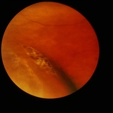
This large and complicated retinal tear in a 51-year-old man resulted from an acute posterior vitreous detachment which concentrated its tractional forces around this area of lattice degeneration. Because of the powerful traction, there is an additional central tear splitting the large retinal flap and almost severing one of its arms. The traction was strong enough to completely rupture the blood vessel just to the left of the flap. Marking the ruptured peripheral end of the blood vessel is a yellow depigmented thrombus.
Condition/keywords: acute posterior vitreous detachment, depigmented thrombus, lattice degeneration, retinal tear, tractional retinal detachment
-
 Acute Retinal Detachment
Nov 9 2012 by Norman Byer
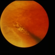
This 54-year-old man was referred because of sudden symptoms in his opposite eye in which he had suffered an acute retinal detachment secondary to a horseshoe tear around lattice degeneration. During the examination, the fellow eye shown here was also found to have this large horseshoe tear about 1 o’clock hour (4 disc diameters) in size. A tear occurred around a lattice lesion which is present on the flap but is out of focus. This tear had been asymptomatic even though it was caused by a posterior vitreous detachment and illustrates that even very large tears may produce no symptoms or mild symptoms that are easily overlooked.
Condition/keywords: lattice degeneration, posterior vitreous detachment
-
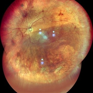
 Chronic Retinal Detachment with Proliferative Vitreoretinopathy
Jan 25 2024 by Isaac Agranoff
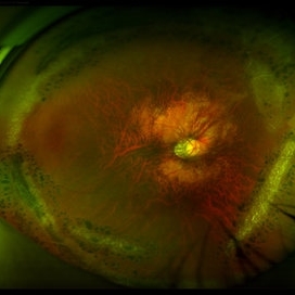
Widefield fundus photography of a 24 year old male presenting with subtotal retinal detachment with circumferential anterior proliferative vitreoretinopathy. The detachment is bullous inferiorly with atrophic retina and subretinal bands. There are also scattered patches of lattice with atrophic holes and associated detachment in the periphery. Patient presented with flashes for 2 years with worsening vision over the past 6-8 months, measured at 20/100 ph 20/60 OS.
Photographer: Isaac Agranoff, Ashley Rigdon
Imaging device: Optos California
Condition/keywords: atrophic hole, chronic retinal detachment, lattice degeneration, proliferative vitreoretinopathy (PVR), subretinal bands
-
 ERMageddon - Wrinkle in the Space-time Fabric of Macula
Oct 29 2025 by SHRADDHA RAJ SHRIVASTAVA
38 year old female with Epiretinal Membrane (ERM) over macula, post laser barrage for multiple symptomatic Horse-shoe Tears (HSTs) and Lattice Degenerations (seen on wide-field image). Posterior pole revealed tilted disc with peripapillary atrophy. There is thick opaque epiretinal membrane obscuring the underlying superior arcade vessels and causing foveal ectopia with distortion of perimacular vasculature. Patient was planned for Right Eye pars plana vitrectomy for ERM peeling.
Photographer: Dr. Shraddha Raj Shrivastava
Imaging device: Nidek Mirante SLO/OCT (Confocal scanning/Spectral domain OCT
Condition/keywords: BARRAGE LASER, ectopic fovea, epiretinal membrane (ERM), horseshoe tear, lattice degeneration, vitreomacular traction (VMT)
-
 Flat Lattice Lesion
Nov 9 2012 by Norman Byer
This 24-year-old woman had a flat lattice lesion without holes observed with no change for six years. She then developed two tiny retinal holes in this lesion and three years later the clinical retinal detachment shown here. She responded well to surgery. Even though such atrophic holes and lattice lesions may occasionally lead to a clinical detachment, it is important to understand that the mere presence of such holes is not an indication for prophylactic treatment. The reason for this is that we now know statistically that fewer than 1 percent of such cases lead to a retinal detachment.
Condition/keywords: lattice degeneration, retinal hole, scleral depression
-
 Flat Lattice Lesion
Nov 9 2012 by Norman Byer
This 34 year-old man had a flat lattice lesion with no hole at this location for five years. Then he developed this round hole with a small subclinical retinal detachment which has not changed in appearance for four years. Note the tiny glial tuft just to the left of the hole and superimposed against the dark background.
Condition/keywords: glial tuft, lattice degeneration, round hole
-
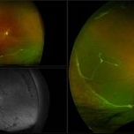
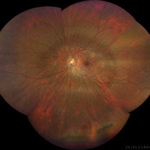 High Myopia with Posterior staphyloma
Nov 7 2023 by Harsh Vardhan Singh, MS
27-year old with both eyes high myopia & posterior staphyloma with left eye peripheral lattice degeneration & white without pressure
Photographer: Harsh Vardhan Singh
Imaging device: Clarus 700
Condition/keywords: lattice degeneration, myopia, peripheral lattice degeneration, posterior staphylomaloma, white without pressure
-
 Inferior Retinal Detachment with Lattice
Sep 30 2020 by Sham Talati, DOMS
A patient of inferior retinal detachment with lattice inferiorly.
Photographer: Dr. Sham Talati,Retina Foundation,Ahmedabad
Imaging device: Nidek Mirante
Condition/keywords: lattice degeneration
-
 Laser Treated Lattice Degeneration
Jul 12 2021 by Gabriel Costa Andrade, PhD
Fundus photograph of an 22-year-old man with peripheral lattice retinal degeneration treated with photocoagulation.
Photographer: Gabriel Andrade
Condition/keywords: lattice degeneration, retina
-
 Lattice Combined with Tiny Round Hole
Nov 9 2012 by Norman Byer
This 45 year-old man shows the snail track form of lattice combined with a tiny round hole. There is a tiny subclinical retinal detachment confined to the lesion itself.
Condition/keywords: glial vitreous tuft, lattice degeneration, round hole, snail track
-
 Lattice Degeneration
Nov 9 2012 by Norman Byer
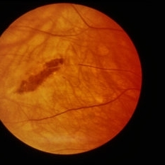
This 16-year-old girl has lattice degeneration and also this large oval retinal hole with a surrounding narrow zone of subretinal fluid. This lesion illustrates how large the atrophic holes of lattice degeneration may be. Occasionally the hole can be as large as the initial lattice lesion and can therefore obliterate all other evidence of its true identity. This was almost true in this case, but there does remain a small whitish remnant of the original lattice lesion at the lower end of the oval hole.
Condition/keywords: lattice degeneration, retinal hole, subretinal fluid, white lattice lines
-
 Lattice Degeneration
Nov 9 2012 by Norman Byer
This is a very subtle example of lattice degeneration showing the mildest possible changes in a 27-year-old man. In the upper left there is a vein directed toward the center of the slide. Just above and to the right of the pigment spot it veers to the right and then abruptly disappears as it passes through the lattice lesion. As it leaves the lesion, it resumes its normal appearance going down to the right. In a similar manner, the arteriole in the lower left enters the lesion just to the right of the pigment spot, then disappears as it passes through the lesion and reappears later as it emerges. The only change in this lesion in 12 years was the appearance of the pigment spot.
Condition/keywords: lattice degeneration
-
 Lattice Degeneration
Nov 9 2012 by Norman Byer
This is a more typical classical example of lattice degeneration in a 42-year-old woman in a photograph taken without scleral indentation. It shows much more marked vascular changes than the previous case. Note the tapering of the blood columns as the vessels approach the lesion and also the white sheathing of the vessel walls. Note also the continuity of the blood vessels on opposite sides of the lesion with the characteristic white lattice lines. More than 45 years ago Vogt pointed this out as a proof that these white lines were actually caused by changed blood vessels. Note also that this lesion shows a combination of several individual features of lattice degeneration. In addition to the white lines, there is a reddish crater-like area beneath the main horizontal white line. There is a prominent horizontal zone below this white line showing a snailtrack appearance. Also, there are two tiny atrophic retinal holes outside the photograph on the right end of this lesion. This eye contained five such retinal holes and they have all remained unchanged for more than 10 years of observation without treatment.
Condition/keywords: atrophic retinal hole, lattice degeneration, moderate snail track, tapering blood columns, white lattice lines, white sheath vessel
-
 Lattice Degeneration
Nov 9 2012 by Norman Byer
This is lattice degeneration in a 10-year-old boy showing an almost pure snailtrack feature with only a hint of a reddish crater in the center. It has not changed over 10 years. The photograph was taken with scleral indentation.
Condition/keywords: lattice degeneration, reddish crater, scleral indentation, snail track
-
 Lattice Degeneration
Nov 9 2012 by Norman Byer
This lesion in a 51-year-old woman is also an example of lattice degeneration but shows only a uniform reddish crater with no other features. This lesion has remained exactly the same for 9 years but such red craters sometimes give rise to punched-out atrophic retinal holes which may lead to subclinical retinal detachment. This sequence of events will be shown in the next two slide pairs.
Condition/keywords: lattice degeneration, lattice lesion, reddish crater
-
 Lattice Degeneration
Apr 27 2018 by Carolyn Daley
Fundus photograph of a 15-year-old woman with lattice degeneration and atrophic holes. Patient will have laser treatment at her next appointment.
Photographer: Carolyn Daley, Retina Specialists of Michigan
Imaging device: OPTOS Ultra-Wide Field Camera
Condition/keywords: atrophic retinal hole, lattice degeneration, Optos
-
---thumb.JPG/image-square;max$300,300.ImageHandler) Lattice Degeneration
Lattice Degeneration
Jul 12 2013 by Jason S. Calhoun
Composite of HD-OCT and fundus photo showing demarcation line of lattice degeneration inferiorly temporally at 4-o'clock in a young black female.
Photographer: Jason S. Calhoun, Department of Ophthalmology, Mayo Clinic Jacksonville, Florida
Condition/keywords: lattice degeneration
-
 Lattice Degeneration
Nov 9 2012 by Norman Byer
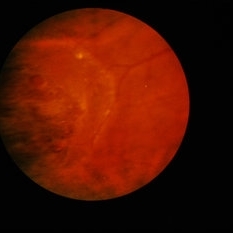
In this 54-year-old woman, lattice degeneration has led to a large horseshoe tractional tear around the posterior side on one end of the lesion resulting in a clinical retinal detachment. Note the very attenuated blood column passing through the white sheath vessel that crosses the tear. This demonstrates that the white blood vessels and a fragment of attached tissue are the only structures which have escaped the tearing effect of the strong vitreoretinal traction which occurred. This usually is true, although, in some cases this bridging vessel may bleed.
Condition/keywords: bridging vessel, lattice degeneration, tractional retinal tear, white sheath vessel
-
 Lattice Degeneration
Nov 9 2012 by Norman Byer
Lattice degeneration in a 42-year-old man which has produced four atrophic holes in a linear arrangement surrounded by a subclinical retinal detachment of unknown duration. By age 63, 21 years later, a posterior vitreous detachment was diagnosed in this eye, which was not present four years earlier. Nevertheless, the appearance seen here has remained exactly the same for 30 years, more than eight years with a concurrent PVD.
Condition/keywords: atrophic retinal hole, lattice degeneration, posterior vitreous detachment
-
 Lattice Degeneration
Nov 9 2012 by Norman Byer
Lesion immediately adjacent to the ora serrata in an 18-year-old boy probably represents lattice degeneration characterized primarily by a reddish crater. It has remained unchanged for more than three years.
Condition/keywords: lattice degeneration, ora serrata, reddish crater
-
 Lattice Degeneration
Aug 22 2023 by Vaidehi Sathaye
Wide field Montage of RE of a 46 year male with Lattice degeneration
Photographer: Dr. Vaidehi Sathaye
Imaging device: Mirante
Condition/keywords: lattice degeneration
-
 Lattice Degeneration
Jan 5 2015 by H. Michael Lambert, MD
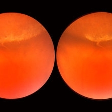
Parallel tracks of peripheral lattice (stereo pair A).
Condition/keywords: lattice degeneration
-
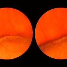
 Lattice Degeneration
Jan 5 2015 by H. Michael Lambert, MD
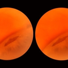
Parallel tracks of peripheral lattice (stereo pair B).
Condition/keywords: lattice degeneration
-
 Lattice Degeneration
Jan 5 2015 by H. Michael Lambert, MD
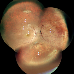
Lattice degeneration with round holes.
Condition/keywords: lattice degeneration
-
 Lattice Degeneration
Jan 5 2015 by H. Michael Lambert, MD
Perivascular lattice degeneration (stereo pair A).
Condition/keywords: lattice degeneration
 Loading…