 Initializing download.
-
 By Norman Byer
From Dr. Norman E. Byer’s “The Peripheral Retina in Profile” - Uploaded on Nov 9, 2012.
- Last modified by Suber S. Huang, MD, MBA, FASRS on Feb 10, 2013.
- Reviewed by Chayal Patel
- Rating
- Appears in
- Miscellaneous
- Condition/keywords
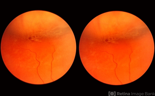
- retinal hole, lattice degeneration, scleral depression
- Description
- This 24-year-old woman had a flat lattice lesion without holes observed with no change for six years. She then developed two tiny retinal holes in this lesion and three years later the clinical retinal detachment shown here. She responded well to surgery. Even though such atrophic holes and lattice lesions may occasionally lead to a clinical detachment, it is important to understand that the mere presence of such holes is not an indication for prophylactic treatment. The reason for this is that we now know statistically that fewer than 1 percent of such cases lead to a retinal detachment.
---thumb.jpg/image-square;max$79,0.ImageHandler "Lattice Degeneration")


