Search results (502 results)
-
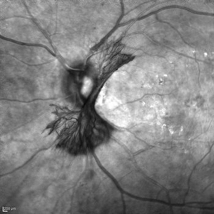 "NVD Flower"
Oct 20 2023 by Daniel Davis, OCT-C
Infrared image of NVD (52F)
Imaging device: Heidelberg Spectralis
Condition/keywords: neovascularization of the disc (NVD)
-
 Acquired Optic Pit Maculopathy
Aug 20 2014 by Andree Henaine-Berra, MD
Optical coherence tomography of the left eye of a 60-year-old man with an acquired optic pit maculopathy and glaucoma. The image shows an enlarged optic disc cup and a macular serous detachment.
Photographer: Andree Henaine-Berra. Asociacion Para Evitar la Ceguera en Mexico. Mexico City.
Imaging device: Heidelberg Spectralis
Condition/keywords: glaucoma, maculopathy, optic pit
-
 Acquired Optic Pit Maculopathy
Aug 20 2014 by Andree Henaine-Berra, MD
Fundus photograph of the left eye of a 60-year-old man with an acquired optic pit maculopathy and glaucoma. The image shows an enlarged optic disc cup and a macular serous detachment.
Photographer: Andree Henaine-Berra. Asociacion Para Evitar la Ceguera en Mexico. Mexico City.
Imaging device: Heidelberg Spectralis
Condition/keywords: glaucoma, maculopathy, optic pit
-
 Acquired Optic Pit Maculopathy
Aug 20 2014 by Andree Henaine-Berra, MD
Autofluorescence image of the left eye of a 60-year-old man with an acquired optic pit maculopathy and glaucoma.
Photographer: Andree Henaine-Berra. Asociacion Para Evitar la Ceguera en Mexico. Mexico City.
Imaging device: Heidelberg Spectralis
Condition/keywords: glaucoma, maculopathy, optic pit
-
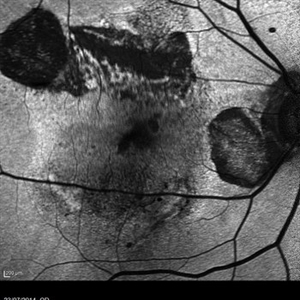
 Acquired Optic Pit Maculopathy
Aug 20 2014 by Andree Henaine-Berra, MD
Optical coherence tomography of the left eye of a 60-year-old man with an acquired optic pit maculopathy and glaucoma. The image shows subretinal fluid extending to the optic nerve and schisis of the outer retinal layers.
Photographer: Andree Henaine-Berra. Asociacion Para Evitar la Ceguera en Mexico. Mexico City.
Imaging device: Heidelberg Spectralis
Condition/keywords: glaucoma, maculopathy, optic pit
-
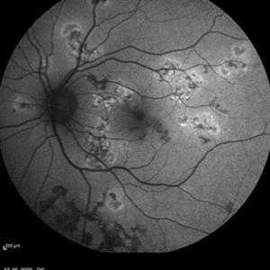 Active multifocal choroiditis
May 26 2025 by Moazzam Parvez
Auto fluorescence photograph of an 43 year old man with active choroiditic lesion present in the left eye with recurrence
Photographer: Dr Moazzam Parvez , Netralayam , Kolkata
Imaging device: Heidelberg Spectralis
Condition/keywords: active choroididtis, choroiditi
-
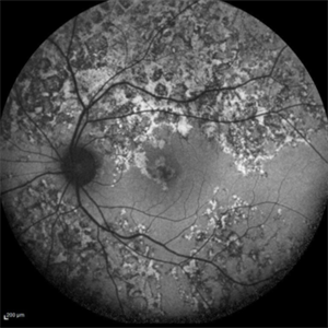 Active Multi Focal Choroiditis
Jun 21 2025 by Moazzam Parvez
Auto fluorescence image of a 28 year old gentleman with active multifocal choroiditis in his left eye and healed choroiditic patches in the right eye.
Photographer: Moazzam Parvez , Netralayam , Kolkata
Imaging device: Heidelberg Spectralis
Condition/keywords: active, multifocal choroiditis
-
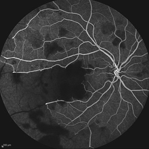
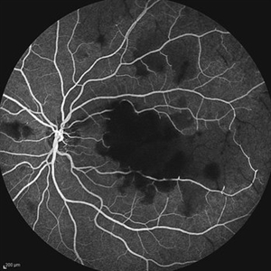
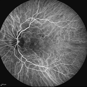
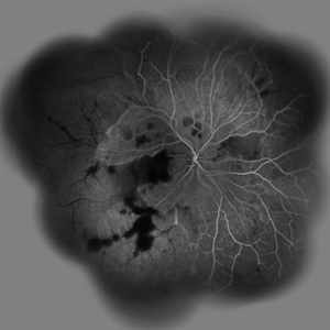 Acute Idiopathic Occlusive Retinal Vasculitis
May 31 2014 by Hamid Ahmadieh, MD
Wide- field fluorescein angiogram of the right eye of a 28-year-old woman with acute drop of vision due to occlusive retinal vasculitis leading to extensive capillary nonperfusion and macular infarction.
Photographer: Naghmeh Nozhat, Negah Eye Center, Tehran
Imaging device: Heidelberg Spectralis
Condition/keywords: capillary nonperfusion, retinal infarction, retinal vasculitis
-
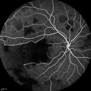
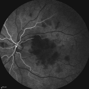
 Acute Idiopathic Occlusive Retinal Vasculitis
May 31 2014 by Hamid Ahmadieh, MD
Mid phase fluorescein angiogram of the right eye of a 28-year-old woman with acute drop of vision due to occlusive retinal vasculitis leading to extensive capillary nonperfusion and macular infarction.
Photographer: Naghmeh Nozhat, Negah Eye Center, Tehran
Imaging device: Heidelberg Spectralis
Condition/keywords: capillary nonperfusion, retinal vasculitis
-
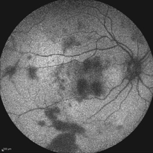
 Acute Idiopathic Occlusive Retinal Vasculitis
May 31 2014 by Hamid Ahmadieh, MD
Early phase fluorescein angiogram of the right eye of a 28-year-old woman with acute drop of vision due to occlusive retinal vasculitis leading to extensive capillary nonperfusion and macular infarction.
Photographer: Naghmeh Nozhat, Negah Eye Center, Tehran
Imaging device: Heidelberg Spectralis
Condition/keywords: retinal vasculitis
-
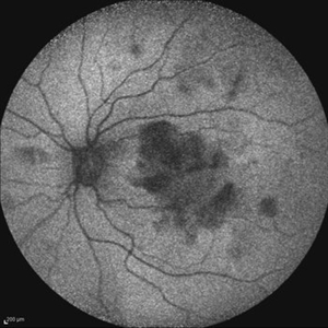
 Acute Idiopathic Occlusive Retinal Vasculitis
May 31 2014 by Hamid Ahmadieh, MD
Fundus autofluorescence image of the right eye of a 28-year-old woman with acute drop of vision due to occlusive retinal vasculitis leading to extensive nerve fiber layer infarction and retinal hemorrhages.
Photographer: Naghmeh Nozhat, Negah Eye Center, Tehran
Imaging device: Heidelberg Spectralis
Condition/keywords: fundus autofluorescence (FAF), retinal vasculitis
-
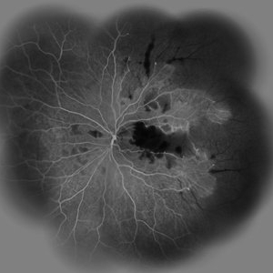 Acute Idiopathic Occlusive Retinal Vasculitis
May 31 2014 by Hamid Ahmadieh, MD
Wide- field fluorescein angiogram of the left eye of a 28-year-old woman with acute drop of vision due to occlusive retinal vasculitis leading to extensive capillary nonperfusion and macular infarction.
Photographer: Naghmeh Nozhat, Negah Eye Center, Tehran
Imaging device: Heidelberg Spectralis
Condition/keywords: capillary nonperfusion, retinal infarction, retinal vasculitis
-
 Acute Idiopathic Occlusive Retinal Vasculitis
May 31 2014 by Hamid Ahmadieh, MD
Mid- phase fluorescein angiogram of the left eye of a 28-year-old woman with acute drop of vision due to occlusive retinal vasculitis leading to extensive capillary nonperfusion and macular infarction.
Photographer: Naghmeh Nozhat, Negah Eye Center, Tehran
Imaging device: Heidelberg Spectralis
Condition/keywords: capillary nonperfusion, retinal vasculitis
-
 Acute Idiopathic Occlusive Retinal Vasculitis
May 31 2014 by Hamid Ahmadieh, MD
Early phase fluorescein angiogram of the left eye of a 28-year-old woman with acute drop of vision due to occlusive retinal vasculitis leading to extensive capillary nonperfusion and macular infarction.
Photographer: Naghmeh Nozhat, Negah Eye Center, Tehran
Imaging device: Heidelberg Spectralis
Condition/keywords: retinal vasculitis
-
 Acute Idiopathic Occlusive Retinal Vasculitis
May 31 2014 by Hamid Ahmadieh, MD
Fundus autofluorescence image of the left eye of a 28-year-old woman with acute drop of vision due to occlusive retinal vasculitis leading to extensive nerve fiber layer infarction and retinal hemorrhages.
Photographer: Naghmeh Nozhat, Negah Eye Center, Tehran
Imaging device: Heidelberg Spectralis
Condition/keywords: fundus autofluorescence (FAF), retinal vasculitis
-
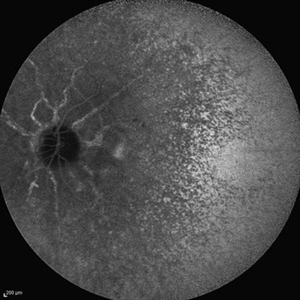
 Acute Macular Neuroretinopathy
Dec 11 2019 by Lauren Whaley
34-year-old female patient presented with changes in vision after recent upper respiratory infection. Referring doctor originally thought it was a blood pressure issue. She noticed a "C" shape in her vision. Infrared image was captured showing exactly what patient was describing! Doctor confirmed with this image that it was AMN.
Photographer: Lauren R. Whaley, COA
Imaging device: Heidelberg Spectralis
Condition/keywords: 30 degrees, acute macular neuroretinopathy, Heidelburg Spectralis, left eye, macula, near infrared autofluorescence (NIRAF)
-
 AMD
AMD
Jul 26 2014 by Avris Romario Diparaja Siahaan
An autofluorescence image of a 78-year-old-man with an age-related macular degeneration on his both eyes.
Photographer: Avris Romario Diparaja Siahaan, Klinik Mata Nusantara
Imaging device: Heidelberg Spectralis
Condition/keywords: age-related macular degeneration (AMD), autofluorescence imaging
-
 Angioid Streaks
Sep 29 2012 by Hamid Ahmadieh, MD
Late phase ICG angiography image of the right eye of a 59-year-old man with angioid streaks.
Photographer: Hamid Ahmadieh, MD; Ophthalmic Research Center, Labbafinejad Medical Center, Shahid Beheshti University of Medical Sciences
Imaging device: Heidelberg Spectralis
Condition/keywords: angioid streaks, indocyanine green (ICG) angiography
-
 Angioid Streaks
Sep 29 2012 by Hamid Ahmadieh, MD
Late phase ICG angiography image of the left eye of a 59-year-old man with angioid streaks.
Photographer: Hamid Ahmadieh, MD; Ophthalmic Research Center, Labbafinejad Medical Center, Shahid Beheshti University of Medical Sciences
Imaging device: Heidelberg Spectralis
Condition/keywords: angioid streaks, indocyanine green (ICG) angiography
-
Angioid Streaks & CNV (Fig 1)
Aug 25 2012 by Hamid Ahmadieh, MD
Fundus autofluorescence (FAF) of a 53-year-old woman with a juxtafoveal CNV secondary to angioid streaks.
Photographer: Hamid Ahmadieh, Ophthalmic Research Center, Labbafinejad Medical Center
Imaging device: Heidelberg Spectralis
Condition/keywords: angioid streaks, choroidal neovascularization (CNV), fundus autofluorescence (FAF)
-
 Angioid Streaks & CNV (Fig 3)
Aug 25 2012 by Hamid Ahmadieh, MD
Early phase ICG angiography imaging of a 53-year-old woman with a juxtafoveal CNV secondary to angioid streaks.
Photographer: Hamid Ahmadieh, Ophthalmic Research Center, Labbafinejad Medical Center
Imaging device: Heidelberg Spectralis
Condition/keywords: angioid streaks, choroidal neovascularization (CNV), indocyanine green (ICG) angiography
-
 Angioid Streaks & CNV (Fig 4)
Aug 25 2012 by Hamid Ahmadieh, MD
Late phase ICG angiography imaging of a 53-year-old woman with a juxtafoveal CNV secondary to angioid streaks.
Photographer: Hamid Ahmadieh, Ophthalmic Research Center, Labbafinejad Medical Center
Imaging device: Heidelberg Spectralis
-
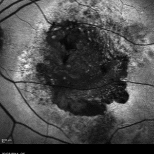
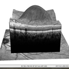 APMPPE With Serous Macular Detachment 3D SD-OCT
Jun 2 2014 by Rameez N Hussain, MD
3D SD-OCT of acute posterior multifocal placoid pigment epitheliopathy (APMPPE) with serous macular detachment.
Photographer: Rameez N Hussain MD, Vitreo Retinal Services, Giridhar Eye Institute, Cochin, India
Imaging device: Heidelberg Spectralis
Condition/keywords: acute posterior multifocal placoid pigment epitheliopathy (APMPPE), serous retinal detachment
-
 APMPPE With Serous Macular Detachment SD-OCT
Jun 2 2014 by Rameez N Hussain, MD
SD OCT image of acute posterior multifocal placoid pigment epitheliopathy (APMPPE) with serous macular detachment.
Photographer: Rameez N Hussain MD, Vitreo Retinal Services, Giridhar Eye Institute, Cochin, India
Imaging device: Heidelberg Spectralis
Condition/keywords: acute posterior multifocal placoid pigment epitheliopathy (APMPPE), serous retinal detachment
 Loading…