Search results (39 results)
-
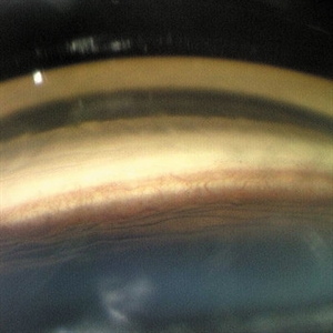
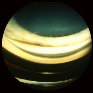
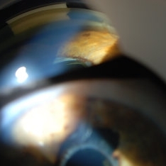
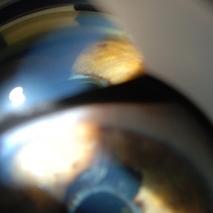
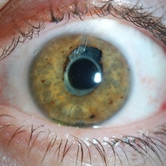
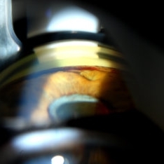
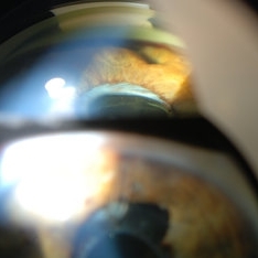
 Angle Closure/ UBM and Gonioscopy
Jul 8 2013 by Jason S. Calhoun
Patient came in a day after dilation with severe headache and nausea. Patient's IOP was 64 in the right eye. Patient ended up having angle closure attack. Promoted severe nausea and eye pain. Peripheral iridotomy was performed and IOP dropped slowly. Patient was followed up the next morning and will be seen 2-days after that.
Photographer: Jason S. Calhoun, Department of Ophthalmology, Mayo Clinic Jacksonville, Florida
Condition/keywords: angle closure, gonioscopy, ultrasound
-
 Angle Neovascularization
Mar 21 2013 by Yusuke Oshima, MD, PhD
Angle neovascularization due to ischemic CRVO.
Photographer: Yusuke Oshima, MD, PhD, Osaka University Graduate School of Medicine
Condition/keywords: angle neovascularization, gonioscopy
-
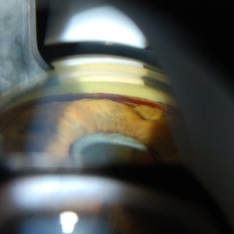
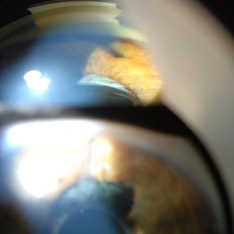
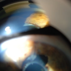
 Angle Recession, Gonioscopy
Oct 1 2012 by Jeffrey G. Gross, MD, FASRS
Angle recession, gonioscopy
Condition/keywords: angle recession, gonioscopy
-
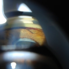
 Angle Recession, Gonioscopy
Oct 1 2012 by Jeffrey G. Gross, MD, FASRS
Angle recession, gonioscopy
Condition/keywords: angle recession, gonioscopy
-
 Ciliary body melanoma
Jul 4 2021 by Gerardo Rivera Arroyo
Clinical image taken in a slit lamp with a gonioscopy of a 39-year-old female patient with ciliary body melanoma before enucleation and pathological study.
Condition/keywords: ciliary body melanoma, gonioscopy
-
 Ciliary Body Melanoma
Jul 4 2021 by Gerardo Rivera Arroyo
Clinical image taken in a slit lamp with a gonioscopy of a 39-year-old female patient with ciliary body melanoma before enucleation and pathological study.
Condition/keywords: ciliary body melanoma, gonioscopy
-
 Ciliochoroidal Melanoma Photograph with Gonioscopy Lens
May 14 2020 by Anfisa Ayalon, MD
Gonioscopy photograph of a 71-year-old woman with ciliochoroidal melanoma. Note a melanoma-associated exudative retinal detachment and feeding vessels.
Photographer: Anfisa Ayalon, MD., Meir Medical Center, Kfar Saba, Israel.
Condition/keywords: ciliary body melanoma, exudative retinal detachment, gonioscopy
-
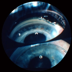
 Gonioscopy of Blood in the Angle
Jul 12 2013 by Jason S. Calhoun
Young female who has a history of uveitis, glaucoma, and juvenile rheumatoid arthritis. VA is hand motion in the left eye. Pigment changes with some blood in the anterior chamber seen here on gonioscopy.
Photographer: Jason S. Calhoun, Department of Ophthalmology, Mayo Clinic Jacksonville, Florida
Condition/keywords: gonioscopy
-
 Gonioscopy of Blood in the Angle
Jul 12 2013 by Jason S. Calhoun
Young female who has a history of uveitis, glaucoma and juvenile rheumatoid arthritis. VA is hand motion in the left eye. Pigment changes with some blood in the anterior chamber seen here on Gonioscopy.
Photographer: Jason S. Calhoun, Department of Ophthalmology, Mayo Clinic Jacksonville, Florida
Condition/keywords: gonioscopy
-
 Gonioscopy of Blood in the Angle
Jul 12 2013 by Jason S. Calhoun
Young female who has a history of uveitis, glaucoma and juvenile rheumatoid arthritis. VA is hand motion in the left eye. Pigment changes with some blood in the anterior chamber seen here on gonioscopy.
Photographer: Jason S. Calhoun, Department of Ophthalmology, Mayo Clinic Jacksonville, Florida
Condition/keywords: gonioscopy
-
 Gonioscopy of Blood in the Angle
Jul 12 2013 by Jason S. Calhoun
Young female who has a history of uveitis, glaucoma, and juvenile rheumatoid arthritis. VA is hand motion in the left eye. Pigment changes with some blood in the anterior chamber seen here on gonioscopy.
Photographer: Jason S. Calhoun, Department of Ophthalmology, Mayo Clinic Jacksonville, Florida
Condition/keywords: gonioscopy
-
 Gonioscopy of Blood in the Angle
Jul 12 2013 by Jason S. Calhoun
Young female who has a history of uveitis, glaucoma, and juvenile rheumatoid arthritis. VA is hand motion in the left eye. Pigment changes with some blood in the anterior chamber seen here on gonioscopy.
Photographer: Jason S. Calhoun, Department of Ophthalmology, Mayo Clinic Jacksonville, Florida
Condition/keywords: gonioscopy
-
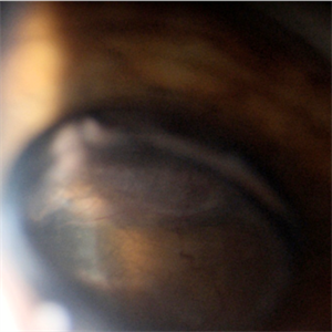
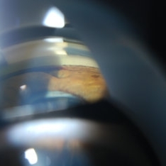
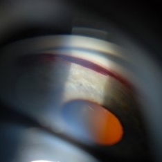 Gonioscopy, Blood in the Anterior Chamber from Hyphema
Jul 8 2013 by Jason S. Calhoun
Patient with blunt trauma to the right eye due to a BB gun incident. Patient was present with a hyphema at 8-o'clock about 1mm thick. Gonioscopy photos were then taken to show blood from the hyphema entered into the anterior chamber. Patient had no angle recession in the right eye.
Photographer: Jason S. Calhoun, Department of Ophthalmology, Mayo Clinic Jacksonville, Florida
Condition/keywords: angle recession, gonioscopy
-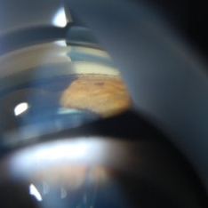
 Gonioscopy, Blood in the Anterior Chamber from Hyphema
Jul 8 2013 by Jason S. Calhoun
Patient with blunt trauma to the right eye due to a BB gun incident. Patient was present with a hyphema at 8-o'clock about 1mm thick. Gonioscopy photos were then taken to show blood from the hyphema entered into the anterior chamber. Patient had no angle recession in the right eye.
Photographer: Jason S. Calhoun, Department of Ophthalmology, Mayo Clinic Jacksonville, Florida
Condition/keywords: angle recession, gonioscopy
 Loading…