Search results (39 results)
-
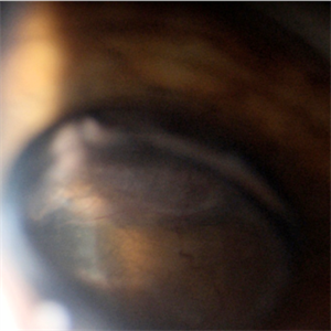
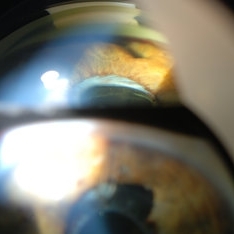
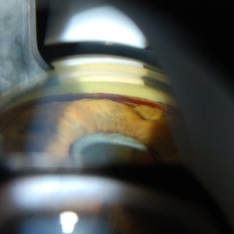
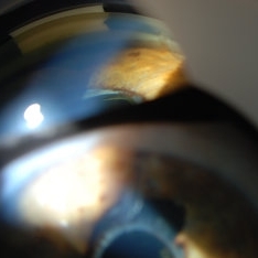
 Ciliary Body Melanoma
Jul 4 2021 by Gerardo Rivera Arroyo
Clinical image taken in a slit lamp with a gonioscopy of a 39-year-old female patient with ciliary body melanoma before enucleation and pathological study.
Condition/keywords: ciliary body melanoma, gonioscopy
-
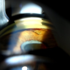
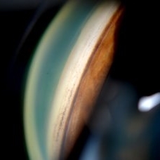
 Ciliary body melanoma
Jul 4 2021 by Gerardo Rivera Arroyo
Clinical image taken in a slit lamp with a gonioscopy of a 39-year-old female patient with ciliary body melanoma before enucleation and pathological study.
Condition/keywords: ciliary body melanoma, gonioscopy
-
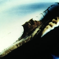
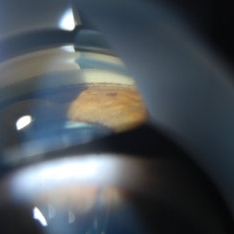
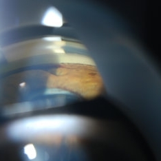
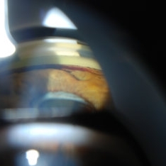
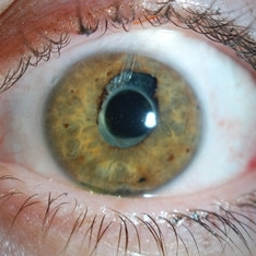
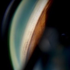
 Ciliochoroidal Melanoma Photograph with Gonioscopy Lens
May 14 2020 by Anfisa Ayalon, MD
Gonioscopy photograph of a 71-year-old woman with ciliochoroidal melanoma. Note a melanoma-associated exudative retinal detachment and feeding vessels.
Photographer: Anfisa Ayalon, MD., Meir Medical Center, Kfar Saba, Israel.
Condition/keywords: ciliary body melanoma, exudative retinal detachment, gonioscopy
-
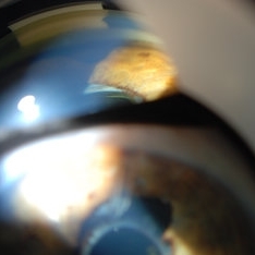
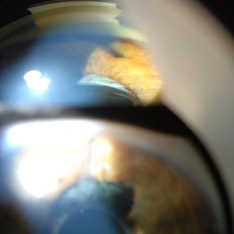
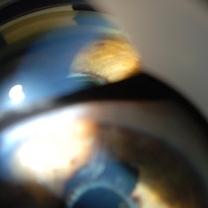
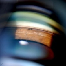
 Severe Rubeosis and Angle Neovascularization
Nov 21 2019 by Anfisa Ayalon, MD
Patient with proliferative diabetic retinopathy, severe retinal ischemia, rubeosis and angle neovascularization with IOP elevation.
Photographer: Anfisa Ayalon,MD., Meir Medical Center, Kfar Saba, Israel.
Imaging device: with gonioscopy lens
Condition/keywords: angle neovascularization, neovascular glaucoma, proliferative diabetic retinopathy (PDR), rubeosis
-
 Slide 7-33
Feb 25 2019 by Lancaster Course in Ophthalmology
Gonioscopy of Axenfeld's syndrome.
Condition/keywords: Axenfeld-Rieger syndrome, gonioscopy
-
 Wilson's Disease
Feb 20 2015 by H. Michael Lambert, MD
Kayser-Fleischer ring (copper deposition in Decemet's membrane in the peripheral cornea). Seen on gonioscopy.
Condition/keywords: Kayser-Fleischer ring, Wilson's disease
-
 Wilson's Disease
Feb 20 2015 by H. Michael Lambert, MD
Kayser-Fleischer ring (copper deposition in Decemet's membrane in the peripheral cornea). Seen on gonioscopy.
Condition/keywords: Kayser-Fleischer ring, Wilson's disease
-
 Gonioscopy of Blood in the Angle
Jul 12 2013 by Jason S. Calhoun
Young female who has a history of uveitis, glaucoma, and juvenile rheumatoid arthritis. VA is hand motion in the left eye. Pigment changes with some blood in the anterior chamber seen here on gonioscopy.
Photographer: Jason S. Calhoun, Department of Ophthalmology, Mayo Clinic Jacksonville, Florida
Condition/keywords: gonioscopy
-
 Gonioscopy of Blood in the Angle
Jul 12 2013 by Jason S. Calhoun
Young female who has a history of uveitis, glaucoma and juvenile rheumatoid arthritis. VA is hand motion in the left eye. Pigment changes with some blood in the anterior chamber seen here on gonioscopy.
Photographer: Jason S. Calhoun, Department of Ophthalmology, Mayo Clinic Jacksonville, Florida
Condition/keywords: gonioscopy
-
 Gonioscopy of Blood in the Angle
Jul 12 2013 by Jason S. Calhoun
Young female who has a history of uveitis, glaucoma and juvenile rheumatoid arthritis. VA is hand motion in the left eye. Pigment changes with some blood in the anterior chamber seen here on Gonioscopy.
Photographer: Jason S. Calhoun, Department of Ophthalmology, Mayo Clinic Jacksonville, Florida
Condition/keywords: gonioscopy
-
 Gonioscopy of Blood in the Angle
Jul 12 2013 by Jason S. Calhoun
Young female who has a history of uveitis, glaucoma, and juvenile rheumatoid arthritis. VA is hand motion in the left eye. Pigment changes with some blood in the anterior chamber seen here on gonioscopy.
Photographer: Jason S. Calhoun, Department of Ophthalmology, Mayo Clinic Jacksonville, Florida
Condition/keywords: gonioscopy
-
 Gonioscopy: Pigment Dispersion Glaucoma
Jul 8 2013 by Jason S. Calhoun
Patient with no family history of glaucoma, came in with elevated IOP. During gonioscopy exam. brown pigment overlying the trabecular meshwork. Also, trans-illumination defects on the iris.
Photographer: Jason S. Calhoun, Department of Ophthalmology, Mayo Clinic Jacksonville, Florida
Condition/keywords: gonioscopy, pigment dispersion syndrome of iris
-
 Gonioscopy; Scattered Peripheral Anterior Synechiae
Jul 8 2013 by Jason S. Calhoun
Patient came in for an evaluation for glaucoma. Patient also had a history of uveitis. Last flare up was back in 1990. Patient's VA was 20/30, Right eye and 20/40-1, Left eye. Slit lamp gonioscopy reveals iris bow with scattered PAS around the angles of the anterior chamber. You can also see pigmentation in the trabecular meshwork. Patient will follow up in 3 months.
Photographer: Jason S. Calhoun, Department of Ophthalmology, Mayo Clinic Jacksonville, Florida
Condition/keywords: gonioscopy, goniosynechiae
-
 Gonioscopy; Scattered Peripheral Anterior Synechiae
Jul 8 2013 by Jason S. Calhoun
Patient came in for evaluation for glaucoma. Patient also has a history of uveitis. Last flare up was back in 1990. Patient's VA was 20/30, right eye and 20/40-1, left eye. Slit Lamp gonioscopy reveals iris bow with scattered PAS around the angles of the anterior chamber. You can also see pigmentation in the trabecular meshwork. Patient will follow up in 3 months.
Photographer: Jason S. Calhoun, Department of Ophthalmology, Mayo Clinic Jacksonville, Florida
Condition/keywords: gonioscopy, goniosynechiae
-
 Gonioscopy; Scattered Peripheral Anterior Synechiae
Jul 8 2013 by Jason S. Calhoun
Patient came in for evaluation for glaucoma. Patient also has a history of uveitis. Last flare up was back in 1990. Patient's VA was 20/30, Right eye and 20/40-1, Left eye. Slit Lamp Gonioscopy reveals iris bow with scattered PAS around the angles of the anterior chamber. You can also see pigmentation in the trabecular meshwork. Patient will follow up in 3 months.
Photographer: Jason S. Calhoun, Department of Ophthalmology, Mayo Clinic Jacksonville, Florida
Condition/keywords: gonioscopy, goniosynechiae
-
 Angle Closure/ UBM and Gonioscopy
Jul 8 2013 by Jason S. Calhoun
Patient came in a day after dilation with severe headache and nausea. Patient's IOP was 64 in the right eye. Patient ended up having angle closure attack. Promoted severe nausea and eye pain. Peripheral iridotomy was performed and IOP dropped slowly. Patient was followed up the next morning and will be seen 2-days after that.
Photographer: Jason S. Calhoun, Department of Ophthalmology, Mayo Clinic Jacksonville, Florida
Condition/keywords: angle closure, gonioscopy, ultrasound
 Loading…