Search results (11 results)
-
 Slide 9-40
Feb 26 2019 by Lancaster Course in Ophthalmology
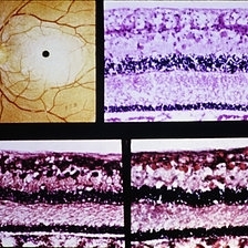
Tay-Sachs disease. Cherry-red spot (upper left). Retinal ganglion cells are distended by material which stains positively with the periodic acid-Schiff reaction (upper right) and with stains for lipid (Nile blue sulfate, lower left) (oil red-O, lower right).
Condition/keywords: ganglion cell, Tay-Sachs
-
 Slide 9-7
Feb 25 2019 by Lancaster Course in Ophthalmology
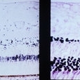
Inner retinal ischemia of central retinal artery occlusion. Early changes with edema and pyknosis of the nuclei of ganglion cells and inner aspect of the inner nuclear layer (left). With time, all of the inner retinal layers supplied by the central retinal artery disappear, including the nerve fiber, the ganglion cell, the inner plexiform layers, and the inner aspect of the inner nuclear layer (right).
Condition/keywords: edema, ganglion cell, pyknosis, retinal ischemia
-
 Slide 9-9
Feb 25 2019 by Lancaster Course in Ophthalmology
Localized area of inner ischemic infarction of the nerve fiber layer, presumably the site of a previous cotton-wool exudate. There is loss of the ganglion cell layer and thinning of the inner nuclear layer (between arrows).
Condition/keywords: ganglion cell, inner ischemic infarction
-
 Histopathology Mouse Retina - Normal
Apr 25 2013 by Suber S. Huang, MD, MBA, FASRS
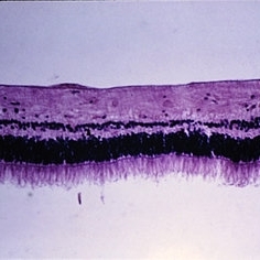
Mouse retinal structure is presented. The retina consists of seven layers, ganglion cell layer, inter plexiform layer, inner nuclear layer, outer plexiform layer, outer nuclear layer, photoreceptor layer and the retinal pigmented epithelium layer. Nuclei were stained with dapi (blue). Two kinds of photoreceptor cells; cone photoreceptors were stained with PNA (green) and rod photoreceptors were stained with anti-rhodopsin antibody (red).
Photographer: Akiko Maeda, Tadao Maeda
Imaging device: Fluorescence microscope
Condition/keywords: histopathology, retina
-
 Myelinated Nerve Fibers
Dec 31 2014 by Thomas A. Ciulla, MD, MBA, FASRS
Myelinated nerve fibers well depicted in this image as a distinct white patch on the inner retina (retinal ganglion cell axons as they sweep towards the optic nerve). This is often asymptomatic and noted as an incidental finding.
Photographer: Thomas Steele
Condition/keywords: myelinated nerve fiber layer, myelinated nerve fibers
-
 Representative Pattern Electroretinography Responses
May 13 2024 by Gabrielle Hallai
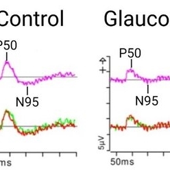
The pattern ERG response on the left is from a control individual with no known retinal pathology. There is a clearly discernable P50 and N95 peak. On the right, there is a representative image from a patient with end-stage glaucoma. In advanced glaucoma, there is often a lack of a clearly discernable N95 peak along with a diminished N95:P50 amplitude ratio, due to retinal ganglion cell dysfunction or degeneration. The top traces are averages from two independent trials shown on the bottom. Pattern ERG testing was completed using the Diagnosys pattern ERG protocol on a CRT monitor.
Photographer: Gabrielle Hallai, PhD, Cleveland Clinic Cole Eye Institute
Condition/keywords: electroretinography, glaucoma, pattern ERG (PERG)
-
 Sialidosis
Jul 10 2025 by Jessilla Phou
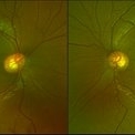
These are fundus photographs capturing an 18 year old male with Type 1 Sialidosis, a rare inherited lysosomal storage disorder caused by a deficiency in the neuraminidase 1 (Neu1) enzyme. Currently, there are fewer than 1,000 people in the USA who have this disorder. It is characterized by a cherry red spot in the macula which occurs when lipids accumulate in the retinal ganglion cells. This causes the macula to appear red as seen in these fundus images. The patient presented at our office with ataxia, depth perception issues, and slow reaction time. His visual acuity was 20/40, suggestive of early stage Sialidosis.
Photographer: Jessilla Phou
Imaging device: Optos California
Condition/keywords: cherry red spot, fundus photograph, Sialidosis
-
 Slide 11-30
Feb 26 2019 by Lancaster Course in Ophthalmology
Primary or descending optic atrophy. Horizontal section of the optic disk ( x 16). Note the increase in thickness of the pial septa in the nerve, the loss of ganglion cell and nerve fiber layers in the adjacent retina, and the loss of the optic cup.
Condition/keywords: optic atrophy, pial septa
-
 Slide 9-38
Feb 26 2019 by Lancaster Course in Ophthalmology
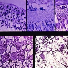
Type III systemic mucopolysaccharidosis (San Filippo syndrome). The retinal ganglion cells are distended (upper left) and contain mucopolysaccharide (middle, top: colloidal iron), lipid (upper right: Sudan-black B). Phase-contrast (lower left) shows numerous vacuoles, and by electron microscopy (lower right) there are numerous inclusions of the fibrillogranular and multimembranous types.
Condition/keywords: mucopolysaccharidoses, Sanfilippo syndrome, Type III systemic mucopolysaccharidosis
-
 Slide 9-89
Feb 26 2019 by Lancaster Course in Ophthalmology
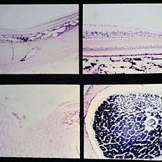
Nutritional amblyopia. The nerve fiber and ganglion cell layers are absent in the macular area (upper views). The temporal side of the optic nerve head (lower left) is partially atrophy, with marked reduction in the size of the nerve fiber bundles and secondary gliosis.
Condition/keywords: amblyopia, atrophy, gliosis, macular
 Loading…