Search results (247 results)
-
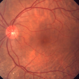
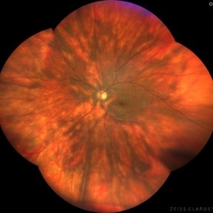
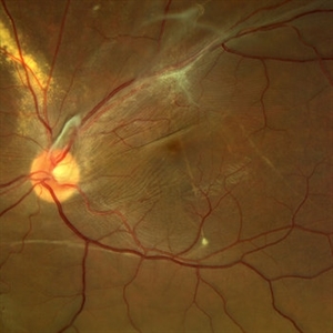 Displaced & folded macula
Oct 10 2022 by Ricardo Leitão Guerra
Tractional retinal detachment due to sickle cell retinopathy leading to a displaced and folded appearance of the macula in this 36-yo male. Subretinal bands are also noticed crossing the macula towards inferior retinal detachment area.
Photographer: Ricardo Leitão Guerra
Imaging device: Clarus 700 - Zeiss
Condition/keywords: folds, sickle cell retinopathy, subretinal bands, tractional retinal detachment
-
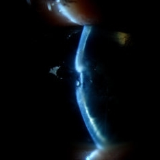
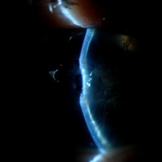
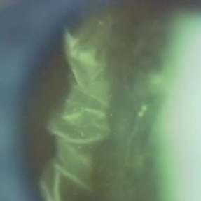 Folds in Detached Posterior Vitreous Cortex
May 31 2022 by Joshua Friedman
Slit lamp (video) image showing folds in the posterior vitreous cortex in an eye with PVD.
Photographer: Martin Snead, MD, Cambridge, England
Condition/keywords: folds, posterior vitreous cortex, PVD, vision degrading myodesopsia, vitreous
-
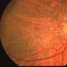
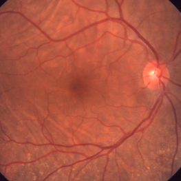
 A Classic Case of Retinal Ora Serrata Imaging
Jan 16 2025 by yuan duo
A 5-year-old girl, born full-term with no history of systemic disease, presented with poor vision since early childhood and underwent fundus examination. Anterior segments of both eyes showed no significant abnormalities. Fundus examination revealed retinal folds extending from the optic disc to the temporal peripheral retina, with blood vessels coursing through the folds (A, B). Avascular zones were observed in the peripheral retina, and the ora serrata’s boundaries were clearly visible, displaying dentate processes and bays (C, D). Retinal pigmentation was evident. Genetic testing confirmed the final diagnosis of bilateral Familial Exudative Vitreoretinopathy (FEVR).
Condition/keywords: Retinal Ora Serrata
-
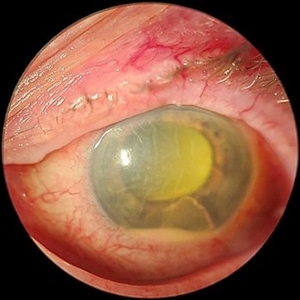 Acute Endophthalmitis
Nov 14 2023 by Virginia Gebhart
85 year old female with acute endophthalmitis 14 days s/p IVEylea injection. 3+ injection and descemets folds, contracted fibron, 3mm Hypopyon, and 3+ cell. No view posteriorly, vision CF. Visual prognosis unknown at this time
Photographer: Virginia Gebhart
Condition/keywords: endophthalmitis
-
 Advanced Proliferative Diabetic Retinopathy
Apr 9 2025 by Gustavo Uriel Fonseca Aguirre
B-mode ultrasound of a patient with long-standing poorly controlled diabetes demonstrates characteristic findings of advanced proliferative diabetic retinopathy. The examination reveals moderate vitreous hemorrhage appearing as diffuse hyperechoic opacities throughout the vitreous cavity, along with a posterior hyaloid membrane densely infiltrated by hemorrhagic material, showing irregular thickening and increased reflectivity. A mild subhyaloid hemorrhage is visible as a subtle hyphema-like space anterior to the retinal surface. The study documents a total tractional retinal detachment, evidenced by rigid retinal folds with clear insertion points of vitreous strands, accompanied by a significant subretinal hemorrhage seen as a prominent hyperechoic collection beneath the elevated retina. These findings collectively illustrate the severe vitreoretinal interface pathology characteristic of end-stage diabetic eye disease, with predominant tractional components and distinct echographic stratification of hemorrhagic layers - from anterior vitreous involvement to deeper subretinal blood accumulation.
Photographer: Gustavo U. Fonseca Aguirre, Hospital Conde de Valenciana, Ciudad de México
Condition/keywords: diabetic retinopathy, tractional retinal detachment, Vitreous hemorrhage
-
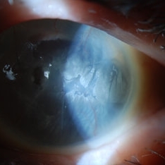
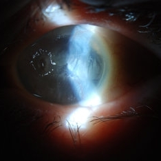 Band Keratopathy/Neurotrophic Ulcer
Nov 29 2013 by Jason S. Calhoun
Patient comes in with blind painful left eye. Slit lamp photos shows corneal diffuse scarring, descemets folds, corneal striae, band keratopathy, left eye. Proceed with Jupiter contact lens fitting on the left eye.
Photographer: Jason S. Calhoun, Ophthalmic Photographer, Department of Ophthalmology, Mayo Clinic Jacksonville
Imaging device: TOPCON D-90 SL NIKON CAMERA
Condition/keywords: band-shaped keratopathy, corneal dystrophy, folds in Descemet's membrane
-
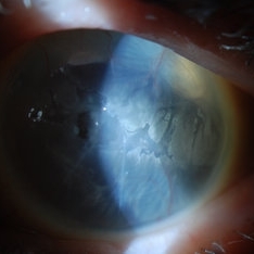
 Band Keratopathy/Neurotrophic Ulcer
Nov 29 2013 by Jason S. Calhoun
Patient comes in with blind painful left eye. Slit lamp photos shows corneal diffuse scarring, descemets folds, corneal striae, band keratopathy, left eye. Proceed with Jupiter contact lens fitting on the left eye.
Photographer: Jason S. Calhoun, Ophthalmic Photographer, Department of Ophthalmology, Mayo Clinic Jacksonville
Imaging device: TOPCON D-90 SL NIKON CAMERA
Condition/keywords: band-shaped keratopathy, corneal dystrophy, folds in Descemet's membrane
-
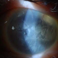
 Band Keratopathy/Neurotrophic Ulcer
Nov 29 2013 by Jason S. Calhoun
Patient comes in with blind painful left eye. Slit lamp photos shows corneal diffuse scarring, descemets folds, corneal striae, band keratopathy, left eye. Proceed with Jupiter contact lens fitting on the left eye.
Photographer: Jason S. Calhoun, Ophthalmic Photographer, Department of Ophthalmology, Mayo Clinic Jacksonville
Imaging device: TOPCON D-90 SL NIKON CAMERA
Condition/keywords: band-shaped keratopathy, corneal dystrophy, folds in Descemet's membrane
-
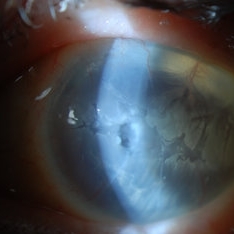
 Band Keratopathy/Neurotrophic Ulcer
Nov 29 2013 by Jason S. Calhoun
Patient comes in with blind painful left eye. Slit lamp photos shows corneal diffuse scarring, descemets folds, corneal striae, band keratopathy, left eye. Proceed with Jupiter contact lens fitting on the left eye.
Photographer: Jason S. Calhoun, Ophthalmic Photographer, Department of Ophthalmology, Mayo Clinic Jacksonville
Imaging device: TOPCON D-90 SL NIKON CAMERA
Condition/keywords: band-shaped keratopathy, corneal dystrophy, folds in Descemet's membrane
-
 Band Keratopathy/Neurotrophic Ulcer
Nov 29 2013 by Jason S. Calhoun
Patient in with blind painful left eye. Slit lamp photos shows corneal diffuse scarring, descemets folds, corneal striae, band keratopathy, left eye. Proceed with Jupiter contact lens fitting on the left eye.
Photographer: Jason S. Calhoun, Ophthalmic Photographer, Department of Ophthalmology, Mayo Clinic Jacksonville
Imaging device: TOPCON D-90 SL NIKON CAMERA
Condition/keywords: band-shaped keratopathy, corneal dystrophy, folds in Descemet's membrane
-
 Bilateral C-R Folds
Mar 26 2019 by Gary R. Cook, MD, FACS
Fundus photo of the left eye of a white male with bilateral C-R folds.
Imaging device: Topcon VT-50
Condition/keywords: bilateral chorioretinal folds, chorioretinal fold
-
 Bilateral C-R Folds
Mar 26 2019 by Gary R. Cook, MD, FACS
Fundus photo of the right eye of a white male with bilateral C-R folds.
Imaging device: Topcon VT-50
Condition/keywords: bilateral chorioretinal folds, chorioretinal fold
-
 Bilateral Idiopathic Choroidal Folds
Jan 11 2013 by Gerardo Garcia-Aguirre, MD
Fundus photograph of the right eye showing choroidal folds.
Imaging device: Zeiss FF4
Condition/keywords: choroidal folds
-
 Bilateral idiopathic choroidal folds
Jan 11 2013 by Gerardo Garcia-Aguirre, MD
Fundus photograph showing choroidal folds.
Imaging device: Zeiss ff4
Condition/keywords: choroidal folds
-
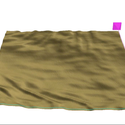 Bilateral Idiopathic Choroidal Folds
Jan 11 2013 by Gerardo Garcia-Aguirre, MD
3D reconstruction of the RPE showing choroidal folds.
Photographer: Gerardo Garcia-Aguirre, MD
Imaging device: Zeiss Cirrus HD OCT
Condition/keywords: choroidal folds
-
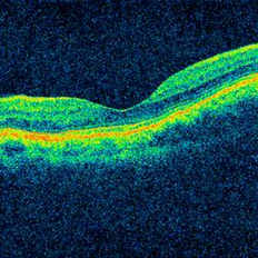 Bilateral Idiopathic Choroidal Folds - OCT
Jan 11 2013 by Gerardo Garcia-Aguirre, MD
OCT of the macula showing choroidal folds.
Photographer: Gerardo Garcia-Aguirre, MD
Imaging device: Zeiss Cirrus HD OCT
Condition/keywords: choroidal folds
-
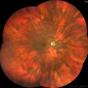 Birdshot Retinopathy
May 9 2023 by JEFFERSON R SOUSA, Tecg.º (Biomedical Systems Technology)
Female patient, 41 years old, with progressive low visual acuity, progressive history of autoimmune disease. In the multimodal retinal fundoscopic evaluation, important characteristics compatible with "Birdshot Retinopathy" were observed. Birdshot retinopathy, also known as birdshot chorioretinopathy or birdshot uveitis, is a rare, chronic inflammatory disorder that affects the retina and the choroid of the eye. It typically develops in adults between the ages of 30 and 60 years, and is more common in women than men. The name "birdshot" refers to the small, round, yellow-white spots that appear on the retina, which resemble the pattern of a shotgun blast. These spots are caused by inflammation in the eye, and can lead to vision loss if left untreated. Symptoms of birdshot retinopathy include blurred vision, floaters, loss of night vision, and difficulty adapting to changes in lighting. The condition can also cause inflammation in other parts of the eye, leading to redness, pain, and sensitivity to light. The exact cause of birdshot retinopathy is unknown, but it is believed to be an autoimmune disorder, in which the body's immune system mistakenly attacks the retina and choroid. Treatment typically involves the use of immunosuppressive medications, such as corticosteroids or biologic agents, to reduce inflammation and preserve vision. Close monitoring by an ophthalmologist is important, as the disease can progress even with.
Photographer: JEFFERSON ROCHA DE SOUSA - Retinal Department at Institute Dr. Suel Abujamra Sao Paulo-Brazil
Imaging device: Clarus 700 - Zeiss, composition of five 135 degree images.
Condition/keywords: bilateral chorioretinal folds, birdshot, birdshot chorioretinopathy, birdshot choroidopathy, birdshot retinochoroidopathy
-
 Birdshot Retinopathy
May 9 2023 by JEFFERSON R SOUSA, Tecg.º (Biomedical Systems Technology)
Female patient, 41 years old, with progressive low visual acuity, progressive history of autoimmune disease. In the multimodal retinal fundoscopic evaluation, important characteristics compatible with "Birdshot Retinopathy" were observed. Birdshot retinopathy, also known as birdshot chorioretinopathy or birdshot uveitis, is a rare, chronic inflammatory disorder that affects the retina and the choroid of the eye. It typically develops in adults between the ages of 30 and 60 years, and is more common in women than men. The name "birdshot" refers to the small, round, yellow-white spots that appear on the retina, which resemble the pattern of a shotgun blast. These spots are caused by inflammation in the eye, and can lead to vision loss if left untreated. Symptoms of birdshot retinopathy include blurred vision, floaters, loss of night vision, and difficulty adapting to changes in lighting. The condition can also cause inflammation in other parts of the eye, leading to redness, pain, and sensitivity to light. The exact cause of birdshot retinopathy is unknown, but it is believed to be an autoimmune disorder, in which the body's immune system mistakenly attacks the retina and choroid. Treatment typically involves the use of immunosuppressive medications, such as corticosteroids or biologic agents, to reduce inflammation and preserve vision. Close monitoring by an ophthalmologist is important, as the disease can progress even with.
Photographer: JEFFERSON ROCHA DE SOUSA - Retinal Department at Institute Dr. Suel Abujamra Sao Paulo-Brazil
Imaging device: Clarus 700 - Zeiss, composite of four 135 degree images.
Condition/keywords: bilateral chorioretinal folds, birdshot, birdshot chorioretinopathy, birdshot choroidopathy, birdshot retinochoroidopathy
-
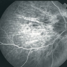 C-R Folds
Mar 26 2019 by Gary R. Cook, MD, FACS
Mid-phase FA image of the right eye of a white male with bilateral C-R folds showing alternating hyper- and hypofluorescent bands.
Imaging device: Topcon VT-50
Condition/keywords: bilateral chorioretinal folds, chorioretinal fold, FA mid phase, fluorescein angiogram (FA)
-
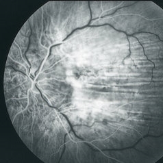 C-R Folds
Mar 26 2019 by Gary R. Cook, MD, FACS
Early phase FA frame of the left eye of a WM with bilateral C-R folds showing alternating hyper- and hypofluorescent bands.
Imaging device: Topcon VT-50
Condition/keywords: bilateral chorioretinal folds, chorioretinal fold, FA early phase, fluorescein angiogram (FA)
-
 Central Serous Chorioretinopathy (CSCR) With Choroidal Folds
Jun 2 2015 by Mallika Goyal, MD
Right fundus photograph of a 56-year-old male with CSCR with a hypopigmented spot corresponding to the FFA leak with choroidal folds (seen on OCT); subretinal fluid was limited to an area superior to macular centre, and patient presented with complaint of inferior field loss for 4 weeks.
Photographer: Mallika Goyal, MD, Apollo Health City, Jubilee Hills, Hyderabad
Condition/keywords: central serous chorioretinopathy (CSCR)
-
 Central Serous Retinopathy
Mar 6 2014 by Howard Schatz, MD
57-year-old white male. CSR (Chor folds). 20/25 OU. Central serous retinopathy.
Condition/keywords: central serous retinopathy (CSR)
 Loading…