Search results (247 results)
-
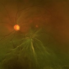
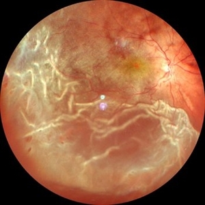
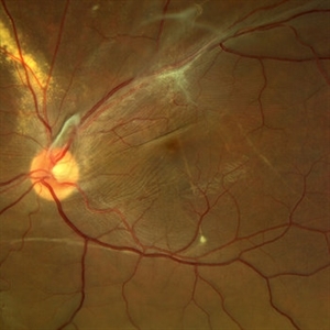 Displaced & folded macula
Oct 10 2022 by Ricardo Leitão Guerra
Tractional retinal detachment due to sickle cell retinopathy leading to a displaced and folded appearance of the macula in this 36-yo male. Subretinal bands are also noticed crossing the macula towards inferior retinal detachment area.
Photographer: Ricardo Leitão Guerra
Imaging device: Clarus 700 - Zeiss
Condition/keywords: folds, sickle cell retinopathy, subretinal bands, tractional retinal detachment
-
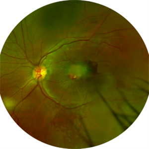
 Idiopathic Choroidal Folds-Multimodal Imaging
Jan 22 2024 by SHILPI H NARNAWARE, ICO ( Retina) , FAICO ( Vitreo-Retina)
45 year female with bilateral idiopathic choroidal folds
Photographer: Shilpi Narnaware, Sarakshi Netralaya , Nagpur, Maharashtra , India
Imaging device: Mirante ( by Nidek)
Condition/keywords: Choroidal Folds
-
 Idiopathic Choroidal Folds
Aug 22 2017 by Carolyn Daley
This is an autofluorescence image of a 77-year-old male with idiopathic choroidal folds in his right eye.
Photographer: Carolyn Daley
Imaging device: Heidelberg Spectralis
Condition/keywords: autofluorescence imaging, choroidal folds
-
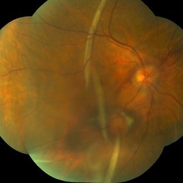
 Long Standing Retinal Detachment with Folds on Macula
Jan 23 2018 by Nilesh K Kanjani, MD
Fundus photograph of 17-year-old male showing long standing retinal detachment with folds on macula.
Photographer: Nilesh K Kanjani
-
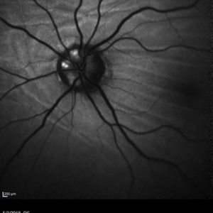
 Normal Nasal Ora Serrata
Nov 9 2012 by Norman Byer
This is the normal nasal ora serrata showing a prominent meridional fold. Such folds are most commonly seen at the lower part of the upper nasal quadrant, and are present in 26% of the population. They are a normal developmental variation and are often bilateral.
Condition/keywords: meridional fold, normal developmental variation, normal nasal ora serrata, upper nasal quadrant
-
 Post Choroidal Folds OD
Mar 12 2014 by Manish Nagpal, MD, FRCS (UK), FASRS
32-year-old male had presented with extensive choroidal folds. Oral and sub tenon steroids resolved the folds and only a few residual stria are seen with good visual recovery.
Photographer: Pooja Barot
Condition/keywords: choroidal folds
-
 Choroidal Folds and Optic Disc Drusen
Aug 1 2018 by Emily Cooper
Fundus autofluorescence photo of a 62-year-old man who presented for evaluation of choroidal folds and optic disc drusen. He is currently following up with neuro-ophthalmology and has suspected intracranial hypertension.
Photographer: Emily Cooper, Retina Specialists of Michigan
Condition/keywords: choroidal folds, drusen of optic disc
-
 Proliferative Vitreoretinopathy
Sep 28 2024 by Anjana Mirajkar, MS Ophthalmology
An intra operative image of right eye showing multiple star folds involving the macular area.
Photographer: Dr. Anjana Mirajkar -Retina Foundation, Ahmedabad
Condition/keywords: Starfolds
-
 Star Fold
Nov 1 2021 by Gregory R. Blaha, MD, PhD
Star fold in retinal detachment with 1 month of decreased vision.
Photographer: Jonathan Rosen
Imaging device: Optos California
Condition/keywords: star folds
-
 Choroidal Folds in NVAMD
Sep 10 2012 by James B. Soque, CRA, OCT-C, COA, FOPS
75 y/o Female, FA with Superior and Inferior Choroidal Folds, and Neovascular Age Related Macular Degeneration of the Right Eye.
Photographer: James B Soque, CRA, COA
Imaging device: TRC-50DX
Condition/keywords: choroidal folds
-
 Combined Hamartoma
Feb 29 2016 by Andrea Arriola-Lopez, MD MSc
40 year-old man with diminished VA since 6 month ago. Fundus examination revealed macular folds, yellow-whitish elevated lesion at the fovea and a subretinal hemorrhage.
Photographer: Andrea Elizabeth Arriola-Lopez MD, MSc
Imaging device: OPTOS Dakota
Condition/keywords: combined hamartoma, macula, subretinal hemorrhage
-
 Familial Exudative Vitreoretinopathy (FEVR)
Apr 24 2021 by Alexandre Grandinetti, MD, PhD
6-year-old girl with retinal folds on both eyes secondary to FEVR.
Photographer: Corina Szrek
Imaging device: Optos California
Condition/keywords: familial exudative vitreoretinopathy (FEVR)
-
 Ocular Hypotony Due to Leaking Bleb
Apr 1 2019 by Anfisa Ayalon, MD
81-year-old male who had trabeculectomy in his right eye 4 years ago, presented to the emergency room with complains of decreased vision in that eye for two months. Slit-lamp examination showed cystic bleb with leakage, intraocular pressure was 0 MMHg. Fundus examination showed hypotony maculopathy, peripheral choroidal detachments, multiple chorioretinal folds with subretinal fluid.
Photographer: Anfisa Ayalon, MD., Meir Medical Center, Kfar Saba, Israel.
Imaging device: California, Optos 200 DTX
Condition/keywords: choroidal detachment, hypotonous retinopathy, hypotony maculopathy
-
 PRE CF OD June 5, 2013
Mar 12 2014 by Manish Nagpal, MD, FRCS (UK), FASRS
Fundus photo of a 32-year-old male presenting with post traumatic choroidal folds and hypotony.
Photographer: Pooja Barot
Condition/keywords: choroidal folds
-
 Retinal Detachment with Vitreous Hemorrhage
Jun 10 2020 by Manish Nagpal, MD, FRCS (UK), FASRS
Retinal detachment with folds and minimal vitreous hemorrhage.
Photographer: Gayathri Mohan, Retina Foundation
Imaging device: NIDEK SLO MIRANTE
Condition/keywords: vitreous hemorrhage
-
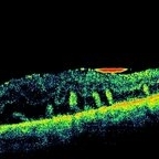 Retinal Folds Following Retinal Reattachment Surgery
Nov 22 2015 by Mallika Goyal, MD
Multiple retinal folds 4 weeks following vitreous surgery (perfluorodecalin assisted) for retinal detachment with giant retinal tear. OCT shows residual subretinal fluid and outer retinal folds (ORFs) seen as vertical hyperreflective lesions consisting of folded inner segment/outer segment of photoreceptors band and external limiting membrane band.
Photographer: Mallika Goyal, MD, Apollo Health City, Jubilee Hills, Hyderabad, India
Condition/keywords: retinal fold
-
 Chorioretinal Folds
-
 Posterior Retinal Folds
Feb 9 2015 by Leandro C. Zacharias, MD, PhD
Fundus photograph of a 59-year-old woman 3 weeks after buckle for a macula-off retinal detachment.
Photographer: Leandro Cabral Zacharias
Imaging device: Zeiss Visucam
Condition/keywords: retinal fold
-
 A Classic Case of Retinal Ora Serrata Imaging
Jan 16 2025 by yuan duo
A 5-year-old girl, born full-term with no history of systemic disease, presented with poor vision since early childhood and underwent fundus examination. Anterior segments of both eyes showed no significant abnormalities. Fundus examination revealed retinal folds extending from the optic disc to the temporal peripheral retina, with blood vessels coursing through the folds (A, B). Avascular zones were observed in the peripheral retina, and the ora serrata’s boundaries were clearly visible, displaying dentate processes and bays (C, D). Retinal pigmentation was evident. Genetic testing confirmed the final diagnosis of bilateral Familial Exudative Vitreoretinopathy (FEVR).
Condition/keywords: Retinal Ora Serrata
-
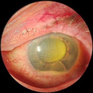 Acute Endophthalmitis
Nov 14 2023 by Virginia Gebhart
85 year old female with acute endophthalmitis 14 days s/p IVEylea injection. 3+ injection and descemets folds, contracted fibron, 3mm Hypopyon, and 3+ cell. No view posteriorly, vision CF. Visual prognosis unknown at this time
Photographer: Virginia Gebhart
Condition/keywords: endophthalmitis
-
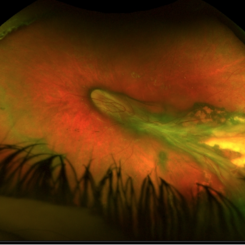
 Advanced Proliferative Diabetic Retinopathy
Apr 9 2025 by Gustavo Uriel Fonseca Aguirre
B-mode ultrasound of a patient with long-standing poorly controlled diabetes demonstrates characteristic findings of advanced proliferative diabetic retinopathy. The examination reveals moderate vitreous hemorrhage appearing as diffuse hyperechoic opacities throughout the vitreous cavity, along with a posterior hyaloid membrane densely infiltrated by hemorrhagic material, showing irregular thickening and increased reflectivity. A mild subhyaloid hemorrhage is visible as a subtle hyphema-like space anterior to the retinal surface. The study documents a total tractional retinal detachment, evidenced by rigid retinal folds with clear insertion points of vitreous strands, accompanied by a significant subretinal hemorrhage seen as a prominent hyperechoic collection beneath the elevated retina. These findings collectively illustrate the severe vitreoretinal interface pathology characteristic of end-stage diabetic eye disease, with predominant tractional components and distinct echographic stratification of hemorrhagic layers - from anterior vitreous involvement to deeper subretinal blood accumulation.
Photographer: Gustavo U. Fonseca Aguirre, Hospital Conde de Valenciana, Ciudad de México
Condition/keywords: diabetic retinopathy, tractional retinal detachment, Vitreous hemorrhage
-
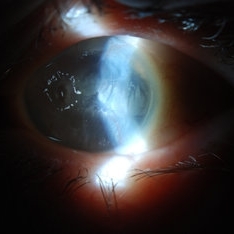 Band Keratopathy/Neurotrophic Ulcer
Nov 29 2013 by Jason S. Calhoun
Patient comes in with blind painful left eye. Slit lamp photos shows corneal diffuse scarring, descemets folds, corneal striae, band keratopathy, left eye. Proceed with Jupiter contact lens fitting on the left eye.
Photographer: Jason S. Calhoun, Ophthalmic Photographer, Department of Ophthalmology, Mayo Clinic Jacksonville
Imaging device: TOPCON D-90 SL NIKON CAMERA
Condition/keywords: band-shaped keratopathy, corneal dystrophy, folds in Descemet's membrane
-
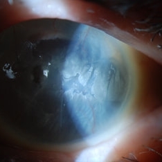 Band Keratopathy/Neurotrophic Ulcer
Nov 29 2013 by Jason S. Calhoun
Patient comes in with blind painful left eye. Slit lamp photos shows corneal diffuse scarring, descemets folds, corneal striae, band keratopathy, left eye. Proceed with Jupiter contact lens fitting on the left eye.
Photographer: Jason S. Calhoun, Ophthalmic Photographer, Department of Ophthalmology, Mayo Clinic Jacksonville
Imaging device: TOPCON D-90 SL NIKON CAMERA
Condition/keywords: band-shaped keratopathy, corneal dystrophy, folds in Descemet's membrane
-
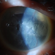 Band Keratopathy/Neurotrophic Ulcer
Nov 29 2013 by Jason S. Calhoun
Patient comes in with blind painful left eye. Slit lamp photos shows corneal diffuse scarring, descemets folds, corneal striae, band keratopathy, left eye. Proceed with Jupiter contact lens fitting on the left eye.
Photographer: Jason S. Calhoun, Ophthalmic Photographer, Department of Ophthalmology, Mayo Clinic Jacksonville
Imaging device: TOPCON D-90 SL NIKON CAMERA
Condition/keywords: band-shaped keratopathy, corneal dystrophy, folds in Descemet's membrane
-
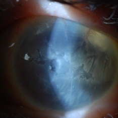 Band Keratopathy/Neurotrophic Ulcer
Nov 29 2013 by Jason S. Calhoun
Patient comes in with blind painful left eye. Slit lamp photos shows corneal diffuse scarring, descemets folds, corneal striae, band keratopathy, left eye. Proceed with Jupiter contact lens fitting on the left eye.
Photographer: Jason S. Calhoun, Ophthalmic Photographer, Department of Ophthalmology, Mayo Clinic Jacksonville
Imaging device: TOPCON D-90 SL NIKON CAMERA
Condition/keywords: band-shaped keratopathy, corneal dystrophy, folds in Descemet's membrane
 Loading…