Search results (100 results)
-
 Anterior Capsular Contraction Syndrome
Feb 8 2018 by Claire Kiernan, MD
Slit lamp photo of a female with underlying Retinitis Pigmentosa Zonulopathy and new anterior capsular contraction syndrome following cataract surgery.
Photographer: Steve Crow, University of Tennessee Hamilton Eye Institute, Memphis, TN
Condition/keywords: anterior capsule opacification, cataract surgery, zonules
-
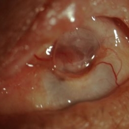
 Anterior Capsular Opacity
Feb 8 2018 by Claire Kiernan, MD
Slit lamp photograph of a 39-year-old female following uncomplicated cataract surgery shown here with dense fibrinous changes of the anterior capsule. This patient underwent Nd:YAG laser anterior capsulotomy with clearing of her visual axis.
Photographer: Steve Crow, University of Tennessee Hamilton Eye Institute, Memphis, TN
Condition/keywords: anterior capsule opacification, cataract extraction, cataract surgery
-
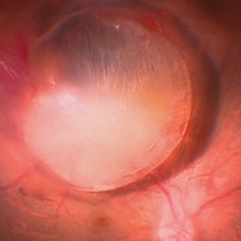
 Capsular Bag Dislocation into the Anterior Chamber
Jan 3 2020 by Manuel Ángel Alcántara Delgado, MD
Slit lamp photograph of a 65-year-old woman with previous history of complicated cataract surgery.
Photographer: Manuel Ángel Alcántara Delgado, CMN SXXI, Mexico City
Condition/keywords: anterior chamber, anterior dislocation of lens, anterior segment, cataract surgery, dropped capsular IOL bag complex
-
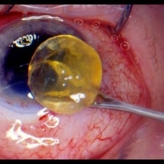 Cataract
Feb 24 2018 by JEFFERSON R SOUSA, Tecg.º (Biomedical Systems Technology)
81-year-old patient, male, in surgical procedure (Facectomy - FEC). Removal of the fully opacified lens is observed.
Photographer: JEFFERSON R SOUSA - Study Center and Ophthalmological Research Dr. Andre M V Gomes, Institute Dr. Suel Abujamra São Paulo-Brazil
Imaging device: Canon / Lens Sigma 35mm F / 1.4 Dg Hsm. without flash user and keeping the limits of safety of the surgeon.
Condition/keywords: cataract surgery
-
 Cataract Surgery Complications
Aug 21 2019 by Narciso F. Atienza, MD, MBA, FASRS, FPCS, FPAO.
57-year-old male patient who underwent phacoemulsification. Pre-op vision was 20/70. Complicated surgery. With vitreous loSS, cataract surgeon decided to place the foldable intraocular lens in the AC. Presented in the clinic with a vision of hand movement, with intra-ocular preSSure of 65 mmHg. He was managed by the same surgeon who gave Cosopt TID, and Alphagan QID.
Photographer: Narciso F. Atienza, Jr. MD, MBA
Condition/keywords: anterior segment, cataract surgery
-
 Elschnig's Pearls
Sep 1 2015 by René Hernán Parada Vásquez
Fundus photograph of 58-year-old male with Elschnig's pearls, you can see the transparent clusters formed by proliferation of epithelial lens cells found in the remains of the capsule of the crystalline lens following cataract surgery.
Photographer: Parada René, ESO, Guatemala.
Condition/keywords: cataract surgery
-
 Endophthalmitis
Apr 9 2014 by Aleksandra V. Rachitskaya, MD, FASRS
Slit lamp photo of a patient with endophthalmitis after cataract surgery. An infectious infiltrate is noted next to the clear corneal incision.
Photographer: Bascom Palmer Eye Institute
Condition/keywords: cataract surgery, endophthalmitis
-
 Large Ciliochoroidal Melanoma Diagnosed During Cataract Surgery
Jun 1 2020 by Sophia El Hamichi, MD
A 62-year-old male referred for a large ciliochoroidal melanoma OS noticed during cataract surgery. Visual acuity OS was 20/20.
Photographer: Belinda Rodriguez, Murray Ocular Oncology and Retina, Miami
Condition/keywords: cataract surgery, ciliochoroidal melanoma
-
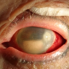
 Panophthalmitis
Jul 12 2014 by Philip J. Polkinghorne, MD
A 85-year-old lady who presented with an eroding intraocular lens. She had been initially treated with herbal medicines which failed to control the infection.
Photographer: Philip Polkinghorne
Condition/keywords: cataract surgery, endophthalmitis, panophthalmitis
-
 360 Degrees Choroidal Detachment
Sep 22 2024 by Anand Temkar
A 52 year old male came with chief complaints of diminution of vision in RE since past 15 days. He gave history of ( RE ) cataract surgery + IOL about 2 months ago. His vision was 6/9 in RE and PL +ve, PR inaccurate in LE. His IOP was 10 mm of Hg in RE and 20 mm of Hg in LE.
Photographer: Dr.Anand Temkar- Retina Foundation, Ahmedabad
Imaging device: Mirante
Condition/keywords: choroidal detachment, choroidals, serous choroidal detachment
-
 Autofluorescence of Ocular Hypotony
May 29 2013 by Zofia Anna Nawrocka (vel Michalewska), MD, PhD
Autofluorescence image of a 75-year-old patient with hypotony, 2 weeks after trauma, 2 years after extracapsular cataract surgery.
Photographer: Zofia Michalewska, Ophthalmic Clinic "Jasne Blonia
Imaging device: Spectralis
Condition/keywords: hypotony
-
 AV Anastomosis
Sep 19 2017 by Purva Patwari
54-year-old female post cataract surgery.
Photographer: Dr Purva Patwari, Patwari Retina Center,Ahmedabad
Imaging device: Zeiss visu 500
Condition/keywords: arteriovenous anastomosis
-
 Bullous Keratopathy
Jan 4 2025 by Mosab Salah
Corneal Slit photograph of an 84-year-old man underwent uneventful cataract surgery 1 year ago elsewhere, with a multiple fluid filled Bullae, not responding on conservative management and planned for KP.
Photographer: Abu-Ismail, Luai MD, The Islamic Hospital, Amman, Jordan
Imaging device: smartphone photography through SLB
Condition/keywords: bullous keratopathy, corneal edema
-
 Choroidal detachment
May 8 2022 by Adel Al Akeely, MD MBA
Fundus UWF photograph of a 71-year-old man with choroidal detachment with a distant history of trabeclectomy and a recent cataract surgery
Photographer: Adel AlAkeely, Magrabi Center, Riyadh Saudi Arabia
Imaging device: Optos Silverstone
Condition/keywords: choroid, detachment
-
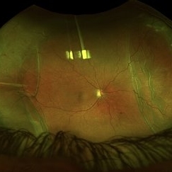
 Chronic Open Funnel Retinal Detachment With Horse Shoe Tear
Feb 7 2024 by Harsh Vardhan Singh, MS
67 year old male with history of cataract surgery 1 year presented with old chronic retinal detachment with open funnel configuration with multiple breaks.
Photographer: Harsh Vardhan Singh
Imaging device: Clarus 700
Condition/keywords: chronic retinal detachment, Retinal Detachment, Retinal Detachment with multiple breaks
-
 Chronic Regmatogenous Retinal Detachment
Mar 21 2024 by Mauricio Bayram-Suverza, MD
A 65-year-old man came to our department with a complaint of chronic visual loss in his right eye. He had undergone cataract surgery in the same eye at another facility 8 years ago. During the examination, it was observed that the patient had no light perception in the affected eye. Further, slit lamp examination revealed a chronic anteriorized retinal detachment.
Photographer: Mauricio Bayram-Suverza, Fundación Hospital Nuestra Señora de la Luz
Imaging device: iphone
Condition/keywords: proliferative vitreoretinopathy (PVR), vitreoretinal surgery
-
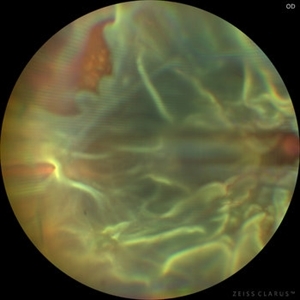
 Closed Funnel Retinal Detachment
Apr 9 2017 by Aliya Sultana
Fundus phtograph of an 51-year-old man with closed funnel rhegmatogenous retinal detachment presented to our department 6 weeks after cataract surgery. Posterior capsule rent noticed with vitreous in anterior chamber, condensed vitreous tag is incarcerated in side port wound.
Photographer: Dr Aliya Sultana , Assistant Professor,Sarojini Devi Eye Hospital, Hyderabad, Telangana. India.
-
 CME DME After CE
Aug 27 2014 by Susanna S. Park, MD, PhD
Macular OCT of a 62-year-old diabetic woman with severe vision loss 2 weeks after cataract surgery due to severe worsening of macular edema. Exam also showed new proliferative diabetic retinopathy.
Photographer: chandra
Condition/keywords: diabetic macular edema, optical coherence tomography (OCT)
-
 CME-FP
Apr 28 2015 by Neha Goel, MS DNB FRCS (Glasg)
Fundus photograph of the right eye of a 60-year-old male complaining of decreased vision following cataract surgery, that was performed one month earlier.
Photographer: Neha Goel
Imaging device: Zeiss visucam
Condition/keywords: cystoid macular edema (CME)
-
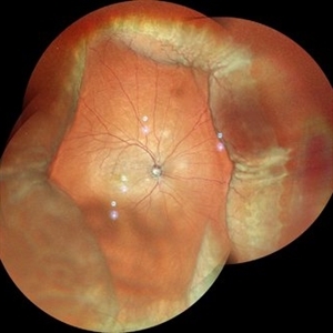
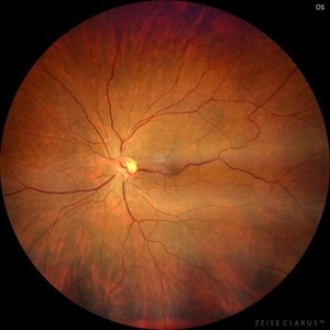
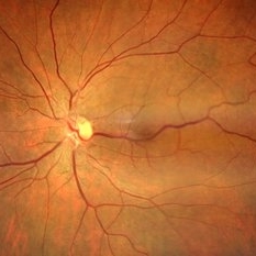 Congenital Retinal Macro Vessel
May 15 2024 by KANWALJEET HARJOT MADAN, M.S. (Ophthalmology); FAICO (Vitreous - Retina)
This is the Fundus Picture of a 56 years Female, who had come for Cataract Surgery of her left eye. Her best corrected vision in left eye was 6/12. She was diagnosed to have Congenital Retinal Macro vessel in her left eye on fundus exam. She underwent cataract surgery and vision improved to 6/9. She is kept under regular follow up.
Photographer: Dr. Kanwaljeet Harjot Madan
Condition/keywords: congenital, RETINAL MACROVESSEL
-
 Congenital Retinal Macro Vessel
May 15 2024 by KANWALJEET HARJOT MADAN, M.S. (Ophthalmology); FAICO (Vitreous - Retina)
This is the Fundus Picture of a 56 years Female, who had come for Cataract Surgery of her left eye. Her best corrected vision in left eye was 6/12. She was diagnosed to have Congenital Retinal Macro vessel in her left eye on fundus exam. She underwent cataract surgery and vision improved to 6/9. She is kept under regular follow up.
Photographer: Dr Kanwaljeet Harjot Madan
Condition/keywords: congenital, RETINAL MACROVESSEL
-
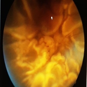
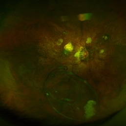 Dislocated Capsular Tension Ring in Vitreous Cavity
Dec 21 2019 by Pablo Baquero Ospina, MD
Fundus photograph of an 52-year-old woman with pseudoexfoliation glaucoma and previous cataract surgery with capsular tension ring. 5 years later she refers floaters.
Photographer: Pablo Baquero, Asociacion Para Evitar la Ceguera en Mexico, Mexico city
Imaging device: Optos/Daytona
Condition/keywords: fundus photograph, pseudoexfoliation glaucoma
-
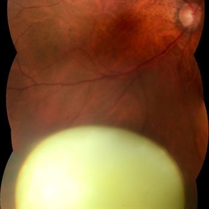
 Dislocated Cataractous Lens
Jun 16 2024 by Anjana Mirajkar, MS Ophthalmology
An intra operative still image of a 65 year old male showing an dislocated cataractous lens during cataract surgery which was removed during vitrectomy and and secondary IOL was placed.
Photographer: Dr. Anjana Mirajkar -Retina Foundation, Ahmedabad
Condition/keywords: dislocated crystalline lens
-
 dislocated crystalline lens
Jul 25 2019 by JEFFERSON R SOUSA, Tecg.º (Biomedical Systems Technology)
Male patient 54-years-old. In the preoperative follow-up of cataract surgery, he suffered blunt trauma to the right eye, with a total dislocation of the lens.
Photographer: JEFFERSON R SOUSA - Study Center and Ophthalmological Research Dr. Andre M V Gomes, Institute Dr. Suel Abujamra São Paulo-Brazil
Imaging device: Topcon TRC-50 DX, Imaginet 5.0, angle de 50 graus. Flash 18W-S
Condition/keywords: dislocated crystalline lens
-
 Dislocated Intraocular Lens (IOL)
Aug 2 2019 by JEFFERSON R SOUSA, Tecg.º (Biomedical Systems Technology)
A 53-year-old male patient suffered blunt trauma 15 days after cataract surgery. Note total dislocation of the intraocular lens. No glass reaction.
Photographer: JEFFERSON R SOUSA - Study Center and Ophthalmological Research Dr. Andre M V Gomes, Institute Dr. Suel Abujamra São Paulo-Brazil
Imaging device: Topcon TRC-50 DX, Imaginet 4.0, angle de 50 graus. Flash 18w-s
Condition/keywords: dislocated intraocular lens (IOL)
 Loading…