Search results (181 results)
-
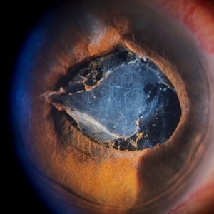
 Anterior segment
Feb 19 2013 by From the Collections of Thomas M. Aaberg, MD and Thomas M. Aaberg Jr., MD
Anterior segment photo taken with fundus camera.
Condition/keywords: fundus photograph, unknown
-
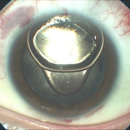
 Capsular Bag Dislocation into the Anterior Chamber
Jan 3 2020 by Manuel Ángel Alcántara Delgado, MD
Slit lamp photograph of a 65-year-old woman with previous history of complicated cataract surgery.
Photographer: Manuel Ángel Alcántara Delgado, CMN SXXI, Mexico City
Condition/keywords: anterior chamber, anterior dislocation of lens, anterior segment, cataract surgery, dropped capsular IOL bag complex
-
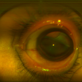
 Cataract Surgery Complications
Aug 21 2019 by Narciso F. Atienza, MD, MBA, FASRS, FPCS, FPAO.
57-year-old male patient who underwent phacoemulsification. Pre-op vision was 20/70. Complicated surgery. With vitreous loSS, cataract surgeon decided to place the foldable intraocular lens in the AC. Presented in the clinic with a vision of hand movement, with intra-ocular preSSure of 65 mmHg. He was managed by the same surgeon who gave Cosopt TID, and Alphagan QID.
Photographer: Narciso F. Atienza, Jr. MD, MBA
Condition/keywords: anterior segment, cataract surgery
-
 Iris Vascular Tuft
Jul 5 2022 by Olivia Rainey
Anterior segment imaging of a 66-year-old male with Vascular Disorders of Iris and Ciliary Body affecting his right eye. The physician stated that the findings are most consistent with a benign vascular tuft at the pupillary margin. The patient presented at the office with 20/20 vision in both eyes and had no ocular complaints at the time of his appointment.
Photographer: Olivia Rainey, OCT-C, COA
Imaging device: Heidelberg Spectralis, Slit Lamp with Samsung Galaxy 7
Condition/keywords: anterior segment, fluorescein angiogram (FA), heidelberg spectralis, infrared image, near infrared autofluorescence (NIRAF), slit lamp photo, vascular anomaly, vascular disorders of iris and ciliary body, vascular tuft
-
 Left -Anterior Segment
Aug 10 2020 by RITESH VERMA
Normal anterior segment of the left eye.
Photographer: Dr. Ritesh Verma, Regional institute of Ophthalmology, Rohtak, Haryana, India
Imaging device: CR-2AF CANON
Condition/keywords: anterior segment, normal eye
-
 Metastatic Breast Carcinoma
Jan 21 2021 by Jamin S. Brown, MD
This anterior segment photograph was taken with a smartphone camera attached to a regular Haag Streit slit lamp ocular demonstrates unusual clustering of white cells on the posterior surface of the intraocular lens. The clinical diagnosis is metastatic breast carcinoma to the vitreous, which is very rare.
Photographer: Stefanie Palmer CRA, Retina Vitreous Surgeons of CNY
Imaging device: Cell phone camera
Condition/keywords: anterior segment, breast cancer, cell phone camera, slit lamp photo
-
 Plateau Iris from Aqueous Misdirection
Jan 18 2018 by Olivia Rainey
Anterior segment OCT of a 90-year-old female with plateau iris from aqueous misdirection affecting her right eye.
Photographer: Olivia Rainey
Imaging device: Heidelberg Spectralis
Condition/keywords: anterior segment, aqueous misdirection, Heidelburg Spectralis, optical coherence tomography (OCT), plateau iris
-
 Right Eye Anterior-Segment
Aug 10 2020 by RITESH VERMA
Anterior segment photograph of right eye showing a silent anterior chamber.
Photographer: Dr. Ritesh Verma, Regional institute of Ophthalmology, Rohtak, Haryana, India
Imaging device: CR-2AF CANON
Condition/keywords: anterior chamber, anterior segment
-
 Total Rhegmatogenous and Tractional Retinal Detachment Following Choroidal Melanoma Laser Ablation Treatment
Sep 22 2020 by Sophia El Hamichi, MD
A 69-year-old female, with a history of choroidal melanoma in her left eye with exudative detachment, underwent tumor laser ablation. She then developed a complex combined tractional and rhegmatogenous retinal detachment with a giant retinal tear.
Photographer: Belinda Rodriguez, Murray Ocular Oncology and Retina, Miami
Condition/keywords: anterior segment, melanoma
-
 Waardenberg Syndrome
Jan 21 2021 by Jamin S. Brown, MD
9-year-old African American female, fellow eye color blue.
Photographer: Stefanie Palmer CRA, Retina Vitreous Surgeons of CNY
Condition/keywords: anterior segment, iris, waardenburg syndrome
-
 4 Point Scleral Fixation Akreos AO60 With Gore Tex Suture
May 21 2021 by Jesus Lozano, MD
Anterior segment photo of a 54-year-old man after 4 point scleral fixation Akreos AO60 with Gore Tex suture plus PPV who had a severe traumatic iris defect and was aphakic after ocular trauma.
Photographer: Luigi Zinn, Hadassah Medical Center, Jerusalem.
Condition/keywords: aphakia, cornea rupture, lens, penetrating trauma
-
 6 mm Canula
Jun 30 2012 by Stanislao Rizzo, MD
The 6 mm cannula is important in anterior segment opacity
Condition/keywords: anterior segment opacity, canula
-
 Acute Anterior Uveitis
Apr 14 2022 by Divya Jain
Anterior Segment Slit Lamp photograph of a 33 year old woman with first episode of acute granulomatous anterior uveitis showing circumcorneal congestion, mutton fat KP'S, 3+ cells, 2+ flare and Koeppe's nodules at pupillary margin.
Photographer: Divya Jain
Condition/keywords: acute anterior uveitis
-
 Anterior Chamber Gas and PFC Migration
Jun 21 2018 by Maria Stephanie R. Jardeleza, MD
Anterior segment photographs of 30-year-old male who underwent superior rhegmatogenous retinal detachment repair with intraocular gas tamponade. Perfluorocarbon was used to flatten the macula to prevent a macular fold and was removed during PFC/air exchange. Post operative week two visit shows gas migration into the anterior chamber with retained PFC layered in a tear drop shape posterior to the gas bubble and anterior to the lens. Patient had been maintaining face down positioning.
Photographer: Andy Zepeda, COA, Retina Clinic, San Antonio Eye Center, San Antonio, TX
Condition/keywords: retained perfluorocarbon, retina surgery complications, vitreous substitutes
-
 Anterior Iris Claw Artisan Lens
May 14 2025 by Moazzam Parvez
Anterior segment image of a 40 year old gentleman with a anteriorly placed iris claw lens post retinal detachment surgery.
Photographer: Dr Moazzam Parvez, Netralayam , Kolkata
Imaging device: Topcon DC-4
Condition/keywords: Anteriorly placed iris claw lens
-
 Anterior Segment Gas Bubble and PFC Interface
Jun 21 2018 by Maria Stephanie R. Jardeleza, MD
Anterior segment photographs of 30-year-old male who underwent superior rhegmatogenous retinal detachment repair with intraocular gas tamponade. Perfluorocarbon was used to flatten the macula to prevent a macular fold and was removed during PFC/air exchange. Post operative week two visit shows gas migration into the anterior chamber with retained PFC on the posterior aspect of the gas bubble/anterior surface of the lens. Patient had been maintaining face down positioning.
Photographer: Andy Zepeda, COA, Retina Clinic, San Antonio Eye Center, San Antonio, TX
Condition/keywords: retained perfluorocarbon, vitreous substitutes
-
000---thumb.jpg/image-square;max$300,300.ImageHandler) Anterior Segment Photo of Emulsified Silicone Oil
Anterior Segment Photo of Emulsified Silicone Oil
Dec 25 2013 by Dong Yoon Kim, MD
47-year-old woman underwent vitrectomy and silicone oil tampoande for tractional retinal detachment due to proliferative diabetic retinopathy. 8 months after silicone oil tamponade, silicone oil was emulsified. And emulsified silicone oil was observed at anterior chamber.
Condition/keywords: silicone oil, tractional retinal detachment
-
 Choroidal Melanoma
Apr 12 2018 by SUSHIL BHATT
Anterior segment fundus photograph of a 30-year-old woman with a left eye choroidal melanoma observed.
Photographer: Bhatt Sushil PGIMER chandigarh, India
Imaging device: OPTOS ultra-widefield (UWF™)
-
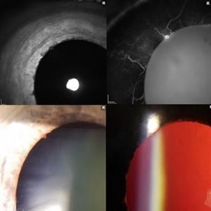
 Coexistent Morning Glory Syndrome and Persistent Hyperplastic Primary Vitreous
Jul 15 2024 by Arthi Mohankumar , MS,MRCS ED, FICO,FAICO
Anterior segment and fundus photograph of a 9-year-old female revealing PHPC changes (Images A,B,C) and morning glory syndrome (image D).
Photographer: Arthi Mohankumar
Condition/keywords: Morning Glory Anomaly, Morning Glory Syndrome, persistent hyperplastic primary vitreous (PHPV)
-
 Corneal Dystrophy
Jan 3 2017 by Jason Griffith
Anterior segment photo of corneal dystrophy using a fundus imaging camera.
Photographer: Jason Griffith
Imaging device: Topcon TRC-50EX
Condition/keywords: cornea dystrophy
-
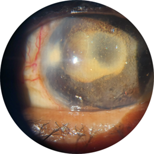 Crystallized silicone oil particles in the anterior chamber
Oct 26 2023 by Anmol Naik, MS, DNB, FMRF, FICO, MNAMS
Anterior segment image of a 67-year-old Indian woman who had proliferative diabetic retinopathy with traction retinal detachment with neovascular glaucoma. Patient underwent vitrectomy with membrane peeling with endolaser followed by silicone oil injection 1 year back. Patient was lost to follow up and presented a year later with this picture. She had crystallized silicone oil particles in the anterior chamber rendering a polychromatic lustre like appearance; a unique and rare finding.
Photographer: Anmol Naik
Condition/keywords: Polychromatic lustre in Anterior Chamber
-
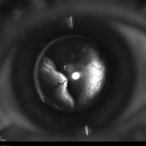
 Ectopia Lentis et Pupillae
Sep 24 2024 by Christian A Leal, MD
Anterior segment photo during exam under anesthesia in a child with spontaneous lens dislocation and history of ectopia lentis et pupillae. The lens is not visible. The pupil shown here has been pharmacologically dilated.
Photographer: Baker Hubbard, MD; Emory Eye Center
Condition/keywords: spontaneous lens dislocation
-
 Hydrophilic IOL With Fine Bubble Opacities Post-PPV for RD
Aug 21 2018 by Russell Pokroy, MD
Anterior segment photograph of 68-year-old woman 2 years after PPV for RD shows fine bubble-like opacities in the central part of this hydrophilic intraocular lens (IOL). These opacities are thought to be due to prolonged contact of the IOL with a large gas bubble in the vitreous cavity, particularly hydrophilic acrylic IOLs exposed to C3F8 gas. In addition, perfluorocarbon liquid exposure during surgery may put the IOL at risk of gas-induced opacity development. Minor Elschnig pearls are seen anterior to the wide optic-haptic junction in the capsule bag at 6-o-clock. A large symmetric capsulotomy with a rolled edge is seen behind the IOL. The slightly decreased yet stable vision of 20/30 was thought to be due to the central opacification of the IOL.
Photographer: Russell Pokroy, Assaf Harofe Medical Center, Israel
Condition/keywords: hydrophilic intraocular lens, IOL opacification
-
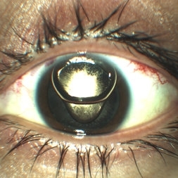
 ICC-8 Pinhole IOL
Nov 5 2024 by Kimberly Wakester
Anterior segment photograph of a 74-year-old woman with an ICC-8 pinhole IOL with stellate areas on the pinhole in the right eye.
Photographer: Kimberly Wakester, COA
Imaging device: Topcon TRC-50DX
Condition/keywords: Pinhole IOL
-
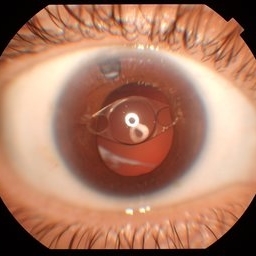
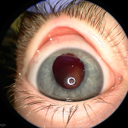
 Inflammatory pupillary membrane in patient with endophthalmitis
Jan 28 2023 by Kingston Rodolfo Ureña-Wong, MD, Opht, MSc
Anterior segment photography of a 54-year-old woman with post phacoemulsification endophthalmitis. She did not improve after first intravitreal antibiotics injection and develop an inflammatory pupillary membrane. After two vitrectomies, and a complete three intravitreal injections scheme, we decided to remove the intraocular lens and capsules.
Photographer: Marco Antonio Rubio-Atonal,UNAM, Asociación para evitar la ceguera en México
Imaging device: Zeiss Clarus 700
Condition/keywords: endophthalmitis, pupillary membranes
 Loading…