Search results (464 results)
-
 Branch Retinal Vein Occlusion
Dec 15 2022 by Christopher R. Adam, M.D.
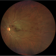
Branch Retinal Vein Occlusion
Condition/keywords: branch retinal vein occlusion (BRVO)BRVOcystoid macular edema (CME)
-
 Branch Retinal Vein Occlusion
Dec 15 2022 by Christopher R. Adam, M.D.
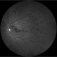
AF
Condition/keywords: branch retinal vein occlusion (BRVO)BRVOcystoid macular edema (CME)
-
 Branch Retinal Vein Occlusion
Dec 15 2022 by Christopher R. Adam, M.D.
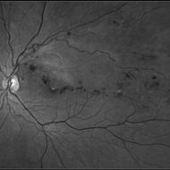
FA 35 sec
Condition/keywords: branch retinal vein occlusion (BRVO)BRVOcystoid macular edema (CME)
-
 Branch Retinal Vein Occlusion
Dec 15 2022 by Christopher R. Adam, M.D.
FA 10 min
Condition/keywords: branch retinal vein occlusion (BRVO)BRVOcystoid macular edema (CME)
-
 Branch Retinal Vein Occlusion
Dec 15 2022 by Christopher R. Adam, M.D.
OCT
Condition/keywords: branch retinal vein occlusion (BRVO)BRVOcystoid macular edema (CME)
-
 BRANCH RETINAL VEIN OCCLUSION
Oct 11 2022 by Akansha Sharma
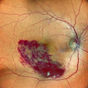
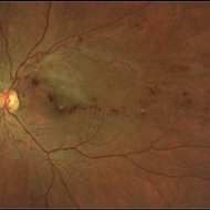
COLOUR FUNDUS PHOTOGRAPH OF A 59 YEAR OLD FEMALE WITH BRACH RETINAL VEIN OCCLUSION
Photographer: Dr. Akansha Sharma-Retina Foundation, Ahmedabad
Condition/keywords: BRVO
-
 Branch Retinal Vein Occlusion
Mar 5 2023 by Kalyan Singh
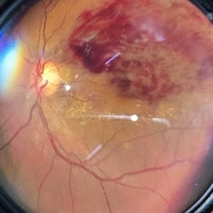
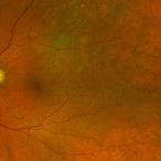
Young female with unilateral chronic diminution of vision presented in our OPD.
Photographer: Kalyan Singh, GSVM medical college, Kanpur
Imaging device: Smartphone (1 plus 10R)
Condition/keywords: BRVO
-
 Branch Retinal Vein Occlusion
Aug 22 2024 by Virginia Gebhart
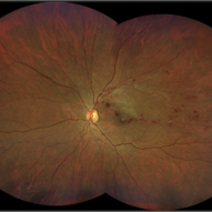
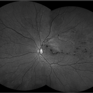
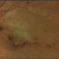
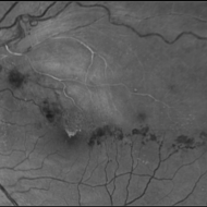
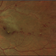
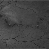
Fluorescein angiogram of branch retinal vein occlusion in 75 year old female. Scattered microaneurysms with late CME and persistent SRF. Pt will consider laser treatment but is hesitant for injections at this time due to possible side effects.
Photographer: Virginia Gebhart
Imaging device: Optos California
Condition/keywords: branch retinal vein occlusion (BRVO)BRVOcystoid macular edema (CME)FAFA late phasefluorescein angiogram (FA)macular edemamicroaneurysmsretinal microaneurysms
-
 Branch Retinal Vein Occlusion
Aug 13 2024 by Shakhzod Muratov
Fundus photograph of a 66 year old woman with a BRVO and Bullous retinoschisis.
Photographer: Shakhzod Muratov, S. Fyodorov Eye Microsurgery Federal State Institution
Imaging device: Zeiss Clarus 500
Condition/keywords: BRVOretinoschisis
-
 Branch retinal vein occlusion - Colour & Red free image - ring shaped collaterals
Jul 18 2023 by Harsh Vardhan Singh, MS
43-year-old woman presented with left eye old STBRVO with chronic CME of duration 6month showing ring shaped collaterals more evident on red free image
Photographer: Harsh Vardhan Singh, AIIMS, Guwahati
Imaging device: Zeiss Clarus 700
Condition/keywords: branch retinal vein occlusion (BRVO)BRVOnon-perfused branch retinal vein occlusion (BRVO)
-
 BRVO
BRVO
Aug 28 2019 by Megan Fanelli
CASE: A 50-year-old male with past medical history significant for hypertension and a branch retinal vein occlusion. He complained of flashing lights and floaters for the past month. The floaters were consistent with red blood cells in the anterior vitreous. His visual acuity was 20/25 -1+2 in the left eye and 20/20 -1 in the right eye. The patient has been followed for BRVO since 2011 and received focal laser treatment and anti-VEGF injections. His last injection was 19 months prior to the vitreous hemorrhage. The plan is to treat the patient with sector pan-retinal photocoagulation. Image Description: Late phase wide field fluorescein angiogram of the left eye shows peripheral non-perfusion with neovascularization elsewhere with a pre-retinal hemorrhage. The image also displays leakage within the macula and previous focal laser treatment.
Condition/keywords: branch retinal vein occlusion (BRVO)
-
 BRVO
BRVO
Sep 16 2021 by Stefanie Palmer
An FA image of a 72 year-old-female with BRVO.
Photographer: Stefanie Palmer, CRA
Condition/keywords: branch retinal vein occlusion (BRVO)ischemia
-
 BRVO
BRVO
Mar 8 2022 by Jeffrey Barker
70 year old Female with stable BRVO. Status Post Laser
Photographer: Jeffrey P. Barker, B.S.
Imaging device: Optos
Condition/keywords: branch retinal vein occlusion (BRVO)
-
 BRVO
BRVO
Mar 8 2022 by Jeffrey Barker
Fluorescein Angiogram picture of 70 year old woman with a stable BRVO. Status Post Laser.
Photographer: Jeffrey P. Barker, B.S.
Condition/keywords: branch retinal vein occlusion (BRVO)
-
 BRVO
BRVO
Mar 12 2025 by T. P . VIGNESH, MBBS,MS
Fundus photograph of a 52-year-old man with a macular branch retinal vein occlusion and hard exudate deposition at fovea.
Photographer: Sivanath
Imaging device: EIDON
Condition/keywords: branch retinal vein occlusion (BRVO)
 Loading…