 Initializing download.
-
 By Megan Fanelli
Co-author(s): Michael F Ward III, DO and Steven J Marks, MD - Uploaded on Aug 28, 2019.
- Last modified by Caroline Bozell on Aug 29, 2019.
- Rating
- Appears in
- Miscellaneous
- Condition/keywords
- branch retinal vein occlusion (BRVO)
- Description
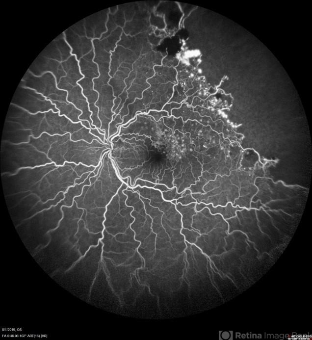
- CASE: A 50-year-old male with past medical history significant for hypertension and a branch retinal vein occlusion. He complained of flashing lights and floaters for the past month. The floaters were consistent with red blood cells in the anterior vitreous. His visual acuity was 20/25 -1+2 in the left eye and 20/20 -1 in the right eye. The patient has been followed for BRVO since 2011 and received focal laser treatment and anti-VEGF injections. His last injection was 19 months prior to the vitreous hemorrhage. The plan is to treat the patient with sector pan-retinal photocoagulation. Image Description: Late phase wide field fluorescein angiogram of the left eye shows peripheral non-perfusion with neovascularization elsewhere with a pre-retinal hemorrhage. The image also displays leakage within the macula and previous focal laser treatment.


 Caused due Branch Retinal Vein Occlusion (BRVO)")
 after Anti VEGF Treatment")
")


