Discover images
-
 "Boat-Shaped" Preretinal Hemorrhage
Feb 21 2019 by Mitzy E Torres Soriano, MD
Color fundus photograph showing preretinal (subhyaloid) hemorrhage in a diabetic patient with proliferative diabetic retinopathy.
Photographer: Andrea Vitale, MD
Condition/keywords: preretinal hemorrhage, proliferative diabetic retinopathy (PDR), subhyaloid hemorrhage
-
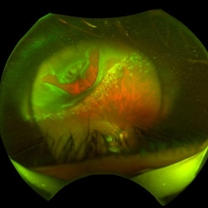
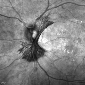
 "Flower Cataract"
Jul 11 2013 by Jason S. Calhoun
Patient presents with a cataract shaped like a flower. Patient had surgery to remove cataract.
Photographer: Jason S. Calhoun, Department of Ophthalmology, Mayo Clinic Jacksonville, Florida
Condition/keywords: cataract
-
 "Flower Cataract"
Jul 11 2013 by Jason S. Calhoun
Patient presents with a cataract shaped like a flower. Patient had surgery to remove cataract.
Photographer: Jason S. Calhoun, Department of Ophthalmology, Mayo Clinic Jacksonville, Florida
Condition/keywords: cataract
-
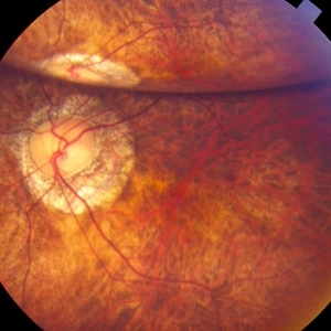
---thumb.JPG/image-square;max$300,300.ImageHandler) "Flower" Macular Degeneration (Wet)
"Flower" Macular Degeneration (Wet)
Jul 13 2013 by Jason S. Calhoun
Patient with (wet) macular degeneration in the left eye. Notice the "flower" shape abnormal blood vessels staining.
Photographer: Jason S. Calhoun, Department of Ophthalmology, Mayo Clinic Jacksonville, Florida
Imaging device: TOPCON TRC 50-EX
Condition/keywords: choroidal neovascularization (CNV)
-
 "Hang in There"
Apr 20 2021 by Tomas Minelli, MD
Fundus wide field photograph of a 50-year-old man with a macular detachment associated with a big temporal superior tear. The laser is firmly holding the progression of the tear in the 14th day post- laser. BCVA 20/20
Photographer: Livia Conci, Universtity of São Paulo
Imaging device: Optos Daytona
Condition/keywords: giant retinal tear
-
 "Internal Mirroring" Effect by Intraocular Gas
Mar 25 2014 by Homayoun Tabandeh, MD, FASRS
"Internal mirroring" by residual intraocular gas in a highly myopic patient 3 weeks post repair of retinal detachment with pars plana vitrectomy and C3F8 gas.
Photographer: Danny Rivas
Condition/keywords: high myopia, intraocular gas
-
 "Mud-Splatter" of Posterior Pole and Peripheral Radial Streaks in a Carrier of Ocular Albinism
Jan 22 2019 by John S. King, MD
14-year-old healthy white female with family history of ocular albinism was seen by Dr. Hruby for a second opinion. Father and some of his brothers were positive for a history of ocular albinism. Va cc 20/30 J1+ OU; no nystagmus; no TIDs; no foveal hypoplasia. A "mud-spatter" appearance to the posterior pole was present, along with peripheral alternating streaks (photo). Dr. Hruby agreed that this was most likely a carrier of Ocular Albinism Type-1 (XR; GPR143 mutation), and possible genetic testing/counselling was discussed.
Photographer: Gretchen Harper
Imaging device: Optos California
Condition/keywords: Nettleship-Falls ocular albinism, ocular albinism
-
 "Mud-Splatter" of Posterior Pole and Peripheral Radial Streaks in a Carrier of Ocular Albinism
Jan 22 2019 by John S. King, MD
14-year-old healthy white female with family history of ocular albinism was seen by Dr. Hruby for a second opinion. Father and some of his brothers were positive for a history of ocular albinism. Va cc 20/30 J1+ OU; no nystagmus; no TIDs; no foveal hypoplasia. A "mud-spatter" appearance to the posterior pole was present, along with peripheral alternating streaks, which are very prominent in this late phase FA of the right eye. Dr. Hruby agreed that this was most likely a carrier of Ocular Albinism Type-1 (XR; GPR143 mutation), and possible genetic testing/counselling was discussed.
Photographer: Gretchen Harper
Imaging device: Optos California
Condition/keywords: Nettleship-Falls ocular albinism, ocular albinism
-
 "Mud-Splatter" of Posterior Pole and Peripheral Radial Streaks in a Carrier of Ocular Albinism
Jan 22 2019 by John S. King, MD
14-year-old healthy white female with family history of ocular albinism was seen by Dr. Hruby for a second opinion. Father and some of his brothers were positive for a history of ocular albinism. Va cc 20/30 J1+ OU; no nystagmus; no TIDs; no foveal hypoplasia. A "mud-spatter" appearance to the posterior pole was present, along with peripheral alternating streaks that are very prominent on this late phase FA OS. Dr. Hruby agreed that this was most likely a carrier of Ocular Albinism Type-1 (XR; GPR143 mutation), and possible genetic testing/counselling was discussed.
Photographer: Gretchen Harper
Imaging device: Optos California
Condition/keywords: Nettleship-Falls ocular albinism, ocular albinism
-
 "Mud-Splatter" of Posterior Pole and Peripheral Radial Streaks in a Carrier of Ocular Albinism
Jan 22 2019 by John S. King, MD
14-year-old healthy white female with family history of ocular albinism was seen by Dr. Hruby for a second opinion. Father and some of his brothers were positive for a history of ocular albinism. Va cc 20/30 J1+ OU; no nystagmus; no TIDs; no foveal hypoplasia. A "mud-spatter" appearance to the posterior pole was present, along with peripheral alternating streaks (photo). Dr. Hruby agreed that this was most likely a carrier of Ocular Albinism Type-1 (XR; GPR143 mutation), and possible genetic testing/counselling was discussed.
Photographer: Gretchen Harper
Imaging device: Optos California
Condition/keywords: Nettleship-Falls ocular albinism, ocular albinism
-
 "Mud-Splatter" of Posterior Pole and Peripheral Radial Streaks in a Carrier of Ocular Albinism
Jan 22 2019 by John S. King, MD
14-year-old healthy white female with family history of ocular albinism was seen by Dr. Hruby for a second opinion. Father and some of his brothers were positive for a history of ocular albinism. Va cc 20/30 J1+ OU; no nystagmus; no TIDs; no foveal hypoplasia. A "mud-spatter" appearance to the posterior pole was present, along with peripheral alternating streaks. Hypoautofluorescent areas correspond to hyperpigmented areas of retinal pigment epithelium, and vice versa (see photo). Dr. Hruby agreed that this was most likely a carrier of Ocular Albinism Type-1 (XR; GPR143 mutation), and possible genetic testing/counselling was discussed.
Photographer: Gretchen Harper
Imaging device: Optos California
Condition/keywords: Nettleship-Falls ocular albinism, ocular albinism
-
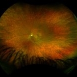
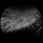
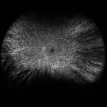
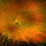
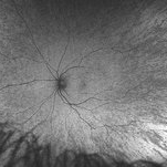
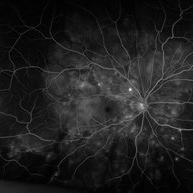
 "NVD Flower"
Oct 20 2023 by Daniel Davis, OCT-C
Infrared image of NVD (52F)
Imaging device: Heidelberg Spectralis
Condition/keywords: neovascularization of the disc (NVD)
-
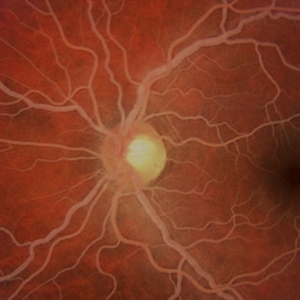 "Salmon-Pink" Fundus in Hypertriglyceridemia
Mar 2 2014 by Homayoun Tabandeh, MD, FASRS
"Salmon-pink" fundus in a patient with severe hypertriglyceridemia.
Condition/keywords: hyperlipidemia
-
 "Smoke Stack" Hyperfluorescence in Central Serous Chorioretinopathy
Mar 2 2014 by Homayoun Tabandeh, MD, FASRS
"Smoke Stack" hyperfluorescence in central serous chorioretinopathy.
Condition/keywords: central serous chorioretinopathy (CSCR)
-
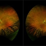
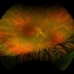
---thumb.JPG/image-square;max$300,300.ImageHandler) "Snowy Vessels"
"Snowy Vessels"
Jul 11 2013 by Jason S. Calhoun
Young female with white sheathing of retina vessels.
Photographer: Jason S. Calhoun, Department of Ophthalmology, Mayo Clinic Jacksonville, Florida
Condition/keywords: vessel sheathing, white sheath vessel
-
---thumb.jpg/image-square;max$300,300.ImageHandler) "Solarization"
"Solarization"
Feb 20 2013 by From the Collections of Thomas M. Aaberg, MD and Thomas M. Aaberg Jr., MD
FA "eradic response to premature sxposure to room light;" extensive vascular occlusions.
Condition/keywords: solarization, vascular occlusions
-
---thumb.jpg/image-square;max$300,300.ImageHandler) "Solarization"
"Solarization"
Feb 20 2013 by From the Collections of Thomas M. Aaberg, MD and Thomas M. Aaberg Jr., MD
FA "eradic response to premature sxposure to room light."
Condition/keywords: solarization, vascular occlusions
-
---thumb.jpg/image-square;max$300,300.ImageHandler) "Spots" In The Central Visual Zone
"Spots" In The Central Visual Zone
Oct 14 2013 by Maurice F. Rabb
A 26 year old healthy female who had been aware of decreased vision in OS for 5 days before the initial examination. When questioned specifically about OD, she did admit to being aware of some "spots" in the central visual zone. Her past ocular history is negative for eye disease and the family history is negative for retinal and macular disease. The patient is in excellent general health. She had a recent upper respiratory infection and is presently disabled because of a herniated disc. Uncorrected vision OD is 20/20 and OS is 20/400, improving to 20/100- with pinhole. The findings of significance are noted in the posterior poles.
Condition/keywords: spots in the central visual zone
-
 "Starry Sky" Fundus in Vogt-Koyanaki-Harada Syndrome
Jan 10 2018 by Peter H. Tang, MD, PhD
Fluorescein angiography imaging of a 27-year-old male with acute inflammation as part of Vogt-Koyanagi-Harada Syndrome.
Imaging device: Optos California
Condition/keywords: chorioretinal inflammations, retina, uveitis, Vogt-Koyanagi-Harada
 Loading…