Search results (253 results)
-
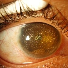 Synchysis Scintillans
Sep 17 2015 by Jessica G Lee, MD
24-year-old male with history of chronic retinal detachment.
Photographer: Bob Masini
Condition/keywords: cholesterol crystals, refractile bodies, synchysis scintillans, trauma, vitreous hemorrhage
-
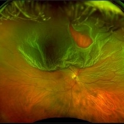 Optos Giant Tear within Retinal Detachment
Apr 30 2019 by Lauren Whaley
Noticed an inferior visual field defect on a patient with history of vitreous hemorrhage. Decided to take an Optos image and this is what we found. Doctor performed pneumatic retinopexy in office and patient recovering well.
Photographer: Lauren R. Whaley
Imaging device: Optos
Condition/keywords: Optos, retinal tear, subretinal fluid
-
 PDR with Active NVD
Oct 8 2012 by Jeffrey G. Gross, MD, FASRS
PDR with active NVD and preretinal hemorrhage, mild VH and partial PRP.
Condition/keywords: neovascularization of the disc (NVD), preretinal hemorrhage, scatter laser photocoagulation, vitreous hemorrhage
-
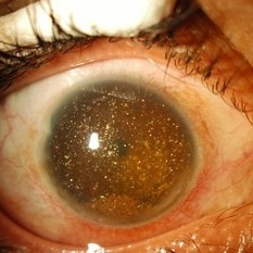 Synchisis Scintillans
Sep 17 2015 by Jessica G Lee, MD
24-year-old male with history of chronic retinal detachment.
Condition/keywords: cholesterol crystals, refractile bodies, synchysis scintillans, trauma, vitreous hemorrhage
-
 Proliferative Diabetic Retinopathy With Vitreous Hemorrhage And Traction Retinal Detachment
Oct 2 2013 by Jerald A. Bovino, MD
There is a moderate vitreous hemorrhage present in this eye with proliferative diabetic retinopathy. An inferonasal traction retinal detachment is present.
Condition/keywords: tractional retinal detachment, vitreous hemorrhage
-
 Vitreous Hemorrhage
Nov 9 2012 by Norman Byer
This 60-year-old man suddenly developed a vitreous hemorrhage from this acute horseshoe tear 3½ years following cataract extraction when a posterior vitreous detachment occurred. The white nubbin identifies this lesion as a preexisting cystic retinal tuft. The pigment spot beneath the flap is evidence of secondary trophic changes in the pigment epithelium. Note the irregular shape of the flap with the narrow tip and broad base. This was caused by vitreous traction which was exerted at two separate points on the retina and which tore the retina at each place.
Condition/keywords: acute posterior vitreous detachment, irregularly shaped flap, trophic pigmented changes, vitreous hemorrhage, vitreous traction, white retinal tuft
-
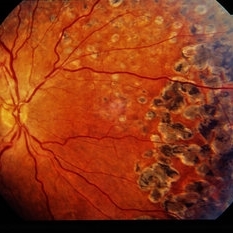
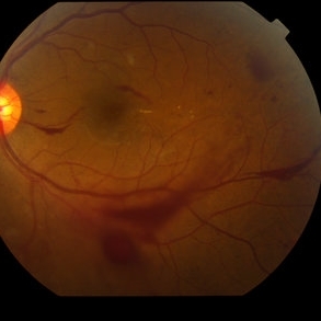 Proliferative Diabetic Retinopathy with Vitreous Hemorrhage - color fundus photo
Oct 18 2012 by Suber S. Huang, MD, MBA, FASRS
30 year old diabetic man with proliferative diabetic retinopathy and vitreous hemorrhage
Photographer: Stacie Hrvatin
Condition/keywords: cotton wool spots, neovascularization (NV), subhyaloid hemorrhage, vitreous hemorrhage
-
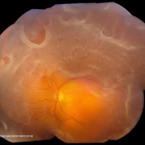
 Bergmeister's Papilla
Sep 29 2020 by Dhaivat Shah
Bergmeister's papilla is a small tuft of glial tissue which arises from the center of the optic disc, and represents a remnant of the fetal hyaloid artery. The hyaloid artery provides nutrition to the lens during development, and runs forward to the lens from the optic disc. At birth the hyaloid artery regresses, and is normally completely regressed by the time of birth. Bergmeister's papilla is frequently observed as an incidental clinical finding if this artery has an incomplete regression posteriorly. However, in the severe forms it can be associated with cataracts, persistence of the primitive vitreous, microphthalmia, vitreous hemorrhages and sometimes tractional retinal detachment, due to contraction of the residual fibro vascular tissue. Therefore, careful monitoring of vitreous thickening in the peripapillary areas, both by examining the ocular fundus, and especially by SD-OCT, is of considerable importance. Here we have one such of a 30 year old young male who came in for a routine checkup, in whom we noted a Bergmeister’s papilla. Due to its benign nature, patient was reassured and was asked to follow up yearly.
Condition/keywords: Bergmeister's Papillae
-
 Proliferative Diabetic Retinopathy With Vitreous Hemorrhage And Traction Retinal Detachment
Oct 2 2013 by Jerald A. Bovino, MD
There is a moderate vitreous hemorrhage present in this eye with proliferative diabetic retinopathy. An inferonasal traction retinal detachment is present.
Condition/keywords: tractional retinal detachment, vitreous hemorrhage
-
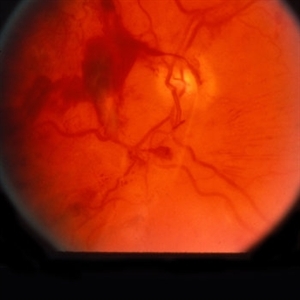
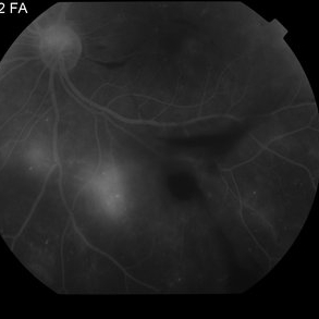
 Proliferative Diabetic Retinopathy with Vitreous Hemorrhage - FA
Oct 18 2012 by Suber S. Huang, MD, MBA, FASRS
30 year old diabetic man with proliferative diabetic retinopathy and vitreous hemorrhage
Photographer: Stacie Hrvatin
Condition/keywords: cotton wool spots, neovascularization (NV), subhyaloid hemorrhage, vitreous hemorrhage
-
 Vitreous Hemorrhage
Jul 10 2018 by Karen Panzegrau
SD-OCT of a 35-year-old female presenting with a vitreous hemorrhage of her left eye. Patient has active proliferative diabetic retinopathy, as well as a completed posterior vitreous detachment in the left eye.
Photographer: Karen Panzegrau
Condition/keywords: diabetes, Heidelburg Spectralis, left eye, optical coherence tomography (OCT), posterior vitreous detachment, proliferative diabetic retinopathy (PDR), vitreous hemorrhage
-
 Proliferative Diabetic Retinopathy with Vitreous Hemorrhage - FA
Oct 18 2012 by Suber S. Huang, MD, MBA, FASRS
30 year old diabetic man with proliferative diabetic retinopathy and vitreous hemorrhage
Photographer: Stacie Hrvatin
Condition/keywords: cotton wool spots, neovascularization (NV), subhyaloid hemorrhage, vitreous hemorrhage
-
 Retinal Detachment With Multiple Retinal Tears
May 18 2017 by Kamal Kishore, MD, MBBS
77-year-old female presented with a report of gradual decreased vision over the span of one week. Vision finger count. Examination showed retinal detachment with multiple retinal tears and vitreous hemorrhage present.
Photographer: Lindsay Shepard, Illinois Retina and Eye Associates, Peru, IL
Imaging device: Topcon TRC- 50 EX
Condition/keywords: retinal tear
-
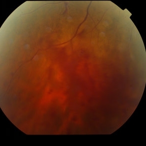
 Proliferative Diabetic Retinopathy with Vitreous Hemorrhage - Red Free
Oct 18 2012 by Suber S. Huang, MD, MBA, FASRS
30 year old diabetic man with proliferative diabetic retinopathy and vitreous hemorrhage
Photographer: Stacie Hrvatin
Condition/keywords: cotton wool spots, neovascularization (NV), subhyaloid hemorrhage, vitreous hemorrhage
-
 Asteroid Hyalosis, Vitreous Face Attached
Dec 10 2012 by Yale L. Fisher, MD
In asteroid hyalosis, accumulations of calcium soaps dispersed throughout the vitreous produce bright echoes in the usually echolucent vitreous. The appearance of asteroid hyalosis should not be confused with that of vitreous hemorrhage or vitritis. Many of the larger aggregates in asteroid hyalosis are easily seen as the gain is reduced to below 60 db, unlike vitreous hemorrhage or vitritis which usually disappears at low gain settings. There is also an area of clear echolucent vitreous between the posterior hyaloid face and the asteroid particles, which is usually not present in vitreous hemorrhage or vitritis.
Condition/keywords: video
-
 Candy Stripe Sign
Mar 30 2023 by pedro fernandes souza neto
Candy Stripe Sign, patient with proliferative diabetic retinopathy progressing to vitreous hemorrhage and subsequently to ghost cell glaucoma.
Photographer: Marlos Henrique Oliveira Junior, Federal University of Bahia.
Condition/keywords: dehemoglobinized hemorrhage, diabetes, diabetic glaucoma
-
 24 Hours Post Scleral Wound Closure+ Scleral Buckle+25 g Vitrectomy+Silicon Oil
Jan 23 2015 by Carlos Quezada-Ruiz, MD, FASRS
24 hours post op fundus photograph of a 43-year-old man who had perforating injury to the right eye with a small piece of plastic while he was hammering. OD LP, subconjunctival hemorrhage, clear cornea, hyphema, irido and ciclodyalisis as well as a luxated lens with traumatic cataract and a dense vitreous hemorrhage. B-US showed rhegmatogenous retinal detachment with a tear and a big inferior hemorrhagic choroidal detachment. 360 peritomy revealed 2-entry scleral wounds were found in zone II (M V and M VI) and closure was performed. 25 G PPV was performed with the infusion canal placed in the AC through the limbus. Lensectomy and removal of a dense recent vitreous hemorrhage revealed a white detached retina with an exit wound through the temporal inferior segment of the optic nerve with a nasal GRT and sub retinal hemorrhage as well as temporal inferior choroidal, PVD was induced and PFOs helped stabilizing the retina while vitrectomy and sub-retinal hemorrhage was removed through the GRT. Fluid air exchange was made and 360 endolaser over the buckle indentation was done and silicon oil was used as endotamponade. This picture was taken 24 hrs after the surgery.
Photographer: Lilibeth Rodriguez, Instituto de la Visión. Torreon, Mexico.
Condition/keywords: central retinal artery occlusion (CRAO), giant retinal tear, trauma
-
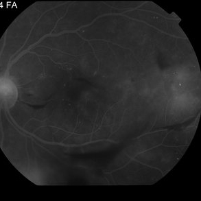
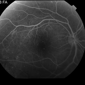
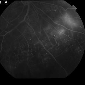 Proliferative Diabetic Retinopathy with Vitreous Hemorrhage - FA late
Oct 18 2012 by Suber S. Huang, MD, MBA, FASRS
30 year old diabetic man with proliferative diabetic retinopathy and vitreous hemorrhage
Photographer: Stacie Hrvatin
Condition/keywords: cotton wool spots, neovascularization (NV), subhyaloid hemorrhage, vitreous hemorrhage
-
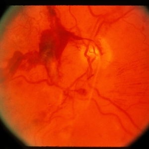
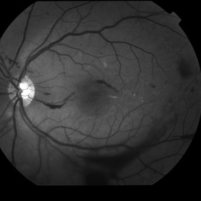
 Venous Loop on the Optic Nerve With Subretinal and Vitreous Hemorrhage
Feb 20 2013 by From the Collections of Thomas M. Aaberg, MD and Thomas M. Aaberg Jr., MD
OD, fellow eye to Venous Loop on the Optic Nerve With Subretinal and Vitreous Hemorrhage.
Condition/keywords: venous loop
-
 Dengue Retinitis
Oct 27 2012 by Mallika Goyal, MD
Right eye of a 37-year-old lady recovering from bilateral dengue retinitis shows dengue foveolitis, neovascularisation disc (NVD) and vitreous hemorrhage.
Photographer: Mallika Goyal, MD
Condition/keywords: Dengue foveolitis, Dengue retinitis, neovascularization of the disc (NVD), vitreous hemorrhage
-
---thumb.jpg/image-square;max$300,300.ImageHandler) Massive Vitreous Hemorrhage 1
Massive Vitreous Hemorrhage 1
Mar 14 2013 by Maurice F. Rabb
This 59 year-old black female was referred with marked decrease in vision in the right eye. She had massive sub-RPE and subretinal hemorrhage. This went on to massive vitreous hemorrhage.
Condition/keywords: decrease in vision, retinal pigment epithelium, subretinal hemorrhage
-
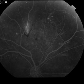 Proliferative Diabetic Retinopathy with Vitreous Hemorrhage - FA mid
Oct 18 2012 by Suber S. Huang, MD, MBA, FASRS
30 year old diabetic man with proliferative diabetic retinopathy and vitreous hemorrhage
Photographer: Stacie Hrvatin
Condition/keywords: cotton wool spots, neovascularization (NV), subhyaloid hemorrhage, vitreous hemorrhage
-
 Sickle Cell Neovascularization and Vitreous Hemorrhage
Oct 30 2015 by David Callanan, MD
Female patient, sickle cell neovascularization and vitreous hemorrhage; pre and post laser.
Condition/keywords: neovascularization (NV), sickle cell, vitreous hemorrhage
 Loading…