Search results (72 results)
-
 Cystic Retinal Tuft
Nov 9 2012 by Norman Byer
This is the same lesion as in the previous slide pair but the photograph was taken nine years later when the patient was 58-years-old soon after an acute posterior vitreous detachment. This demonstrates that posterior vitreous detachment can produce large retinal tears at these sites. However, it is important to emphasize that prophylactic treatment of cystic retinal tufts in the absence of a retinal tear would be very ill-advised because several hundred innocence and harmless lesions would have to be treated in order to prevent one tear of the retina.
Condition/keywords: cystic retinal tuft, posterior vitreous detachment, retinal tear
-
 Peripheral Retinal Lesion
Nov 9 2012 by Norman Byer
This small elevated peripheral retinal lesion in a 48-year-old woman is a cystic retinal tuft. Such tufts are congenital developmental anomalies present from birth and situated behind the vitreous base. They are sites of abnormal vitreoretinal attachment, and can occasionally lead to retinal tears at the time of posterior vitreous detachment. They are present in about 5% of patients.
Condition/keywords: abnormal vitreal retinal attachment, behind the vitreous base, congenital anomaly, cystic retinal tuft, developmental anomaly, peripheral retinal lesion, present from birth
-
 ERM that Spontaneously Peeled
Oct 8 2012 by David R. Chow, MD, FRCS(C)
An ERM that through follow-up sponateously separated with the development of PVD.
Condition/keywords: epiretinal membrane (ERM), posterior vitreous detachment
-
 Weiss Ring
Jan 9 2019 by John S. King, MD
77-year-old white male with ERM and PVD OD; sheet of vitreous with weiss ring in the nasal mid-vitreous cavity.
Photographer: Macey Highfill, RN
Imaging device: Topcon 50
Condition/keywords: posterior vitreous detachment, Weiss ring
-
 Sudden Posterior Vitreous Detachment
Nov 9 2012 by Norman Byer
This is the appearance of the previous lesion three weeks following prophylactic cryotherapy. Continuing vitreal retinal traction has a now torn the flap completely free from the retina. The whitish cystic retinal tuft can be discerned on the upper part of the free operculum. Along the lower half of the operculum superimposed over the dark shadow of the scleral indentation one may observe numerous, delicate, vitreous fibrils actually attaching to the operculum.
Condition/keywords: cystic retinal tuft, free operculum, prophylactic cyrotherapy, retinal flap, scleral indentation, vitreoretinal traction, vitreous fibrils
-
 Sudden Posterior Vitreous Detachment
Nov 9 2012 by Norman Byer
This is the same lesion seen in the previous slide pair. With the scleral indentation performed more posteriorly, a small hemorrhage can be seen on the white tuft. This is proof of the vitreal retinal attachment at this spot. Posterior vitreous detachment can produce a retinal tear at the site of a cystic retinal tuft, but in this case has caused only a small hemorrhage.
Condition/keywords: posterior vitreous detachment, retinal hemorrhage, scleral indentation, vitreoretinal attachment
-
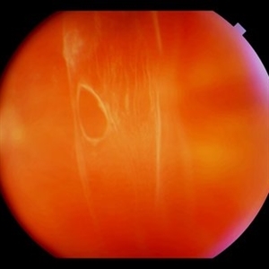
 Lattice Degeneration
Nov 9 2012 by Norman Byer
Lattice degeneration in a 42-year-old man which has produced four atrophic holes in a linear arrangement surrounded by a subclinical retinal detachment of unknown duration. By age 63, 21 years later, a posterior vitreous detachment was diagnosed in this eye, which was not present four years earlier. Nevertheless, the appearance seen here has remained exactly the same for 30 years, more than eight years with a concurrent PVD.
Condition/keywords: atrophic retinal hole, lattice degeneration, posterior vitreous detachment
-
 Posterior vitreous detachment
Jan 11 2013 by Alex P. Hunyor, MD
Posterior vitreous detachment with prominent Weiss ring.
Condition/keywords: posterior vitreous detachment
-
 Posterior Vitreous Detachment
Aug 23 2012 by Gabriela Lopezcarasa Hernandez, MD
Subhyaloid hemorrhage secondary to posterior vitreous detachment
Photographer: Gabriela Lopezcarasa Hernandez, Hospital Angeles Lomas
Imaging device: Zeiss FF4
Condition/keywords: subhyaloid hemorrhage, vitreous detachment
-
 Symptomatic Retinal Tear
Nov 9 2012 by Norman Byer
This is another example of a symptomatic retinal tear which occurred at the site of a cystic retinal tuft two days prior to the photograph when an acute posterior vitreous detachment occurred in this 64-year-old woman. Note the horizontal line of vitreous blood along the lower edge of the flap which demarcates the vitreous attachment to the flap.
Condition/keywords: acute posterior vitreous detachment, cystic retinal tuft, retinal flap, retinal tear, vitreous blood
-
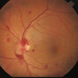
 Acute Posterior Vitreous Detachment
Nov 9 2012 by Norman Byer
This large and complicated retinal tear in a 51-year-old man resulted from an acute posterior vitreous detachment which concentrated its tractional forces around this area of lattice degeneration. Because of the powerful traction, there is an additional central tear splitting the large retinal flap and almost severing one of its arms. The traction was strong enough to completely rupture the blood vessel just to the left of the flap. Marking the ruptured peripheral end of the blood vessel is a yellow depigmented thrombus.
Condition/keywords: acute posterior vitreous detachment, depigmented thrombus, lattice degeneration, retinal tear, tractional retinal detachment
-
 Sudden Posterior Vitreous Detachment
Nov 9 2012 by Norman Byer
This 52-year-old woman suffered a sudden posterior vitreous detachment which caused a large horseshoe tear at 12:00 o’clock in this eye. It also produced another change at 8:45 in this left eye shown in this photograph. Note the small hemorrhage just to the left of the vessel. Immediately to the left of the hemorrhage and lying alongside of the vessel is a yellowish lesion which actually represents a cystic retinal tuft. You will see it better in the next slide pair.
Condition/keywords: cystic retinal tuft, posterior vitreous detachment, retinal hemorrhage, retinal vessel
-
 Sudden Posterior Vitreous Detachment
Nov 9 2012 by Norman Byer
This is the same eye that was described in slide pairs 15 and 16 and shows a large tractional symptomatic retinal tear at 12 o’clock. It was caused by a posterior vitreous detachment which placed sudden traction on a cystic retinal tuft. The whitish tuft is barely visible on the flap because it is not in focus. This tear was treated successfully with cryotherapy. The next slide pair is a postoperative view of the same lesion.
Condition/keywords: cystic retinal tuft, posterior vitreous detachment, tractional retinal tear
-
 Vitreous Hemorrhage
Nov 9 2012 by Norman Byer
This 60-year-old man suddenly developed a vitreous hemorrhage from this acute horseshoe tear 3½ years following cataract extraction when a posterior vitreous detachment occurred. The white nubbin identifies this lesion as a preexisting cystic retinal tuft. The pigment spot beneath the flap is evidence of secondary trophic changes in the pigment epithelium. Note the irregular shape of the flap with the narrow tip and broad base. This was caused by vitreous traction which was exerted at two separate points on the retina and which tore the retina at each place.
Condition/keywords: acute posterior vitreous detachment, irregularly shaped flap, trophic pigmented changes, vitreous hemorrhage, vitreous traction, white retinal tuft
-
 Acute Retinal Detachment
Nov 9 2012 by Norman Byer
This 54-year-old man was referred because of sudden symptoms in his opposite eye in which he had suffered an acute retinal detachment secondary to a horseshoe tear around lattice degeneration. During the examination, the fellow eye shown here was also found to have this large horseshoe tear about 1 o’clock hour (4 disc diameters) in size. A tear occurred around a lattice lesion which is present on the flap but is out of focus. This tear had been asymptomatic even though it was caused by a posterior vitreous detachment and illustrates that even very large tears may produce no symptoms or mild symptoms that are easily overlooked.
Condition/keywords: lattice degeneration, posterior vitreous detachment
-
 Posterior Vitreous Detachment
Nov 9 2012 by Norman Byer
This 68-year-old woman had a recent posterior vitreous detachment which produced this symptomatic horseshoe tear exactly at the site of this cystic retinal tuft. Note the characteristic discrete white nubbin at the apex, which is produced by a cap of glial cells with densely packed cytoplasm.
Condition/keywords: cystic retinal tuft, glial cells, posterior vitreous detachment, white retinal tuft
-
 Sudden Posterior Vitreous Detachment
Nov 9 2012 by Norman Byer
In this view of the previous case, the scleral indentation is being done immediately beneath the lesion. The hemorrhage is now out of sight and the white lesion is seen to be distinctly he elevated and in two parts. The base is white and the apex is translucent with a possible small hole in it. There is also a probable tiny full- thickness tear just behind the flap which cannot be discerned in this view.
Condition/keywords: full thickness retinal tear, peripheral retinal lesion, scleral indentation, translucency of apex, white retinal lesion
-
 Sudden Posterior Vitreous Detachment
Nov 9 2012 by Norman Byer
This 60-year-old man suffered a sudden posterior vitreous detachment which produced a large tractional retinal tear at 11:30 o’clock in this eye. This white cystic retinal tuft located at 9:30 also suffered minor injury at the same time as revealed in the next slide pair.
Condition/keywords: posterior vitreous detachment, white retinal tuft
-
 Scleral Indentation
Nov 9 2012 by Norman Byer
This is the same lesion with scleral indentation. You can see the small discrete preretinal hemorrhage and the sharply circumscribed area of elevated retina with subretinal fluid beneath it. No retinal break is visible, but the posterior vitreous is detached and exerting traction at this site. The area was surrounded with argon laser treatment the same day as the initial examination.
Condition/keywords: posterior vitreous detachment, preretinal hemorrhage, scleral indentation, subretinal fluid, vitreous traction
-
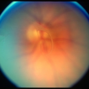
 Peripapillary Glial Proliferation
Oct 18 2012 by Suber S. Huang, MD, MBA, FASRS
61-year-old woman with peripapillary gilal proliferation
Photographer: Stacie Hrvatin
Condition/keywords: glial proliferation, posterior vitreous detachment, Weiss ring
-
 Peripapillary Glial Proliferation
Oct 18 2012 by Suber S. Huang, MD, MBA, FASRS
61-year-old woman with peripapillary gilal proliferation
Photographer: Stacie Hrvatin
Condition/keywords: glial proliferation, posterior vitreous detachment, Weiss ring
-
 Vitreous Hemorrhage
Jul 10 2018 by Karen Panzegrau
SD-OCT of a 35-year-old female presenting with a vitreous hemorrhage of her left eye. Patient has active proliferative diabetic retinopathy, as well as a completed posterior vitreous detachment in the left eye.
Photographer: Karen Panzegrau
Condition/keywords: diabetes, Heidelburg Spectralis, left eye, optical coherence tomography (OCT), posterior vitreous detachment, proliferative diabetic retinopathy (PDR), vitreous hemorrhage
-
 Retinal Tear in Aphakic Fellow Eye
Nov 9 2012 by Norman Byer
This 59-year-old man presented with sudden symptoms of retinal detachment in his opposite aphakic eye secondary to a tiny retinal tear about 1/8th disc diameter in size. During the examination, the fellow eye shown here was found to have this much larger tractional tear approximately 2 disc diameters in total length. If you look carefully, you will see that this is really a series of three separate tears with a common flap. The tears are separated by tiny bridges of remaining tissue which cause the edges of the apparent large tear to be serrated. This was also an aphakic eye with a posterior vitreous detachment but the lesion had produced no symptoms.
Condition/keywords: asymptomatic, bridge of tissue between tears, posterior vitreous detachment, tractional retinal tear
-
 Peripapillary Glial Proliferation
Oct 18 2012 by Suber S. Huang, MD, MBA, FASRS
61-year-old woman with peripapillary gilal proliferation
Photographer: Stacie Hrvatin
Condition/keywords: glial proliferation, posterior vitreous detachment, Weiss ring
-
 Lattice Lesion
Nov 9 2012 by Norman Byer
This lattice lesion in a 44-year-old woman shows an interesting tuft arising from the edge of the lesion and seen well against the background of the shadow of the indentation. It is caused by glial proliferation into the vitreous condensation at the edge of the lesion. Around the borders of each lattice lesion there is an invariable attachment of condensed vitreous. It is this vitreoretinal attachment that comprises the chief danger of lattice lesions where it may lead to acute retinal tears and retinal detachment at the time of posterior vitreous detachment.
Condition/keywords: glial proliferation, lattice degeneration, scleral indentation, vitreoretinal attachment, vitreous condensation, white retinal tuft
 Loading…