 Initializing download.
-
 By Norman Byer
From Dr. Norman E. Byer’s “The Peripheral Retina in Profile” - Uploaded on Nov 9, 2012.
- Last modified by Suber S. Huang, MD, MBA, FASRS on Feb 9, 2013.
- Reviewed by Chayal Patel
- Rating
- Appears in
- Miscellaneous
- Condition/keywords
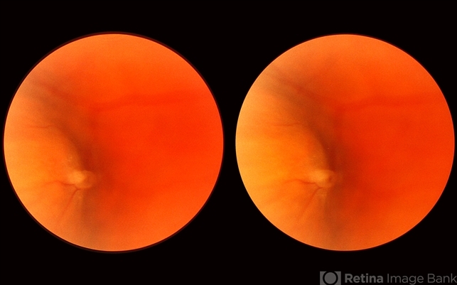
- scleral indentation, white retinal lesion, translucency of apex, peripheral retinal lesion, full thickness retinal tear
- Description
- In this view of the previous case, the scleral indentation is being done immediately beneath the lesion. The hemorrhage is now out of sight and the white lesion is seen to be distinctly he elevated and in two parts. The base is white and the apex is translucent with a possible small hole in it. There is also a probable tiny full- thickness tear just behind the flap which cannot be discerned in this view.


