Search results (249 results)
-
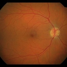
 Punctate Inner Choroidopathy with CNV Treated with Bevacizumab # 6 of 7
Feb 28 2013 by Gregory R. Blaha, MD, PhD
Fundus photo following treatment with bevacizumab in a 31-year-old female with vision loss from a choroidal neovascular membrane (CNV) from punctate inner choroidopathy. The vision improved and was stable following a single injection.
Photographer: Gerard Gauthier, Spindel Eye Assoc., Derry, NH
Imaging device: Zeiss FF 450 Plus
Condition/keywords: bevacizumab, choroidal neovascularization (CNV), punctate inner choroidopathy (PIC)
-
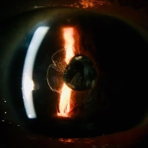
 Ciliary Body Melanoma With Partial Ring Configuration and Diffuse Sentinel Vessels
Feb 26 2014 by Susanna S. Park, MD, PhD
Slit lamp photo of a 57-year-old man with new vision loss from cataract formation. Large ciliary body mass with diffuse sentinel vessels is noted. The eye was removed and the tumor was noted to have a partial ring configuration with predominantly epithelioid cells and early vitreous seeding.
Photographer: Ellen Redenbo, University of California Davis Eye Center
Condition/keywords: ciliary body melanoma, melanoma
-
 Stargardt macular dystrophy slide 1
Oct 22 2012 by Ronald C. Gentile, MD
45-year-old man with progressive central vision loss since the age of 10. Small flecks can be seen surrounding the central area of atrophy.
Photographer: The New York Eye & Ear Infirmary Department of Medical Imaging
Condition/keywords: Stargardt disease, vision loss
-
 Superior Peripapillary Hemorrhage
Jul 13 2013 by Jason S. Calhoun
Patient was seen for acute vision loss in the right eye. Patient has glaucoma. VA was 20/70 in the right eye. Had vitrectomy back in May 2012 for ERM stripping. Also had trabectome with cataract surgery in December of 2012. Fundus photos presents a superior peripapillary Hemorrhage of the optic nerve. Patient will be followed up in one month.
Photographer: Jason S. Calhoun, Department of Ophthalmology, Mayo Clinic Jacksonville, Florida
Imaging device: TOPCON TRC 50-EX
Condition/keywords: peripapillary hemorrhage
-
 Punctate Inner Choroidopathy with CNV Treated with Bevacizumab # 1 of 7
Feb 28 2013 by Gregory R. Blaha, MD, PhD
Fundus photograph in a 31-year-old female with vision loss from a choroidal neovascular membrane (CNV) from punctate inner choroidopathy. Note the CNV and hemorrhage superotemporal to the fovea.
Photographer: Gerard Gauthier, Spindel Eye Associates, Derry, NH
Imaging device: Zeiss FF 450 Plus
Condition/keywords: bevacizumab, choroidal neovascularization (CNV), punctate inner choroidopathy (PIC)
-
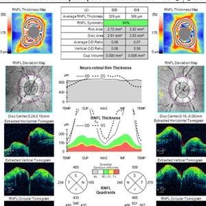 OCT in Patient With IIH Showing Thickened RNFL
Jan 16 2019 by John S. King, MD
18-year-old African American female with increased BMI with a history of headaches, nausea, transient diplopia and vision loss that she notices when getting up from her bed (and goes away after standing upright) for the last two weeks. Went to PCP and was treated for the flu, and after no improvement and visual symptoms known, was sent to ED. MRI did not show any masses and showed empty sella turcia. Vision 20/30 OD and 20/20 OS; no RAPD; IOP 15OU; no anterior segment or vitreous inflammation; discs are elevated with obscuration of the disc margins and some of the smaller vessels; there are no SVPs; there are mild Patton's lines temporally (see Initial Photos). The optic disc cube shows 360 degrees of RNFL thickening (see OCT). Was referred to near-ophthalmologist, Dr. Doyle. She obtained additional work-up, and LP opening pressure was high, and MRV showed bilateral transverse sinus stenosis. Patient showed steady improvement with medical therapy, that included weight loss and oral diamox. On her last visit with Dr. Doyle, vision has remained stable at 20/20-20/25 without an enlarged blindspot; there are SVPs and optic disc edema has resolved (see Post Treatment Photos); she is currently on 1000 mg of diamox and has lost 15 pounds, and no stinting procedure needed.
Imaging device: Cirrus
Condition/keywords: benign idiopatic intracranial hypertension, optic disc edema, papilledema
-
 Cone Dystrophy
Mar 29 2013 by Henry J. Kaplan, MD
Fundus photograph of a patient with progressive vision loss and hemeralopia (cone dystrophy) shows bull`s eye pattern #1.
Condition/keywords: bull's eye maculopathy, cone dystrophy
-
 Bull's Eye Maculopathy
Jun 29 2013 by Jason S. Calhoun
Patient comes in with severe vision loss in her right eye. Patient has been taking plaquenil for about 2 years. Fundus exam shows a bulls eye maculopathy centrally in the right eye.
Photographer: Jason S. Calhoun, Mayo Clinic Jacksonville, Florida
Imaging device: TOPCON TRC 50-EX
Condition/keywords: plaquenil toxicity
-
 Mild Patton's Lines in IIH - Initial Photos
Jan 16 2019 by John S. King, MD
18-year-old African American female with increased BMI with a history of headaches, nausea, transient diplopia and vision loss that she notices when getting up from her bed (and goes away after standing upright) for the last two weeks. Went to PCP and was treated for the flu, and after no improvement and visual symptoms known, was sent to ED. MRI did not show any masses and showed empty sella turcia. Vision 20/30 OD and 20/20 OS; no RAPD; IOP 15OU; no anterior segment or vitreous inflammation; discs are elevated with obscuration of the disc margins and some of the smaller vessels; there are no SVPs; there are mild Patton's lines temporally (see Initial Photos). The optic disc cube shows 360 degrees of RNFL thickening (see OCT). Was referred to near-ophthalmologist, Dr. Doyle. She obtained additional work-up, and LP opening pressure was high, and MRV showed bilateral transverse sinus stenosis. Patient showed steady improvement with medical therapy, that included weight loss and oral diamox. On her last visit with Dr. Doyle, vision has remained stable at 20/20-20/25 without an enlarged blindspot; there are SVPs and optic disc edema has resolved (see Post Treatment Photos); she is currently on 1000 mg of diamox and has lost 15 pounds, and no stinting procedure needed.
Photographer: Gretchen Harper
Imaging device: Topcon 50
Condition/keywords: idiopathic intracranial hypertension, optic disc edema, papilledema, Patton's Lines
-
---thumb.JPG/image-square;max$300,300.ImageHandler) Central retinal artery occlusion
Central retinal artery occlusion
Oct 26 2012 by Mallika Goyal, MD
Fundus photograph of a 55-year-old gentleman one day after sudden vision loss. Shows retinal infarct with "cherry red" spot at macular centre.
Condition/keywords: central retinal artery occlusion (CRAO), cherry red spot
-
 Punctate Inner Choroidopathy with CNV Treated with Bevacizumab # 2 of 7
Feb 28 2013 by Gregory R. Blaha, MD, PhD
Red-free fundus photograph in a 31-year-old female with vision loss from a choroidal neovascular membrane (CNV) from punctate inner choroidopathy. Note the CNV and hemorrhage superotemporal to the fovea.
Photographer: Gerard Gauthier, Spindel Eye Assoc., Derry, NH
Imaging device: Zeiss FF 450 Plus
Condition/keywords: bevacizumab, choroidal neovascularization (CNV), punctate inner choroidopathy (PIC)
-
---thumb.jpg/image-square;max$300,300.ImageHandler) OCT-CMV-Macula-SRF
OCT-CMV-Macula-SRF
Feb 24 2014 by Susanna S. Park, MD, PhD
Macular OCT of a 59-year-old woman on systemic chemotherapy for acute lymphocytic leukemia with new vision loss. Macular infiltration and submacular fluid are noted. Fundus examination showed a hemorrhagic retinitis in the macula from cytomegalovirus infection.
Photographer: Karishma Chandra, University of California Davis Eye Center
Imaging device: Cirrus OCT
Condition/keywords: CMV retinitis, optical coherence tomography (OCT)
-
 Vitreous in AC
Jan 9 2018 by Andrea Arriola-Lopez, MD MSc
78-year-old male. Vision loss in OD. IOP 18 mmHg. Subluxated PCIOL and vitreous in anterior chamber was found.
Photographer: Andrea E. Arriola López MD MS
Condition/keywords: anterior chamber, dislocated intraocular lens (IOL), vitreous
-
 Superior Peripapillary Hemorrhage
Jun 27 2013 by Jason S. Calhoun
Patient was seen for acute vision loss in the right eye. Patient has glaucoma. VA was 20/70 in the right eye. Had vitrectomy back in May 2012 for ERM stripping. Also had trabectome with cataract surgery in December of 2012. Fundus photos presents a superior peripapillary Hemorrhage of the optic nerve. Patient will be followed up in one month.
Photographer: Jason S. Calhoun, Mayo Clinic Jacksonville, Florida
Imaging device: TOPCON TRC 50-EX
Condition/keywords: peripapillary hemorrhage
-
---thumb.jpg/image-square;max$300,300.ImageHandler) Harada's with Exudative RD
Harada's with Exudative RD
Oct 13 2012 by Edwin H. Ryan, MD
OCT of a 35-year-old woman with acute vision loss in one eye.
Condition/keywords: exudative retinal detachment, Harada's disease
-
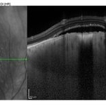
 Paracentral Acute Middle Maculopathy (PAMM)
Mar 21 2019 by Jonathan C. Tsui, MD
26-year-old female with hypertension presenting with chief complaint of "darkening" in her nasal visual field in the right eye. No flashes, floaters, or vision loss. Va 20/60 and nasal VF defect OD. SD-OCT demonstrated hyperreflectivity in the INL consistent with paracentral acute middle maculopathy. She was referred to her PCP for blood pressure optimization and a cardiovascular work-up. She returned for follow-up two months later with 20/80 OD, 20/20 OS. Repeat SD-OCT demonstrated inner retinal atrophy.
Photographer: Zellers, Diane
Condition/keywords: paracentral acute middle maculopathy
-
 Punctate Inner Choroidopathy with CNV Treated with Bevacizumab # 4 of 7
Feb 28 2013 by Gregory R. Blaha, MD, PhD
Mid-phase fluorescein angiogram in a 31-year-old female with vision loss from a choroidal neovascular membrane (CNV) from punctate inner choroidopathy.
Photographer: Gerard Gauthier, Spindel Eye Assoc., Derry, NH
Imaging device: Zeiss FF 450 Plus
Condition/keywords: bevacizumab, choroidal neovascularization (CNV), punctate inner choroidopathy (PIC)
-
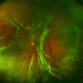 Funnel Retinal Detachment
Feb 2 2015 by Matt Poe, COA
The patient presented with total vision loss for >2months. Patient had history of exudative ARMD with intravitreal injections. No surgical intervention was done due to the long standing detachment and patient health.
Photographer: Matt Poe, COA. Northwest Arkansas Retina Associates, Springdale, AR.
Condition/keywords: retinal defect
-
 Harada's with Exudative RD
Oct 13 2012 by Edwin H. Ryan, MD
Fundus photograph of a 35-year-old woman with acute vision loss in one eye.
Condition/keywords: exudative retinal detachment, Harada's disease
-
 Bulls eye retinopathy FA LE
Nov 20 2012 by Roy Schwartz, MD
75-YEAR-OLD FEMALE presented with bilateral gradual vision loss 6/30 Dx: BE pseudophakia + PCO + BE bulls eye maculopathy per FA VA improved to 6/10 S/P YAG capsulotomy OCT - BE macular subretinal fluid, no history of chloroquine therapy, no drusen or signs of AMD Working Dx - BE chronic CSCR
Condition/keywords: bull's eye maculopathy
-
 Arterial Occlusion
Jul 14 2013 by Jason S. Calhoun
Patient in with vision loss in the lower quadrant of his visual field. Fundus photo shows arterial occlusion superior to the optic nerve
Photographer: Jason S. Calhoun, Department of Ophthalmology, Mayo Clinic Jacksonville, Florida
Imaging device: TOPCON TRC 50-EX
Condition/keywords: branch retinal artery occlusion (BRAO)
-
 Nevoma Leaking EDI OCT
Jul 9 2014 by Susanna S. Park, MD, PhD
EDI OCT imaging of the pigmented choroidal lesion in the right eye of a 28-year-old man with vision loss demonstrating elevation of the choroidal lesion and subretinal fluid over the lesion.
Photographer: Ellen Redenbo
Condition/keywords: choroidal nevus
-
 Example of AREDS Category 1 (Small Drusen But Not Considered AMD)
Feb 11 2013 by Neil M. Bressler, MD
Person in AREDS Category 1 were essentially free of age-related macular abnormalities, with a total drusen area less than 5 small drusen (<63 microns) within 3,000 microns of the center of the macula, and visual acuity of 20/32 or better in both eyes1. These are fundus photographs of a 53-year-old man, with visual acuity 20/20 OD and 20/32 OS presenting for evaluation of any diabetic retinopathy. Reference: 1 Age-Related Eye Disease Study Research Group. A randomized, placebo controlled clinical trial of high-dose supplementation with vitamins C and E, beta carotene, and zinc for age-related macular degeneration and vision loss: AREDS report No. 8. Arch Ophthalmol. 2001;119(10):1417-1436.
Condition/keywords: age-related macular degeneration (AMD)
-
---thumb.jpg/image-square;max$300,300.ImageHandler) Harada's with Exudative RD
Harada's with Exudative RD
Oct 13 2012 by Edwin H. Ryan, MD
OCT of a 35-year-old woman with acute vision loss in one eye.
Condition/keywords: exudative retinal detachment, Harada's disease
-
 OCT CME
Jul 9 2014 by Susanna S. Park, MD, PhD
OCT of a 72-year-old man with vision loss 6 months after cataract surgery showing CME.
Photographer: Ellen Redenbo
Condition/keywords: cystoid macular edema (CME)
 Loading…