Search results (35 results)
-
 Traumatic Macular Hole with Retinal Detachment and PVR - montage
Sep 27 2012 by Pauline T Merrill, MD, FASRS
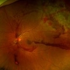
Fundus photo montage of a 13-year-old boy s/p soccer ball injury 1 month previously.
Photographer: Karen Parque, Illinois Retina Associates, Chicago, IL
Condition/keywords: proliferative vitreoretinopathy (PVR), traumatic macular hole
-
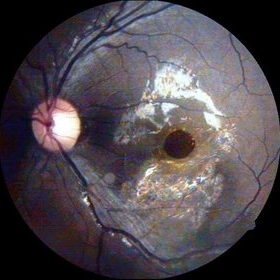
 Traumatic Macular Hole with Retinal Detachment and PVR
Sep 27 2012 by Pauline T Merrill, MD, FASRS
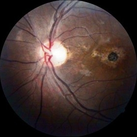
Fundus photo of a 13-year-old boy s/p soccer ball injury 1 month previously. In addition to full-thickness macular hole and total retinal detachment with grade C PVR, note pigment granules visible in vitreous over optic nerve.
Photographer: Karen Parque, Illinois Retina Associates, Chicago, IL
Condition/keywords: proliferative vitreoretinopathy (PVR), traumatic macular hole
-
 Traumatic Macular Hole
Aug 23 2012 by Gabriela Lopezcarasa Hernandez, MD
12-year-old boy with blunt trauma in right eye and central scotoma.
Photographer: Gabriela Lopezcarasa Hernandez, Hospital Angeles Lomas
Imaging device: ZEISS F4
Condition/keywords: blunt trauma, central scotoma, macular hole
-
 Traumatic Macular Hole
Aug 23 2012 by Gerardo Garcia-Aguirre, MD
Fundus photograph of a large macular hole with an area of pigment migration secondary to blunt trauma.
Photographer: Noemí Hernández, Asociación para Evitar la Ceguera en México
Imaging device: Zeiss FF4
Condition/keywords: deformity, macular hole
-
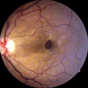
 Traumatic macular hole
Dec 19 2012 by Eric A. Postel, MD
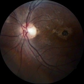
Color fundus photograph of a young male with a traumatic macular hole
Condition/keywords: blunt trauma, macular hole
-
---thumb.jpg/image-square;max$300,300.ImageHandler) Traumatic Choroidal Rupture and Macular Hole
Traumatic Choroidal Rupture and Macular Hole
Jan 1 2013 by John T. Thompson, MD
Traumatic macular hole and choroidal rupture following blunt trauma to globe.
Condition/keywords: blunt trauma, choroidal rupture, traumatic macular hole
-
 Traumatic Macular Hole
Sep 14 2014 by Mehul A Shah
20-year-old presented with cricket ball injury.
Photographer: Drashti Netralaya,Dahod
Imaging device: Zeiss ff450
Condition/keywords: traumatic macular hole
-
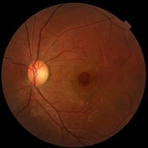 Traumatic Macular Hole and Choroidal Ruptures
Mar 15 2017 by Hamid Ahmadieh, MD
Color fundus photograph of the left eye of a 30 -year-old woman with a history of closed eye injury leading to a large traumatic macular hole and two concentric choroidal ruptures.
Photographer: Solmaz Shahmohammad, Negah Eye Center, Tehran,Iran
Condition/keywords: choroidal rupture, color fundus photograph, traumatic macular hole
-
 Choroidal Rupture, Subretinal and Vitreous Hemorrhage Secondary to Blunt Trauma
Dec 29 2012 by Humberto Ruiz-Garcia, MD
SD-OCT obtained at 72 hours which shows neurosensory macular detachment and severe thinning (impending macular hole).
Photographer: Humberto Ruiz-Garcia
Imaging device: Cirrus HD OCT
Condition/keywords: traumatic macular hole
-
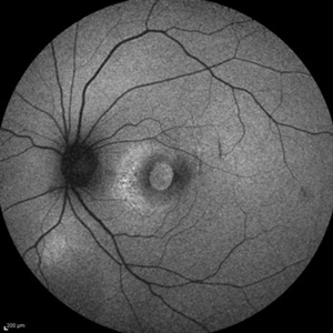 Traumatic Macular Hole and Choroidal Ruptures
Mar 15 2017 by Hamid Ahmadieh, MD
Fundus autofluorescence ( FAF) image of the left eye of a 30 -year-old woman with a recent history of closed eye injury leading to a large traumatic macular hole and two concentric choroidal ruptures.
Photographer: Solmaz Shahmohammad, Negah Eye Center, Tehran,Iran
Condition/keywords: choroidal rupture, fundus autofluorescence (FAF), traumatic macular hole
-
 Traumatic Macular Hole
Sep 14 2014 by Mehul A Shah
15-year-old boy presented with cricket ball injury.
Photographer: Drashti Netralaya,Dahod
Imaging device: Zeiss ff450
Condition/keywords: traumatic macular hole
-
 Traumatic Macular Hole
Sep 14 2014 by Mehul A Shah
17-year-old presented with blunt trauma.
Photographer: Drashti Netralaya,Dahod
Imaging device: Zeiss ff450
Condition/keywords: traumatic macular hole
-
 Traumatice Macular Hole
Oct 4 2014 by Mehul A Shah
A 27-year-old patient presented with history of blunt trauma and patient was examined few months later.
Photographer: Drashti Netralaya,Dahod
Imaging device: Zeiss ff450
Condition/keywords: traumatic macular hole
-
---thumb.jpg/image-square;max$300,300.ImageHandler) Traumatic Macular Hole
Traumatic Macular Hole
Mar 9 2013 by Gabriela Lopezcarasa Hernandez, MD
Traumatic macular hole.
Photographer: Gabriela Lopezcarasa Hdz. MD. Hospital Angeles Lomas
Imaging device: zEISS FF4
Condition/keywords: macular hole
-
 Blunt Ocular Trauma with Commotio Retinae
Nov 5 2019 by Nichole Lewis
11-year-old male with blunt ocular trauma from a soccer ball. Commotio Retinae, retinal hemorrhages, vitreous hemorrhage, multiple retinal tears and a traumatic macular hole. VA 20/70.
Photographer: Nichole Lewis
Imaging device: Optos
Condition/keywords: blunt trauma, commotio retinae, retinal hemorrhage, retinal tear, traumatic macular hole, vitreous hemorrhage
-
 Traumatic Macular Hole
Oct 2 2017 by Mehul A Shah
A 43-year-old male presented with history of blunt trauma before 6 months. Clinical picture was presented to us.
Photographer: Mehul Shah
Condition/keywords: traumatic macular hole
-
 Traumatic Macular Hole
Feb 6 2019 by awaneesh m upadhyay, MBBS, DNB
Fundus photograph of an 8-year-old boy with vision of <20/200 in OD following blunt trauma of 10 days duration show macular hole with Berlin's edema.
Photographer: Dr. Awaneesh Upadhyay
Condition/keywords: Berlin's edema
-
 Traumatic Macular Hole with OCT
Jun 29 2018 by Gareth Lema, MD, PhD
Traumatic macular hole caused by the screw cap of a baby formula bottle. Pressure had built up in the container and propelled the cap into the patient's eye after she partially unscrewed it.
Photographer: Sandra Boglione, Ross Eye Institute, University at Buffalo Jacobs School of Medicine, Buffalo, NY
Imaging device: Optos
Condition/keywords: blunt trauma, traumatic macular hole
-
 Traumatic Macular Hole
Mar 27 2019 by Gary R. Cook, MD, FACS
7-year-old white male with a traumatic macular hole and secondary epiretinal membrane formation OS; hit in the eye with a rock; V.A.= counting fingers.
Imaging device: Topcon VT-50
Condition/keywords: epiretinal membrane formation, full thickness macular hole, macular hole, traumatic macular hole
-
 Very Large Choroidal Melanoma in Monocular Patient - BScan
Feb 13 2020 by Michael Seider, MD
Very large choroidal melanoma in the left eye of a 51-year-old man with long-standing poor vision in the right eye from a childhood injury (with traumatic macular hole and chorioretinal scarring). Note the large superior choroidal tumor with overlying subretinal hemorrhage and extensive inferior exudative retinal detachment. B-Scan ultrasound shows the collar-stud shape of the lesion and the overlying subretinal hemorrhage which is hyper-reflective compared to the vitreous and slightly hypo-reflective compared to the tumor. The patient has optic disk drusen in both eyes. He elected enucleation.
-
 OCT - Traumatic Full Thickness Macular Hole
Feb 6 2019 by awaneesh m upadhyay, MBBS, DNB
Right eye OCT image of an 8-year-old boy shows full thickness macular hole following blunt trauma of 1 week duration.
Photographer: Awaneesh Upadhyay
Imaging device: HEILDERBERG - HRA
Condition/keywords: traumatic macular hole
-
 Traumatic Macular Hole
Mar 27 2019 by Gary R. Cook, MD, FACS
14-year-old white male with a traumatic macular hole OS 23 days post hockey puck injury; V.A.= 20/200.
Imaging device: Topcon VT-50
Condition/keywords: full thickness macular hole, macular hole, trauma, traumatic macular hole
-
 Traumatic Macular Hole And Bruch Membrane Rupture
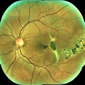
Jan 22 2021 by Renata Garcia Franco, Md
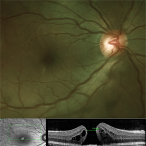
Male with history of ocular blunt injury, full-thickness macular hole, RPE changes and Bruch membrane rupture.
Photographer: Fatima Hernandez, Instituto de la Retina del Bajio SC
Imaging device: Zeiss
Condition/keywords: traumatic macular hole
-
 Very Large Choroidal Melanoma in Monocular Patient - Widefield Color (Eye with Tumor)
Feb 13 2020 by Michael Seider, MD
Very large choroidal melanoma in the left eye of a 51-year-old man with long-standing poor vision in the right eye from a childhood injury (with traumatic macular hole and chorioretinal scarring). Note the large superior choroidal tumor with overlying subretinal hemorrhage and extensive inferior exudative retinal detachment. B-Scan ultrasound shows the collar-stud shape of the lesion and the overlying subretinal hemorrhage which is hyper-reflective compared to the vitreous and slightly hypo-reflective compared to the tumor. The patient has optic disk drusen in both eyes. He elected enucleation.
-
 Traumatic Macular Hole And Bruch Membrane Rupture
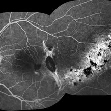
Jan 22 2021 by Renata Garcia Franco, Md
FA shows hypofluorescence in early frames due to a break in choriocapillaris and choroidal vessels at the rupture site with staning at late phases.
Photographer: Fatima Hernandez, Instituto de la Retina del Bajio SC
Imaging device: Zeiss
Condition/keywords: traumatic macular hole
 Loading…