Search results (35 results)
-
 Sub ILM Hemorrhage / CPR
Mar 3 2014 by David Callanan, MD
31-year-old female, undergoing hand surgery and suffered cardiac arrest; had CPR in coma for 2 days; noted field defect when she awoke; 20/40 initially 20/20 at end.
Condition/keywords: internal limiting membrane (ILM) peeling
-
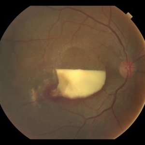
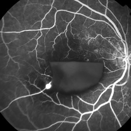
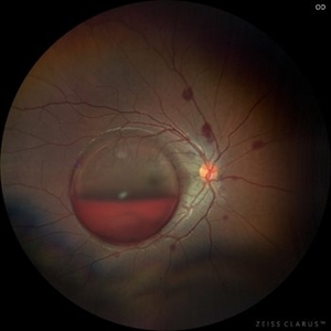
 Sub ILM Hemorrhage
Jul 29 2014 by Mallika Goyal, MD
OCT of the left eye of a 26-year-old lady who presented with sudden vision drop (VA 20/40) reveals sub-ILM haemorrhage. There was no history of trauma, valsalva maneuvre or other contributing factors. This heme cleared within 10 days following gas injection and prone positioning with visual recovery to 20/20.
Photographer: Mallika Goyal, MD, Apollo Health City, Jubilee Hills, Hyderabad-500033
Condition/keywords: sub-inner limiting membrane hemorrhage
-
 Sub ILM Hemorrhage / CPR
Mar 3 2014 by David Callanan, MD
31-year-old female, undergoing hand surgery and suffered cardiac arrest; had CPR in coma for 2 days; noted field defect when she awoke; 20/40 initially 20/20 at end.
Condition/keywords: internal limiting membrane (ILM) peeling
-
 Sub ILM Hemorrhage
Jul 29 2014 by Mallika Goyal, MD
Fundus photograph of the left eye of a 26-year-old lady who presented with sudden vision drop (VA 20/40) reveals sub-ILM hemorrhage. There was no history of trauma, valsalva maneuvre or other contributing factors. This heme cleared within 10 days following gas injection and prone positioning with visual recovery to 20/20.
Photographer: Mallika Goyal, MD, Apollo Health City, Jubilee Hills, Hyderabad-500033
Condition/keywords: sub-inner limiting membrane hemorrhage
-
 Sub ILM Hemorrhage / CPR
Mar 3 2014 by David Callanan, MD
31-year-old female, undergoing hand surgery and suffered cardiac arrest; had CPR in coma for 2 days; noted field defect when she awoke; 20/40 initially 20/20 at end.
Condition/keywords: internal limiting membrane (ILM) peeling
-
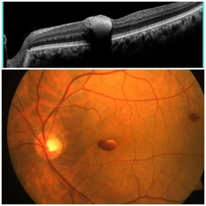
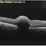
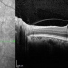 Horizontal OCT Scan of Sub ILM Hemorrhage
Mar 8 2017 by Manish Nagpal, MD, FRCS (UK), FASRS
Patient with a macroaneurysm leading to a sub ILM hemorrhage near fovea showing an interesting horizontal scan passing through the central area.
Photographer: pranita chaudhary
Condition/keywords: hemorrhage, macroaneurysm
-
 Sub ILM Hemorrhage / CPR
Mar 3 2014 by David Callanan, MD
31-year-old female, undergoing hand surgery and suffered cardiac arrest; had CPR in coma for 2 days; noted field defect when she awoke; 20/40 initially 20/20 at end.
Condition/keywords: internal limiting membrane (ILM) peeling
-
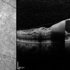 Vertical OCT Scan of Sub ILM Hemorrhage
Mar 8 2017 by Manish Nagpal, MD, FRCS (UK), FASRS
Patient with a macroaneurysm leading to a sub ILM hemorrhage near fovea showing an interesting vertical scan passing through the central area.
Photographer: pranita chaudhary
Condition/keywords: hemorrhage, macroaneurysm
-
 Sub ILM Hemorrhage / CPR
Mar 3 2014 by David Callanan, MD
31-year-old female, undergoing hand surgery and suffered cardiac arrest; had CPR in coma for 2 days; noted field defect when she awoke; 20/40 initially 20/20 at end.
Condition/keywords: internal limiting membrane (ILM) peeling
-
 Sub ILM Hemorrhage / CPR
Mar 3 2014 by David Callanan, MD
31-year-old female, undergoing hand surgery and suffered cardiac arrest; had CPR in coma for 2 days; noted field defect when she awoke; 20/40 initially 20/20 at end.
Condition/keywords: internal limiting membrane (ILM) peeling
-
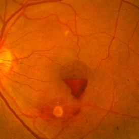
 RAMA with Sub ILM Hemorrhage
Jan 31 2018 by John S. King, MD
73-year-old with well controlled diabetes and hypertension presented with a month onset of acute central scotoma; CF 5'
Photographer: Stacey
Imaging device: Cirrus
Condition/keywords: ruptured macroaneurysm, sub-inner limiting membrane hemorrhage
-
 Subhyaloid Hemorrhage With Vitreous Hemorrhage
Sep 12 2025 by Akansha Sharma
Color fundus photograph of a 56 year old hypertensive and diabetic female who presented with subhyaloid hemorrhage along with vitreous hemorrhage after being administered high dose anti-platelet therapy pre- and post a cardiac procedure.
Photographer: DR. AKANSHA SHARMA
Condition/keywords: SHH, sub ILM hemorrhage, subhyaloid hemorrhage, VH, vitreous hemorrhage
-
 Small subILM Hemorrhage
Oct 26 2019 by Navneet Mehrotra, DNB
44-year-old hypertensive male with sudden decrease in vision showing small sub ILM hemorrhage at macula.
Photographer: Navneet Mehrotra
Imaging device: NidekRS330
Condition/keywords: hypertension, subILM hemorrhage
-
 RAMA with Sub ILM Hemorrhage
Jan 31 2018 by John S. King, MD
73 -year-old with well controlled diabetes and hypertension presented with a month onset of acute central scotoma; CF 5'; FA shows pooling in the aneurysm, blockage by dehemoglobinized heme, some diabetic changes and some IRMAs likely from old vein occlusion (s)
Photographer: Stacey
Imaging device: Cirrus
Condition/keywords: ruptured macroaneurysm, sub-inner limiting membrane hemorrhage
-
 RAMA with Sub ILM Hemorrhage
Jan 31 2018 by John S. King, MD
73-year-old with well controlled diabetes and hypertension presented with a month onset of acute central scotoma; CF 5'; SUB-ILM vs subyaloid elevation
Photographer: Stacey
Imaging device: Cirrus
Condition/keywords: ruptured macroaneurysm, sub-inner limiting membrane hemorrhage
-
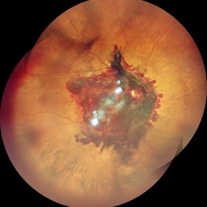
 Macroaneurysm With Sub ILM Hemorrhage
Mar 8 2017 by Manish Nagpal, MD, FRCS (UK), FASRS
Patient with a macroaneurysm leading to a sub ILM hemorrhage near fovea.
Photographer: Pranita Chaudhary
Condition/keywords: hemorrhage, macroaneurysm
-
 Sub ILM Hemorrhage
May 2 2019 by S. Natarajan, MD, FASRS, FRCS (GLASGOW) , FICO, D.Sc, FELA
Fundus photograph of an 56-year-old anemic male who presented with sub ILM hemorrhage at the macula in left eye.
Photographer: Ashwini borde
Imaging device: Carl Zeiss 450 Plus IR
Condition/keywords: hemorrhage, internal limiting membrane (ILM) peeling
-
Vitrectomy for Sub ILM blood over macula
Jan 2 2023 by Manish Nagpal, MD, FRCS (UK), FASRS
This is a case of non resolving ILM haemorrhage over macula. Vitrectomy is carried out and hyaloid is removed. Cutter is used to try to aspirate the blood from the surface of macula. But due to its location in the sub ILM space i use a forceps to make a opening in the ILM. Through the opening the blood aspirates easily.
Condition/keywords: hyaloid, internal limiting membrane, macula, retina, sub ILM blood, sub ILM hemorrhage, video, vitrectomy
-
 Valsalva Retinopathy
Nov 18 2022 by Niloofar Piri, MD
21 yo female presented with decaresed central vision and scotoma immediately after severe vomiting. Color fundus phtograph demonstrates large sub ILM layered hemorrhage in the macula consistent with valsalva retinopathy. Notice the sacttered blot retinal hemorrhages in mid-periphery.
Photographer: Rocio Bentivegna, MD, Saint Louis University
Condition/keywords: sub ILM hemorrhage, valsalva retinopathy
-
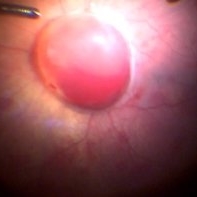
 Sub ILM Hemorrhage
Jan 12 2022 by Manish Nagpal, MD, FRCS (UK), FASRS
Intraoperative view of a non clearing sub ILM hemorrhage over the macula with partly de-hemoglobinized blood.
Photographer: Manish Nagpal, Retinal Foundation, Ahmedabad, India
Imaging device: Sony PMW -10 MD surgical camera
Condition/keywords: sub internal limiting membrane haemorrhage, subILM hemorrhage
-
Vitrectomy for Sub ILM blood over macula
Jan 2 2023 by Manish Nagpal, MD, FRCS (UK), FASRS
This is a case of non resolving ILM hemorrhage over macula. Vitrectomy is carried out and hyaloid is removed after traimcinolone staining. After this brilliant blue dye is injected to stain the ILM. Internal limiting membrane is then removed with a forceps. Once the sub ilm blood is exposed , it easily aspirates with the cutter. The origin is probably from a macroaneurysm and there is a small component of subretinal residual blood noted at the end of the surgery.
Condition/keywords: brilliant blue, hyaloid, internal limiting membrane, macula, microaneurysm, retina, sub ILM blood, sub ILM hemorrhage, triamcinolone, video, vitrectomy
-
 Valsalva Retinopathy
Nov 18 2022 by Niloofar Piri, MD
Sudden vision loss immediately after severe vomiting. Color fundus photo demonstrates large sub ILM hemorrhage consistent with valsalva retinopathy.
Photographer: Sean Kelso, Saint Louis University
Condition/keywords: SUB ILM hemorrhage, sub internal limiting membrane haemorrhage, valsalva retinopathy
-
 Sub ILM Hemorrhage
May 19 2023 by Rahul Bhatia, MS, DNB
A 10-year-old male with Aplastic Anemia presented to Retina Clinic. Fundus Photograph and OCT line scan suggestive of Sub ILM Hemorrhage
Photographer: Dr Rahul Bhatia, LHMC, Delhi, India
Imaging device: Iphone
Condition/keywords: sub internal limiting membrane haemorrhage
-
 Shaken Baby Syndrome
Feb 20 2022 by Imren Akkoyun, MD, FACS
Fundus photograph of an 6 months-old girl. Fundus exam revealed retinal hemorrhages involving multiple layers with subhyaloidal and sub ILM hemorrhage involving optic disc and macula.
Photographer: Imren Akkoyun, MD, FACS;Baskent University Faculty of Medicine Department of Ophthalmology
Imaging device: Zeiss, Operating Microscope
Condition/keywords: Shaken-Baby-Syndrome
-
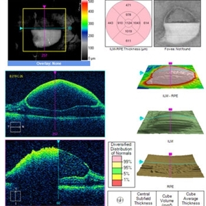
 Sub-Internal Limiting Membrane Hemorrhage
Apr 17 2025 by Malvika Singh
OCT of a 41 year-old, male, with a central retinal vein occlusion and a foveal sub-internal limiting membrane hemorrhage.
Photographer: Dr Malvika Singh, Retina Foundation, Ahmedabad, India
Imaging device: Mirante SLO/OCT
Condition/keywords: optical coherence tomography (OCT), SUB ILM hemorrhage
 Loading…