Search results (91 results)
-
 The Peripheral Retina in Profile: A Stereoscopic Atlas
Mar 12 2013 by Norman Byer
The stereoscopic atlas contains unique stereo photographs vividly portraying the changes in the peripheral fundus and their histopathology, incidence and risks.
Condition/keywords: stereo pair, video
-
 Coloboma, In Stereo
Oct 1 2012 by Michael P. Kelly, FOPS
This is a stereo retinal fundus photograph of a coloboma, with the optic nerve centered, using a Zeiss FF3C retinal fundus camera.
Photographer: Michael P. Kelly, FOPS Director, Duke Eye Labs, Duke University Hospital, Duke Eye Center, Durham, NC
Condition/keywords: coloboma, fundus photograph, stereo pair
-
 Roth Spots
Jul 11 2013 by Jerald A. Bovino, MD
No history, part of stereo pair.
Condition/keywords: stereo pair, white centered retinal hemorrhage (Roth Spot)
-
 Asteroid Hyalosis, In Stereo
Sep 28 2012 by Michael P. Kelly, FOPS
Asteriod hyalosis, stereo.
Photographer: Michael P. Kelly, FOPS Director, Duke Eye Labs Duke University Hospital, Duke Eye Center, Durham, NC
Imaging device: Zeiss FF3C
Condition/keywords: asteroid hyalosis, stereo pair
-
 Choroidal Detachment, In Stereo
Sep 25 2012 by Michael P. Kelly, FOPS
Photographer: Michael P. Kelly, FOPS Director, Duke Eye Labs, Duke University Hospital, Duke Eye Center, Durham, NC
Imaging device: Zeiss FF3C
Condition/keywords: choroidal detachment, stereo pair
-
 Retinal Detachment, in Stereo
Oct 4 2012 by Michael P. Kelly, FOPS
Retinal detachment showing bare RPE, in stereo.
Photographer: Michael P. Kelly, FOPS, Director, Duke Eye Center Labs, Duke University Hospital, Durham, NC
Condition/keywords: stereo pair
-
 Proliferative Diabetic Retinopathy, In Stereo
Sep 28 2012 by Michael P. Kelly, FOPS
Proliferative diabetic retinopathy, PDR, Stereo.
Photographer: Michael P. Kelly, FOPS Director, Duke Eye Labs, Duke University Hospital, Duke Eye Center, Durham, NC
Imaging device: Zeiss FF4
Condition/keywords: stereo pair
-
 Horseshoe Tear, In Stereo
Sep 28 2012 by Michael P. Kelly, FOPS
Horse shoe tear, stereo.
Photographer: Michael P. Kelly, FOPS Director, Duke Eye Labs, Duke University Hospital, Duke Eye Center, Durham, NC
Imaging device: Zeiss FF3C
Condition/keywords: stereo pair
-
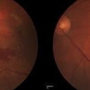
 Subfoveal Choroidal Neovascularization, In Stereo
Sep 28 2012 by Michael P. Kelly, FOPS
Subfoveal Choroidal Neovascularization.
Photographer: Michael P. Kelly, FOPS Director, Duke Eye Labs, Duke University Hospital, Duke Eye Center, Durham, NC
Imaging device: Zeiss FF4
Condition/keywords: pigment epithelial detachment (PED), stereo pair, subfoveal choroidal neovascularization
-
---thumb.JPG/image-square;max$300,300.ImageHandler) Stereo Image of Retinal Tear
Stereo Image of Retinal Tear
Jun 30 2013 by Jason S. Calhoun
Four up image of a stereo 3-D pair of retinal tears.
Photographer: Jason S. Calhoun, Mayo Clinic Jacksonville, Florida
Condition/keywords: retinal tear, stereo pair
-
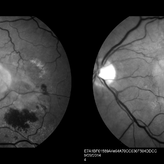
 Inferior Choroidal Coloboma and Tilted Disc
Feb 19 2013 by From the Collections of Thomas M. Aaberg, MD and Thomas M. Aaberg Jr., MD
NLP; Left of stereo pair.
Condition/keywords: coloboma, stereo pair
-
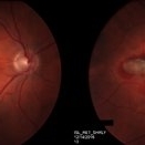
 Optic Pit; two in one nerve
Feb 19 2013 by From the Collections of Thomas M. Aaberg, MD and Thomas M. Aaberg Jr., MD
Color photo, 20/20; left of a stereo pair.
Condition/keywords: color photo, stereo pair
-
 Diabetic Retinopathy Optic Nerve Edema, Fluorescein Angiogram, Stereo
Apr 11 2015 by James B. Soque, CRA, OCT-C, COA, FOPS
Optic Nerve Edema and Leakage on fluorescein angiography in this 48-year-old patient with a 10 year history of diabetes. 50 degree stereo photo fluorescein angiogram.
Photographer: James B. Soque, CRA, COA
Imaging device: Topcon TRC 50 DX, OIS 5 MP Digital Camera, MERGE Software
Condition/keywords: background diabetic retinopathy (BDR), diabetes, disc leakage, fluorescein leakage, optic disc swelling, optic nerve edema, stereo pair
-
 Optic Pit; two in one nerve
Feb 19 2013 by From the Collections of Thomas M. Aaberg, MD and Thomas M. Aaberg Jr., MD
Color photo, 20/20; left of a stereo pair.
Condition/keywords: color photo, stereo pair
-
 Inferior Choroidal Coloboma and Tilted Disc
Feb 19 2013 by From the Collections of Thomas M. Aaberg, MD and Thomas M. Aaberg Jr., MD
NLP; Right of stereo pair.
Condition/keywords: coloboma, stereo pair
-
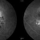
 Neovascular ARMD With Subretinal Hemorrhage, Red-Free Photos - Stereo
Nov 26 2014 by James B. Soque, CRA, OCT-C, COA, FOPS
Stereo FC, RF and FA of a 77-year-old white female with visual acuity CC 20/200-3, with left eye neovascular ARMD, drusen, and subretinal hemorrhage with hard exudates temporally. Peripheral retina reveals cobblestone degeneration.
Photographer: James Soque, CRA, COA, Island Retina, Shirley, NY
Imaging device: Topcon TRC 50 EX, with MERGE software and OIS 5 MP digital Camera
Condition/keywords: neovascular age-related macular degeneration (AMD), red-free, stereo pair
-
---thumb.jpg/image-square;max$300,300.ImageHandler) Pre-Retinal Fibrous Proliferative Membrane
Pre-Retinal Fibrous Proliferative Membrane
Feb 20 2013 by From the Collections of Thomas M. Aaberg, MD and Thomas M. Aaberg Jr., MD
Ultrasound and color photo stereo-pair with next slide demonstrating sever pre-retinal fibrous membrane with contraction, possibly from PDR (second copy of the above pair).
Condition/keywords: color photo, pre-retinal membrane, stereo pair, ultrasound
-
 Neovascular ARMD With Subretinal Hemorrhage, Fluorescein Angiography Photos - Stereo
Oct 14 2014 by James B. Soque, CRA, OCT-C, COA, FOPS
Stereo FC, RF and FA of a 77-year-old white female with visual acuity CC 20/200-3, with left eye neovascular ARMD, drusen, and subretinal hemorrhage with hard exudates temporally. Peripheral retina reveals cobblestone degeneration.
Photographer: James Soque, CRA, COA, Island Retina, Shirley, NY
Imaging device: Topcon TRC 50 EX, with MERGE software and OIS 5 MP digital Camera
Condition/keywords: neovascular age-related macular degeneration (AMD), stereo pair
-
 Stereo View of Retinal Angiomatous Proliferation in Age-Related Macular Degeneration
Jan 21 2016 by James B. Soque, CRA, OCT-C, COA, FOPS
Stereo pair of 75-year-old white male with classic SRN with RAP lesion of right eye, actively receiving anti-VEGF treatment. 50 Degrees, no mag, L and R stereo pair. Single View of OD also visible in this case.
Condition/keywords: age-related macular degeneration (AMD), anti-VEGF, retinal angiomatous proliferation (RAP), stereo pair, subretinal neovascularization (SRNV)
-
---thumb.jpg/image-square;max$300,300.ImageHandler) Recurrent Pseudotumor
Recurrent Pseudotumor
Feb 13 2013 by From the Collections of Thomas M. Aaberg, MD and Thomas M. Aaberg Jr., MD
FA, multiple punctate hyperfluorescent dots stereo.
Condition/keywords: hypofluorescent spots, stereo pair
-
---thumb.jpg/image-square;max$300,300.ImageHandler) Pre-Retinal Fibrous Proliferative Membrane
Pre-Retinal Fibrous Proliferative Membrane
Feb 20 2013 by From the Collections of Thomas M. Aaberg, MD and Thomas M. Aaberg Jr., MD
Ultrasound and color photo stereo-pair with next slide demonstrating sever pre-retinal fibrous membrane with contraction, possibly from PDR.
Condition/keywords: color photo, pre-retinal membrane, stereo pair, ultrasound
-
---thumb.jpg/image-square;max$300,300.ImageHandler) Pre-Retinal Fibrous Proliferative Membrane
Pre-Retinal Fibrous Proliferative Membrane
Feb 20 2013 by From the Collections of Thomas M. Aaberg, MD and Thomas M. Aaberg Jr., MD
Ultrasound and color photo stereo-pair with previous slide demonstrating sever pre-retinal fibrous membrane with contraction, possibly from PDR (second copy of the above pair).
Condition/keywords: color photo, pre-retinal membrane, stereo pair, ultrasound
-
 Subretinal Thickening and Subretinal Hemorrhage – Stereo Color Fundus Photograph
Mar 9 2017 by James B. Soque, CRA, OCT-C, COA, FOPS
Color fundus stereo photograph of a 52-year-old white male with VA loss to 20/200 of unknown etiology. Dilated fundus examination of the right eye reveals a fibrotic scar with subretinal thickening and subretinal hemorrhage.
Photographer: James B Soque, CRA, OCT-C, COA
Imaging device: Topcon TRC 50 DX, MERGE Software
Condition/keywords: blood, color fundus photograph, color photo, stereo pair, subretinal blood, subretinal fibrosis, subretinal thickening
-
 Vascular Loop
Jul 11 2013 by Jerald A. Bovino, MD
No history, slide is labeled vascular loop, this is the same slide, or stereo pair to #475.
Condition/keywords: vascular loop
-
 Neovascular ARMD With Subretinal Hemorrhage, Fundus Color Photos- Stereo
Oct 14 2014 by James B. Soque, CRA, OCT-C, COA, FOPS
Stereo FC, RF and FA of a 77-year-old white female with visual acuity CC 20/200-3, with left eye neovascular ARMD, drusen, and subretinal hemorrhage with hard exudates temporally. Peripheral retina reveals cobblestone degeneration.
Photographer: James Soque, CRA, COA, Island Retina, Shirley, NY
Imaging device: Topcon TRC 50 EX, with MERGE software and OIS 5 MP digital Camera
Condition/keywords: fundus photograph, neovascular age-related macular degeneration (AMD), stereo pair
 Loading…