Search results (14 results)
-
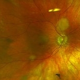
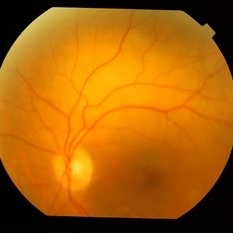
 Femoral Artery Plaque
Sep 9 2020 by John S. King, MD
66-year-old white male, former smoker, with a history of femoral artery stent a plaque removal in 2017 (see picture), triple bypass 2019 (at that time there was no high grade carotid stenosis), diabetes significant for SNPDR OD and PDR OS (NVD). He underwent PRP OS and two months later developed a vitreous hemorrhage and had a PPV OS. Early in the post-operative period vision dropped to LP due to acute CRAO with retinal embolus present. He was found to have progressed to high grade carotid stenosis (versus imaging 6 months ago) and a left carotid endarterectomy was performed . He had a picture of this plaque, and after I asked to use it for image bank, he also showed me this older picture of his femoral plaque.
Condition/keywords: atherosclerosis
-
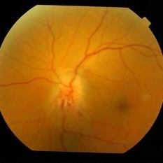
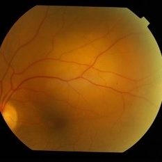
 Central Retinal Artery Occlusion
Apr 20 2018 by Kim Barrett
64-year-old female woke with no vision in her right eye. This image was taken at 6:11 minutes and the vessels have not filled. Patient has been treated with PRP laser and anti-VEGF therapy. Current vision is CF @ 2 ft.
Photographer: Kim Barrett C.O.A.
Imaging device: Heidelberg
Condition/keywords: central retinal artery occlusion (CRAO), diabetes, hypertension, smoker, uncontrolled
-
 Metastatic NSCLCA to the Choroid: Initial Appearance
May 27 2019 by John S. King, MD
60-year-old white male non-smoker presented to Dr. Zocchi with acute transient decreased vision in the right eye. Background history includes metastatic NSCLC (adenocarcinoma). Acuity OD 20/60, and posterior segment had two small, yellow, choroidal lesions, above the nerve and IT arcade (these had a fairly smooth and dome shaped appearance on the OCT, and top lesion had mild SRF) (see photo)
Photographer: Shelly Blair
Imaging device: Optos CA
Condition/keywords: choroidal metastasis, lung cancer metastasis
-
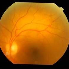
 Choroidal Metastasis
Apr 22 2016 by Mallika Goyal, MD
Right fundus of a 65-year-old male physician, ex-smoker, shows resolved choroidal metastasis nasal to disc from primary lung carcinoma non-responsive to chemotherapy. The metastatic lesion resolved with local radiotherapy.
Photographer: Mallika Goyal, MD, Apollo Health City, Hyderabad, India
Condition/keywords: choroidal metastasis
-
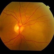
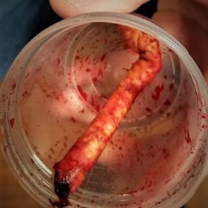 Carotid Artery Plaque
Sep 9 2020 by John S. King, MD
66-year-old white male, former smoker, with a history of femoral artery stent a plaque removal in 2017, triple bypass 2019 (at that time there was no high grade carotid stenosis), diabetes significant for SNPDR OD and PDR OS (NVD). He underwent PRP OS and two months later developed a vitreous hemorrhage and had a PPV OS. Early in the post-operative period vision dropped to LP due to acute CRAO with retinal embolus present. He was found to have progressed to high grade carotid stenosis (versus imaging 6 months ago) and a left carotid endarterectomy was performed (see picture of the large plaque) .
Condition/keywords: carotid artery occlusion
-
 Rare Bilateral Choroidal Metastasis from Occult Primary Lung Cancer
May 5 2021 by Deependra Vikram Singh, MD FASRS
Fundus photographs and OCT scans of a 73-year-old non-smoker Indian male who presented to our retina clinic in 2013 with blurred vision in left eye for past 2 weeks. BCVA was 20/20 in right eye and 20/40 in left eye. Slit lamp exam was unremarkable for both eyes with no cells in aqueous or anterior vitreous. Fundus examination revealed creamy yellow choroidal lesions in both eyes. Lesion in right eye was one disc diameter (DD) in size and was located close to fovea (Fig-1a). Lesion in the left eye was bigger with a size of 2 DD located superior to fovea (Fig-1b). OCT scan for left eye revealed neurosensory detachment involving fovea (Fig-1c). Fundus fluorescein angiography was inconclusive for right eye and showed late hyper fluorescence the choroidal lesion in left eye. Patient underwent detailed systemic work up for malignancy that revealed primary lung non-small cell carcinoma. He had widespread metastasis affecting liver and brain. Palliative chemotherapy and radiotherapy were initiated 4 weeks after he presented to us. The choroidal lesions show progression on fundus picture and OCT scans done at 4 weeks follow up after initial presentation (Fig – 1d, e, f). The lesions in both eyes show regression at 4 weeks and 12 weeks follow up after initiation of therapy. Unfortunately, patient succumbed at 13 weeks follow up due to disease progression. The case demonstrates rare bilateral choroidal metastasis from primary lung cancer and also highlights that lesions can be asymptomatic till they develop neurosensory detachment as evident from asymptomatic lesion in right eye despite proximity to fovea and symptomatic lesion in left eye with NSD.
Photographer: Deependra Vikram Singh, Eye-Q Superspecialty Eye Hospitals, Gurugram
Imaging device: Topcon
Condition/keywords: choroidal mass, choroidal metastasis
-
 Anterior Ischemic Optic Neuropathy in a Smoker
Sep 22 2014 by Mallika Goyal, MD
Left fundus photograph of a 47-year-old male with sudden vision drops 15 days before presentation shows anterior ischemic optic neuropathy with pale disc edema with inferior altitudinal field defect. He is a smoker for several years, diabetic for one year and has no other predisposing factors.
Photographer: Mallika Goyal, MD, Apollo Health City, Jubilee Hills, Hyderabad-500033
Condition/keywords: anterior ischemic optic neuropathy
-
 Choroidal Metastasis
Apr 22 2016 by Mallika Goyal, MD
Left fundus of a 65-year-old male physician, ex-smoker, shows a recurrent choroidal metastasis superior to macula from primary lung carcinoma non-responsive to chemotherapy. The lesion had initially resolved with radiotherapy with recurrence at same site after 2 months.
Photographer: Mallika Goyal, MD, Apollo Health City, Hyderabad, India
Condition/keywords: choroidal metastasis
-
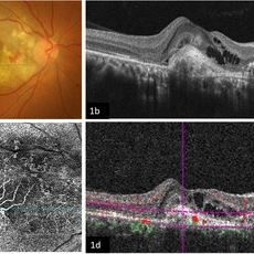 Imaging Feeder Vessels on OCT-A in a case of Retinal Angiomatous Proliferation
Feb 24 2023 by Dhaivat Shah
A 58-year-old male patient, a chronic smoker, came to our OPD with complaints of a diminution of vision in the right eye (BCVA: 2/60). On examination, the following findings were observed in the patient. On fundus examination (Image 1a) - Large areas of exudation with multiple superficial and deep hemorrhages at the macula were noted. On SD-OCT imaging (Image 1b) - Multiple intraretinal spaces were seen along with shallow subretinal fluid and hyperreflective dots (indicative of phagocytosed photoreceptors). On the foveal area- a hyperreflective membrane was noted which seemed to dip down and establish a retino-choroidal anastomosis. On OCT-A imaging (Image 1c) - In the ORCC complex, the neovascularization frown, correlating with the membrane complex on the fundus and structural OCT, was visible which was noted to be supplied by small feeder vessels coming from the superior aspect of the fovea. On OCT-A blood flow analysis imaging (Image 1d) - The blood flow analysis showed increased blood flow signals at the level of the membrane (indicative of increased color signal). Based on the findings of the above investigations and clinical examination, the patient was diagnosed with a Case of CNVM Type 3, also described as RAP, and was managed by Anti VEGF injections. This condition usually requires more injections as compared to Type 1 and 2 CNVMs, and the visual prognosis is guarded. Hence, it's very important to counsel the patient before initiating the treatment that the treatment would be long-term and the aim would be preservation of existing vision.
Photographer: Choithram Netralaya, Indore
Condition/keywords: feeder vessel, OCTA, RAP lesion
-
 Choroidal Metastasis
Apr 22 2016 by Mallika Goyal, MD
Left fundus of a 65-year-old male physician, ex-smoker, shows a recurrent choroidal metastasis superior to macula from primary lung carcinoma non-responsive to chemotherapy. The lesion had initially resolved with radiotherapy with recurrence at same site after 2 months.
Photographer: Mallika Goyal, MD, Apollo Health City, Hyderabad, India
Condition/keywords: choroidal metastasis
-
 Choroidal Metastasis
Apr 22 2016 by Mallika Goyal, MD
Right fundus of a 65-year-old male physician, ex-smoker, shows a choroidal metastasis nasal to disc from primary lung carcinoma non-responsive to chemotherapy. Resolved with radiotherapy with recurrence at same site after 2 months.
Photographer: Mallika Goyal, MD, Apollo Health City, Hyderabad, India
Condition/keywords: choroidal metastasis
-
 Choroidal Metastasis
Apr 22 2016 by Mallika Goyal, MD
Left fundus of a 65-year-old male physician, ex-smoker, shows a recurrent choroidal metastasis superior to macula from primary lung carcinoma non-responsive to chemotherapy. The lesion had initially resolved with radiotherapy with recurrence at same site after 2 months.
Photographer: Mallika Goyal, MD, Apollo Health City, Hyderabad, India
Condition/keywords: choroidal metastasis
-
 Choroidal Metastasis
Apr 22 2016 by Mallika Goyal, MD
Left fundus of a 65-year-old male physician, ex-smoker, shows a choroidal metastasis superior to macula from primary lung carcinoma non-responsive to chemotherapy. Resolved with radiotherapy with recurrence at same site after 2 months.
Photographer: Mallika Goyal, MD, Apollo Health City, Hyderabad, India
Condition/keywords: choroidal metastasis
 Loading…