Search results (76 results)
-
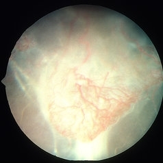
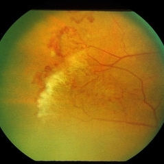 Sickle Cell Sea Fan Retinopathy
Jun 4 2014 by Henry J. Kaplan, MD
Sea fan peripheral retinal neovascularization in sickle cell anemia.
Condition/keywords: sea fan, sickle cell retinopathy
-
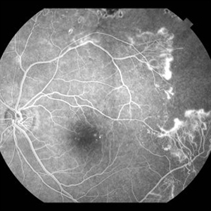
 Sickle Cell Retinopathy
Sep 14 2012 by Michael P. Kelly, FOPS
Fluorescein angiogram image of an individual with sickle cell retinopathy using an Optos P200MA ultra-wide field imaging device.
Photographer: Michael P. Kelly, FOPS Director, Duke Eye Center Labs, Duke University Hospital
Imaging device: Optos P200MA
Condition/keywords: Optos, sea fan, sickle cell retinopathy, ultra-wide field imaging
-
 Sickle Salmon-Patch Hemorrhage
Oct 23 2012 by Larry Halperin, MD
Salmon-patch hemorrhage in sickle cell
Condition/keywords: salmon patch, sickle cell retinopathy
-
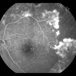
 Sickle Cell Retinopathy with Sea Fans (angiography)
Aug 24 2012 by Geoffrey G. Emerson, MD, PhD, FASRS
Fluorescein angiography (early/mid phase) of a 40-year-old man with African heritage and sickle SC disease. Sea fans are present temporal to the macula (leaking fluorescein).
Photographer: Geoffrey G. Emerson, MD, PhD, Retina Center, Minneapolis
Condition/keywords: sea fan, sickle cell retinopathy
-
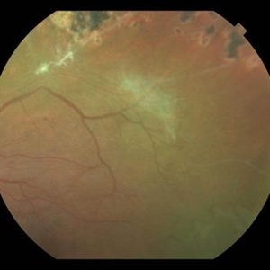 Sickle Cell Retinopathy with Sea Fans
Aug 24 2012 by Geoffrey G. Emerson, MD, PhD, FASRS
Color fundus photograph of a 40-year-old man with African heritage and sickle SC disease. A sea fan (white) is present along the superotemporal arcade adjacent to an area of ischemia.
Photographer: Geoffrey Emerson, MD, PhD, Retina Center, Minneapolis
Condition/keywords: sea fan, sickle cell retinopathy
-
 Sickle Cell Retinopathy with Sea Fans (angiogram)
Aug 24 2012 by Geoffrey G. Emerson, MD, PhD, FASRS
Fluorescein angiography (mid phase) of a 40-year-old man with African heritage and sickle SC disease. Sea fans are present temporal to the macula.
Photographer: Geoffrey Emerson, MD, PhD, Retina Center, Minneapolis
Condition/keywords: sea fan, sickle cell retinopathy
-
 Sickle Cell Retinopathy
Sep 28 2012 by Michael P. Kelly, FOPS
Peripheral non-perfusion in sickle cell retinopathy.
Photographer: Michael P. Kelly, FOPS Director, Duke Eye Center Labs, Duke University Hospital
Imaging device: Zeiss FF3C
Condition/keywords: non-perfusion, sickle cell retinopathy
-
 Sickle Cell Retinopathy
Sep 28 2012 by Raj K. Maturi, MD
Sickle Cell retinopathy
Photographer: Tom Steele, CRA
Imaging device: Optos
Condition/keywords: sickle cell retinopathy
-
 Sea Fan Neovascularization
May 2 2013 by Henry J. Kaplan, MD
Typical sea fan neovascularization in the peripheral retina of a patient with sickle cell disease.
Condition/keywords: sea fan, sickle cell retinopathy
-
 Sickle Cell Retinopathy
Sep 28 2012 by Raj K. Maturi, MD
Photographer: Tom Steele, CRA
Imaging device: Optos
Condition/keywords: sickle cell retinopathy
-
 Proliferative Sickle Retinopathy Stage 3
Oct 9 2012 by Alan D. Letson, MD
21-year-old man with Hgb SC Disease and stage 3 PSR with autoinfarction of sea fans
Photographer: Beverly Radcliffe
Condition/keywords: autoinfarction, sea fan, sickle cell, sickle cell retinopathy
-
 Sickle Cell Retinopathy with Sea Fans (angiography)
Aug 24 2012 by Geoffrey G. Emerson, MD, PhD, FASRS
Fluorescein angiography (early phase) of a 40-year-old man with African heritage and sickle SC disease. Sea fans are present temporal to the macula.
Photographer: Geoffrey Emerson, MD, PhD, Retina Center, Minneapolis
Condition/keywords: sea fan, sickle cell retinopathy
-
 Sickle Cell Retinopathy with Sea Fans (angiogram)
Aug 24 2012 by Geoffrey G. Emerson, MD, PhD, FASRS
Fluorescein angiography (late phase) of a 40-year-old man with African heritage and sickle SC disease. Sea fans are present around the macula (profusely leaking fluorescein dye).
Photographer: Geoffrey Emerson, MD, PhD, Retina Center, Minneapolis
Condition/keywords: sea fan
-
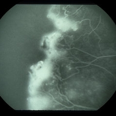 Sickle Cell Sea Fan Retinopathy
Jun 4 2014 by Henry J. Kaplan, MD
Fluorescein angiogram of the same patient shows extensive capillary non-perfusion anterior to the neovascularization area. #2
Condition/keywords: sea fan, sickle cell retinopathy
-
---thumb.JPG/image-square;max$300,300.ImageHandler) Sickle Cell
Sickle Cell
Jul 11 2013 by Jason S. Calhoun
Black female with sickle cell retinopathy in the left eye, temporally.
Photographer: Jason S. Calhoun, Department of Ophthalmology, Mayo Clinic Jacksonville, Florida
Condition/keywords: sickle cell retinopathy
-
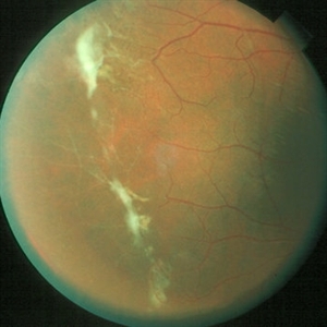
 Proliferative Sickle Cell Retinopathy, Color OD
May 23 2018 by Hosam Attia, MD
45-year-old African American, male with sickle cell anemia (SC disease) with arteriolar attenuation, mild venous tortuosity, Sunburst (S) and large, partially auto-infarcted sea fan with fresh heme, OD.
Imaging device: Optos California Ultra-Wide Field Fundus Camera
Condition/keywords: neovascularization elsewhere (NVE), proliferative retinopathy, sea fan, sickle cell, sickle cell retinopathy
-
 Sickle Cell Retinopathy
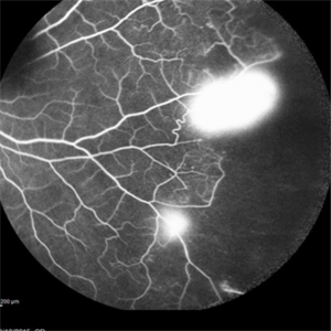
Sep 13 2015 by Thomas A. Ciulla, MD, MBA, FASRS
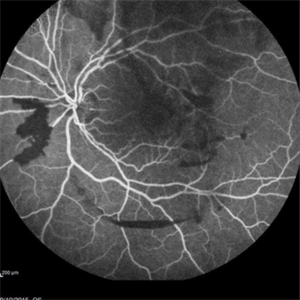
Angiography showed normal vessels posteriorly but severe capillary drop out throughout the periphery OU with scattered severe neovascularization at the edge of the capillary drop out peripherally.
Photographer: Thomas Steele
Condition/keywords: peripheral retinal neovascularization, sea fan, sickle cell retinopathy
-
 Sickle Cell Retinopathy
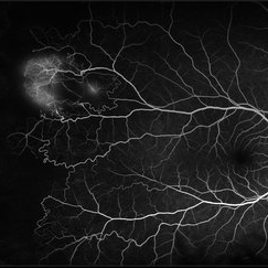
Sep 13 2015 by Thomas A. Ciulla, MD, MBA, FASRS
Angiography showed normal vessels posteriorly but severe capillary drop out throughout the periphery OU with scattered severe neovascularization at the edge of the capillary drop out peripherally.
Photographer: Thomas Steele
Condition/keywords: peripheral retinal neovascularization, sea fan, sickle cell retinopathy
-
 Sickle Cell Retinopathy
Feb 15 2021 by Kim Barrett
24-year-old female with Sickle Cell Retinopathy, stage 3. She confirms she has the trait as well as her grandmother, mother and a sibling. She has seafan neovascularization superotemporal OD. Current VA is 20/20. Photo is pre-PRP laser with areas of non-profusion temporally.
Photographer: Kim Barrett C.O.A. Retina Specialist of Michigan, Grand Rapids, MI
Imaging device: Optos California
Condition/keywords: neovascularization (NV), pan-retinal photocoagulation (PRP), sickle cell retinopathy, stage 3, trait
-
 Proliferative Sickle Cell Retinopathy, Color OS
May 23 2018 by Hosam Attia, MD
45-year-old African American, male with sickle cell anemia (SC disease ) with arteriolar attenuation, mild venous tortuosity, peripheral arterio-venous anastomoses (shown better on red free), multiple small NVEs/ early sea fans (one w/ early auto-infarction) and sunburst (S) - (Not showing very well in photos) OS.
Imaging device: Optos California Ultra-Wide Field Fundus Camera
Condition/keywords: neovascularization elsewhere (NVE), proliferative retinopathy, sea fan, sickle cell, sickle cell retinopathy
-
 Proliferative Sickle Cell Retinopathy, Color OD
May 23 2018 by Hosam Attia, MD
45-year-old African American, male with sickle cell anemia (SC disease) with arteriolar attenuation, mild venous tortuosity, Sunburst (S) and large, partially auto-infarcted Seafan with fresh heme, OD.
Imaging device: Optos California Ultra-Wide Field Fundus Camera
Condition/keywords: neovascularization elsewhere (NVE), proliferative retinopathy, sea fan, sickle cell, sickle cell retinopathy
-
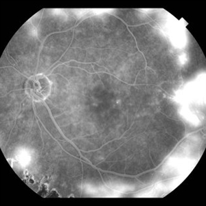
 Proliferative Sickle Cell Retinopathy, Early phase FA OD
May 23 2018 by Hosam Attia, MD
Fluorescein angiogram photograph of a 45-year-old African American, male with sickle cell anemia (SC disease), depicting extensive peripheral capillary non-perfusion, with early hyperfluorescence over the ischemic retina temporally, with late staining and diffuse leakage consistent with partially auto-infarcted, but active NVE/sea fan OD.
Imaging device: Optos California Ultra-Wide Field Fundus Camera
Condition/keywords: neovascularization elsewhere (NVE), proliferative retinopathy, sea fan, sickle cell, sickle cell retinopathy
-
 Sickle Cell Retinopathy
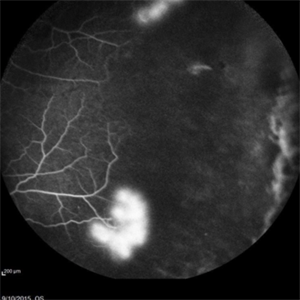
Sep 13 2015 by Thomas A. Ciulla, MD, MBA, FASRS
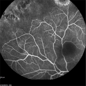
Angiography showed normal vessels posteriorly but severe capillary drop out throughout the periphery OU with scattered severe neovascularization at the edge of the capillary drop out peripherally. Note the preretinal and vitreous hemorrhage obscuring the view of the retinal vasculature.
Photographer: Thomas Steele
Condition/keywords: peripheral retinal neovascularization, sea fan, sickle cell retinopathy
-
 Black Sunburst in Proliferative Sickle Cell Retinopathy
Jul 25 2023 by Kamal Kishore, MD, MBBS
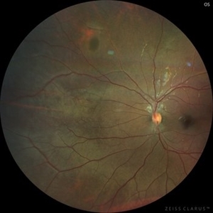
A 17-year-old male with a black sunburst lesion at superonasal periphery.
Photographer: Jessi Wright
Imaging device: Zeiss Clarus
Condition/keywords: Black Sunburst, sickle cell retinopathy
-
 Sickle Cell Retinopathy
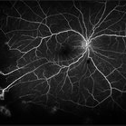
Sep 13 2015 by Thomas A. Ciulla, MD, MBA, FASRS
Angiography showed normal vessels posteriorly but severe capillary drop out throughout the periphery OU with scattered severe neovascularization at the edge of the capillary drop out peripherally.
Photographer: Thomas Steele
Condition/keywords: peripheral retinal neovascularization, sea fan, sickle cell retinopathy
 Loading…