Search results (42 results)
-
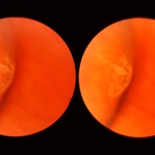 Meridional Fold
Nov 9 2012 by Norman Byer
This is the same lesion as in the previous photograph. With the scleral indentation placed more posterior, we now can see that the fold ends over a small collection of subretinal fluid and that there is a very tiny retinal hole just below the posterior end of the retinal fold.
Condition/keywords: peripheral cystoid degeneration, retinal fold, retinal hole, scleral indentation, subretinal fluid
-
 Cyst of the Pars Plana
Nov 9 2012 by Norman Byer
This is a cyst of the pars plana located just anterior to the ora serrata in the lower temporal quadrant. It illustrates how far anterior one may visualize the fundus with indirect ophthalmoscopy and scleral indentation. Pars plana cysts are common lesions of no particular clinical significance.
Condition/keywords: cyst of the pars plana, lower temporal quadrant, ora serrata, scleral indentation
-
 Lattice Degeneration
Nov 9 2012 by Norman Byer
This is a more typical classical example of lattice degeneration in a 42-year-old woman in a photograph taken without scleral indentation. It shows much more marked vascular changes than the previous case. Note the tapering of the blood columns as the vessels approach the lesion and also the white sheathing of the vessel walls. Note also the continuity of the blood vessels on opposite sides of the lesion with the characteristic white lattice lines. More than 45 years ago Vogt pointed this out as a proof that these white lines were actually caused by changed blood vessels. Note also that this lesion shows a combination of several individual features of lattice degeneration. In addition to the white lines, there is a reddish crater-like area beneath the main horizontal white line. There is a prominent horizontal zone below this white line showing a snailtrack appearance. Also, there are two tiny atrophic retinal holes outside the photograph on the right end of this lesion. This eye contained five such retinal holes and they have all remained unchanged for more than 10 years of observation without treatment.
Condition/keywords: atrophic retinal hole, lattice degeneration, moderate snail track, tapering blood columns, white lattice lines, white sheath vessel
-
 Scleral Indentation In A Normal Eye
Nov 9 2012 by Norman Byer
This shows the appearance of scleral indentation in a normal eye. Note the convex shadow which marks the posterior border of the indented area. It is caused in part by a small angle which separates the viewing axis from the illuminating axis thus allowing the observer to see slightly into the shadow beyond the illuminated crest of the indentation. It is also caused in part by viewing the pigment epithelial layer in a tangential manner. This shadow is of great diagnostic usefulness since it becomes a dark background against which many tiny retinal abnormalities can be seen beautifully by contrast. Two other particular advantages of scleral indentation will be demonstrated in the following photographs: First, the ability to see the extreme anterior part of the retina to the ora serrata and beyond, and second, the ability to examine any abnormality in multiple profiles depending on slight movements of the scleral depressor in various directions.
Condition/keywords: extreme anterior retina, posterior border, scleral indentation, shadow, tangential view of pigment epithelial layer
-
 Lattice Degeneration
Nov 9 2012 by Norman Byer
This is lattice degeneration in a 10-year-old boy showing an almost pure snailtrack feature with only a hint of a reddish crater in the center. It has not changed over 10 years. The photograph was taken with scleral indentation.
Condition/keywords: lattice degeneration, reddish crater, scleral indentation, snail track
-
 Cystic Retinal Tuft
Nov 9 2012 by Norman Byer
This is a rather poor photograph taken in 1969 but is important for comparison with the next slide pair. It shows a cystic retinal tuft in a 49-year-old woman and was taken without scleral indentation. The two pigment spots just inferior to the tuft represent a secondary degenerative change in the pigment epithelium.
Condition/keywords: cystic retinal tuft, degenerative changes of retinal pigment epithelium, pigmented spots
-
 Sudden Posterior Vitreous Detachment
Nov 9 2012 by Norman Byer
This is the appearance of the previous lesion three weeks following prophylactic cryotherapy. Continuing vitreal retinal traction has a now torn the flap completely free from the retina. The whitish cystic retinal tuft can be discerned on the upper part of the free operculum. Along the lower half of the operculum superimposed over the dark shadow of the scleral indentation one may observe numerous, delicate, vitreous fibrils actually attaching to the operculum.
Condition/keywords: cystic retinal tuft, free operculum, prophylactic cyrotherapy, retinal flap, scleral indentation, vitreoretinal traction, vitreous fibrils
-
 Retinal Break at Site of Lattice Degeneration with Scleral Indentation
Nov 9 2012 by Norman Byer
This is the same case as the previous photograph. With scleral indentation slightly more posterior, the flap is seen to be associated with a large retinal tear. This is a tractional tear and it is possible that in this case the cryotherapy itself may have increased the vitreoretinal traction at this site and in this way led to this new tear. The age of the tear is unknown because it was asymptomatic, and even though the eye is aphakic the tear has not caused a clinical retinal detachment.
Condition/keywords: retinal flap, scleral indentation, tractional retinal tear, vitreoretinal traction
-
 Sudden Posterior Vitreous Detachment
Nov 9 2012 by Norman Byer
This is the same lesion seen in the previous slide pair. With the scleral indentation performed more posteriorly, a small hemorrhage can be seen on the white tuft. This is proof of the vitreal retinal attachment at this spot. Posterior vitreous detachment can produce a retinal tear at the site of a cystic retinal tuft, but in this case has caused only a small hemorrhage.
Condition/keywords: posterior vitreous detachment, retinal hemorrhage, scleral indentation, vitreoretinal attachment
-
 Senile Retinoschisis
Nov 9 2012 by Norman Byer
This is the same case as seen in the previous photograph but is a different view with the scleral indentation moved more anterior. The retinoschisis is seen to be very peripheral coming at least grossly right up to the ora serrata. Please notice how clear a view one can get of the ora serrata and pars plana using indirect ophthalmoscopy with scleral indentation.
Condition/keywords: ora serrata, retinoschisis, scleral indentation
-
 Symptomatic Horseshoe Tear
Nov 9 2012 by Norman Byer
This is a fresh, symptomatic horseshoe tear at the site of a cystic retinal tuft in a 63-year-old man. This is really a double horseshoe tear because the two retinal vessels have resisted the vitreal retinal traction and have preserved an intact bridge of tissue between the tears. Note the prominent vitreous condensation attached to the apex of the upper tear and made much more visible because it is seen superimposed over the dark underlying shadow of scleral indentation.
Condition/keywords: bridge of tissue between tears, cystic retinal tuft, scleral indentation, vitreous condensation
-
 Horseshoe Tear
Nov 9 2012 by Norman Byer
This horseshoe tear was the cause of the detachment in this 54-year-old man. The orange area on the right half of the slide represents the area of scleral indentation. Please note that most of the tear lies over the indented area and appears orange. However, the extreme left side of the tear is brownish black in color because it is exactly superimposed over the dark shadow that always lies just beyond the indented area. The ability of scleral indentation to produce this color change combined with a sharp demarcation between the blackish area and the yellowish edge of intact retina is a pathognomonic sign of a full thickness retinal break.
Condition/keywords: scleral indentation
-
 Asymptomatic Lesion
Nov 9 2012 by Norman Byer
This is the same lesion as seen in the previous slide pair. Here the scleral indentation is carried more posterior revealing a tiny, round, full thickness retinal hole. This is not a tear produced by traction even though vitreous is always attached to these flaps. You will note that the hole is round and is separated by a slight distance from the flap itself. It is probably the result of long continued atrophy and devitalization of the retina. A posterior vitreous was not detached. This lesion has not changed its appearance for more than a year of observation, but the age of the hole is actually unknown.
Condition/keywords: asymptomatic, atrophy, full thickness retinal hole, posterior scleral indentation, retinal hole, round hole
-
 Lattice Lesion
Nov 9 2012 by Norman Byer
This lattice lesion in a 30- year-old woman also shows combined features with a reddish crater above and a parallel snailtrack appearance just below it. Please note especially another interesting feature. From the left end of the lesion, there is a faint thin yellow line slanting down toward the right just below the shadow of the scleral indentation. This line identifies the dome of the pocket of liquified vitreous which is present over every lesion of lattice degeneration.
Condition/keywords: lattice degeneration, lattice lesion, liquefied vitreous, reddish crater, scleral indentation, snail track
-
 Sudden Posterior Vitreous Detachment
Nov 9 2012 by Norman Byer
In this view of the previous case, the scleral indentation is being done immediately beneath the lesion. The hemorrhage is now out of sight and the white lesion is seen to be distinctly he elevated and in two parts. The base is white and the apex is translucent with a possible small hole in it. There is also a probable tiny full- thickness tear just behind the flap which cannot be discerned in this view.
Condition/keywords: full thickness retinal tear, peripheral retinal lesion, scleral indentation, translucency of apex, white retinal lesion
-
 Elevated Lesion
Nov 9 2012 by Norman Byer
This photograph and the next are two views of a very interesting elevated lesion in a 45-year-old man. This photograph shows the immense value of closely scrutinizing the profile of the indented area. Note that in the middle of the slide there is a sudden break in the continuity of the dark convex shadow that lies just behind the crest of the scleral indentation. If the elevated tissue is "filmy" or "wispy" or filamentous as in this case, it raises a strong suspicion that a retinal break is present just behind it.
Condition/keywords: elevated retinal lesion, elevated tissue, retinal break, scleral indentation
-
 Scleral Indentation
Nov 9 2012 by Norman Byer
This is the same lesion with scleral indentation. You can see the small discrete preretinal hemorrhage and the sharply circumscribed area of elevated retina with subretinal fluid beneath it. No retinal break is visible, but the posterior vitreous is detached and exerting traction at this site. The area was surrounded with argon laser treatment the same day as the initial examination.
Condition/keywords: posterior vitreous detachment, preretinal hemorrhage, scleral indentation, subretinal fluid, vitreous traction
-
 Lattice Lesion
Nov 9 2012 by Norman Byer
This lattice lesion in a 36-year-old woman has remained unchanged over a period of 13 years. It shows a moderate snailtrack feature with discrete yellow dots visible on the surface of the lesion and especially along the posterior border. One of these can be well seen just below the lesion superimposed over the dark shadow of the scleral indentation. The exact nature of these yellow dots is still not entirely clear.
Condition/keywords: lattice degeneration, moderate snail track, scleral indentation, yellow dots
-
 Scleral Indentation
Nov 9 2012 by Norman Byer
This is the same lesion as in the two previous photographs. With the scleral indentation placed more anterior, this white area is made to lie on the crest of the bulge where it can now be studied in profile. It projects above the retina and has sloping margins. The exact nature of this lesion is not known. This illustrates how the use of scleral indentation with indirect ophthalmoscopy can markedly change the appearance of retinal abnormalities and can bring to light many details not otherwise visible.
Condition/keywords: elevated retinal lesion, scleral indentation, white retinal lesion
-
 Lattice Lesion
Nov 9 2012 by Norman Byer
This is the same lesion as seen in the previous case seen now with scleral indentation. Here you can see directly into the subretinal space through the two retinal holes. The holes appear dark because the shadow of the scleral indentation lies directly beneath them.
Condition/keywords: lattice degeneration, retinal hole, scleral indentation
-
 Retinoschisis Detachment
Nov 9 2012 by Norman Byer
Combined retinoschisis detachment, so-called schisis detachment, in a 47-year-old woman. The large outer layer hole in the center has a posterior yellow border which represents the position of the outer layer. Please observe superior to the hole the dark convexity of the scleral indentation. Just below the hole at the middle of the slide and going to the left the yellow zone comes to lie right against the inner layer and a fluid filled cavity lies deep to the outer layer. At this point, therefore, there is a true neurosensory detachment of the retina. On the right side of the hole, the yellow line slants up and to the right and lies close to the pigment epithelium. On the right side of the photograph, the original schisis cavity can be seen separating the yellow line of the outer layer above from the inner retinal layer below. The mechanism of this detachment is that some of the fluid from the schisis cavity passes through the outer layer hole and detaches the outer layer. This lesion has not been treated and has remained exactly the same for 13 years. A similar symmetrical "schisis-detachment" is present in the fellow eye.
Condition/keywords: neurosensory detachment of retina, outer layer hole, pigment epithelium, retinoschisis, schisis detachment, scleral indentation
-
 Retinoschisis
Nov 9 2012 by Norman Byer
This 53-year-old man has retinoschisis involving the upper temporal quadrant but with no visible yellow dots or white lines to make it obvious. However, with scleral indentation you can see a large convex area showing the so-called white with pressure phenomenon. This area corresponds exactly to the area being indented and therefore must arise either from the outer layer of the retina or from some structure deep to it. White with pressure is an interesting optical phenomenon of uncertain origin but of no definite diagnostic or prognostic significance.
Condition/keywords: retinoschisis, scleral indentation, white with pressure
-
 Lattice Lesion
Nov 9 2012 by Norman Byer
This lattice lesion in a 44-year-old man shows an atrophic retinal hole surrounded by discrete yellowish and pigmented areas. These have been caused by secondary pigment migration and proliferation in the retinal pigment epithelium. There is a small doughnut like elevation of the retina between the edge of the hole and the line of pigment. The lesion and the hole have remained exactly the same for seven years.
Condition/keywords: atrophic retinal hole, elevated retina, lattice degeneration, lattice lesion, proliferation of retinal pigment epithelium, scleral indentation
-
 Scleral Indentation
Nov 9 2012 by Norman Byer
On the crest of this indentation, one can see a small fragment of residual vitreous blood lying close to the retinal surface. The center is becoming typically white as the blood is becoming depigmented.
Condition/keywords: depigmented vitreous blood, scleral indentation, vitreous blood
-
 Lattice Lesion
Nov 9 2012 by Norman Byer
This is a photograph of a lattice lesion in a 23-year-old girl taken without scleral indentation. Just to the left of the center of the slide is a slightly pigmented lesion almost oval in shape with a retinal hole in each end. Ten years earlier at the age of 13 this lesion appeared exactly like the one in the previous case as a pure red crater. Five years later two new round retinal holes were seen, one in each end, with a tiny bit of subretinal fluid within the lattice lesion only. Five years later still the appearance was as shown in this slide pair with the subretinal fluid now extending slightly beyond the lattice lesion as far as the curved row of tiny yellow exudates seen just to the right of the center of the slide. It is now actually a small subclinical retinal detachment. The next slide pair will show this better using scleral indentation.
Condition/keywords: lattice degeneration, lattice lesion, pigmented lesion, reddish crater, retinal hole, subretinal fluid, yellow exudate
 Loading…