Search results (92 results)
-
---thumb.jpg/image-square;max$300,300.ImageHandler) Progressive Outer Retinal Necrosis
Progressive Outer Retinal Necrosis
Feb 15 2013 by From the Collections of Thomas M. Aaberg, MD and Thomas M. Aaberg Jr., MD
Color fundus photograph showing extensive confluent retinal whitening, retinal exudation, intraretinal hemorrhage, and sheathing of retinal vessels consistent with infectious retinitis such as progressive outer retinal necrosis (PORN).
Condition/keywords: occlusive retinitis, retinal necrosis
-
---thumb.jpg/image-square;max$300,300.ImageHandler) Behcet Uveitis.
Behcet Uveitis.
Feb 15 2013 by From the Collections of Thomas M. Aaberg, MD and Thomas M. Aaberg Jr., MD
Color fundus photographs showing peripheral retinal whitening and pigmentary change consistent with early resolution of inflammatory lesions (upper panel) along with persistent exudative retinitis in the post-equatorial retina (lower panels).
Condition/keywords: Behcet's uveitis, retinitis
-
 Berlin's Edema
Apr 8 2019 by Gary R. Cook, MD, FACS
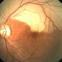
39-year-old white female with geographic area of retinal whitening ( Berlin's edema) without hemorrhage in the midperiphery secondary to blunt trauma; V.A. = 20/25
Imaging device: Topcon VT-50
Condition/keywords: Berlin's edema, blunt trauma, retinal edema
-
 Branch Retinal Artery Occlusion With Calcium Embolus at the Disc - Fundus Photo
Apr 7 2018 by Rameez N Hussain, MD
Acute branch retinal artery occlusion with a calcium embolus at the disc with retinal whitening in the area of retinal edema.
Photographer: DR RAMEEZ N HUSSAIN
Imaging device: zeiss
Condition/keywords: branch retinal artery occlusion (BRAO), embolus, fundus photograph, retinal edema
-
---thumb.jpg/image-square;max$300,300.ImageHandler) Posterior Uveitis
Posterior Uveitis
Feb 15 2013 by From the Collections of Thomas M. Aaberg, MD and Thomas M. Aaberg Jr., MD
Color photograph of the mid-peripheral retina showing scattered intraretinal hemorrhage and foci of retinal whitening consistent with posterior uveitis, such as Behcet disease.
Condition/keywords: posterior uveitis, retinitis
-
 CMV Retinitis with Frosted Branch Angiitis
Sep 23 2020 by Nimesh A. Patel, MD, FASRS
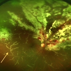
Fundus photo showing peri-vascular inflammation of both arteries and veins with translucent exudation (yellow arrow). Superior nasally, there is classic retinal whitening with retinal hemorrhages superior. This patient was found to have a low CD4 count and a diagnosis of AIDS was made.
Condition/keywords: cytomegalovirus (CMV), HIV, uveitis
-
---thumb.jpg/image-square;max$300,300.ImageHandler) Behcet Uveitis
Behcet Uveitis
Feb 15 2013 by From the Collections of Thomas M. Aaberg, MD and Thomas M. Aaberg Jr., MD
Color fundus photographs of the right eye of a patient suspected to have Behcet Uveitis. Over the course of 11 days, there is progressive optic disc edema, intraretinal whitening, hemorrhage and vessel occlusion.
Condition/keywords: Behcet's uveitis, posterior uveitis, retinitis
-
---thumb.jpg/image-square;max$300,300.ImageHandler) vitreous haze and confluent peripheral retinal whitening consistent with active ocular toxoplasmosis
vitreous haze and confluent peripheral retinal whitening consistent with active ocular toxoplasmosis
Feb 15 2013 by From the Collections of Thomas M. Aaberg, MD and Thomas M. Aaberg Jr., MD
Color fundus photograph showing vitreous haze and confluent peripheral retinal whitening consistent with active ocular toxoplasmosis
Condition/keywords: ocular toxoplasmosis, vitritis
-
 Retinitis Sclopetaria
Jun 29 2018 by Gareth Lema, MD, PhD
Retinal whitening, subretinal hemorrhages, retinal hemorrhages, and vascular tortuosity following blunt trauma from a paintball.
Photographer: Flaum Eye Institute, University of Rochester, Rochester, NY
Condition/keywords: blunt trauma, chorioretinitis sclopetaria
-
---thumb.jpg/image-square;max$300,300.ImageHandler) Posterior Uveitis
Posterior Uveitis
Feb 15 2013 by From the Collections of Thomas M. Aaberg, MD and Thomas M. Aaberg Jr., MD
Schematic diagram depicting Posterior Uveitis characterized by diffuse retinal whitening and exudative retinal detachment.
Condition/keywords: posterior uveitis, retinitis
-
---thumb.jpg/image-square;max$300,300.ImageHandler) Behcet Uveitis
Behcet Uveitis
Feb 15 2013 by From the Collections of Thomas M. Aaberg, MD and Thomas M. Aaberg Jr., MD
Color fundus photographs of the right eye of a patient suspected to have Behcet Uveitis. Over the course of 11 days, there is progressive optic disc edema, intraretinal whitening, hemorrhage and vessel occlusion. Fluorescein angiography confirms impaired retinal perfusion secondary to vessel occlusion.
Condition/keywords: posterior uveitis, retinitis
-
---thumb.jpg/image-square;max$300,300.ImageHandler) Vitreous haze and confluent peripheral retinal whitening
Vitreous haze and confluent peripheral retinal whitening
Feb 15 2013 by From the Collections of Thomas M. Aaberg, MD and Thomas M. Aaberg Jr., MD
Color fundus photograph showing vitreous haze and confluent peripheral retinal whitening consistent with active ocular toxoplasmosis.
Condition/keywords: ocular toxoplasmosis, vitritis
-
---thumb.jpg/image-square;max$300,300.ImageHandler) Acute retinal necrosis
Acute retinal necrosis
Feb 15 2013 by From the Collections of Thomas M. Aaberg, MD and Thomas M. Aaberg Jr., MD
Diffuse intraretinal hemorrhages and whitening in the posterior pole consistent with acute retinal necrosis.
Condition/keywords: macular edema, microangiopathy, retinal necrosis, retinal whitening
-
 Branch Retinal Artery Occlusion With Calcium Embolus at the Disc - Fundus Photo
Apr 7 2018 by Rameez N Hussain, MD
Acute retinal artery occlusion with a calcium embolus at the disc and retinal whitening.
Photographer: DR RAMEEZ N HUSSAIN
Imaging device: zeiss
Condition/keywords: branch retinal artery occlusion (BRAO), embolus, fundus photograph, retinal edema
-
---thumb.jpg/image-square;max$300,300.ImageHandler) Posterior Uveitis
Posterior Uveitis
Feb 15 2013 by From the Collections of Thomas M. Aaberg, MD and Thomas M. Aaberg Jr., MD
Schematic diagram depicting Posterior Uveitis characterized by diffuse retinal whitening and exudative retinal detachment.
Condition/keywords: posterior uveitis, retinitis
-
 Partial Optic Disc Avulsion with Optic Disc Pit
Jul 1 2018 by John S. King, MD
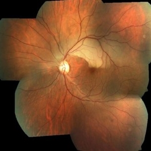
16-year-old with acute loss of vision after blunt finger injury to eye while playing football. This photo is three weeks post-injury. Vision HM. Retinal striae with subhyaloid heme. Decreased retinal whitening. Peripapillary heme clearing, and temporal optic disc avulsion with optic disc pit can be seen.
Photographer: Maisee Yang
Imaging device: Topcon
Condition/keywords: epiretinal membrane (ERM), optic nerve head avulsion, optic nerve pit, traumatic optic neuropathy
-
---thumb.jpg/image-square;max$300,300.ImageHandler) Retinal periphery showing stages of healing following infection
Retinal periphery showing stages of healing following infection
Feb 15 2013 by From the Collections of Thomas M. Aaberg, MD and Thomas M. Aaberg Jr., MD
Color photographs of the retinal periphery showing stages of healing following presumed retinal infection. Areas of vascular sheathing and retinal whitening (left panel) give way to vessel narrowing and dropout and retinal pigmentary changes
Condition/keywords: retinitis
-
---thumb.jpg/image-square;max$300,300.ImageHandler) Vitreous haze and focal peripheral retinal whitening consistent with active ocular toxoplasmosis
Vitreous haze and focal peripheral retinal whitening consistent with active ocular toxoplasmosis
Feb 15 2013 by From the Collections of Thomas M. Aaberg, MD and Thomas M. Aaberg Jr., MD
Color fundus photograph showing vitreous haze and focal peripheral retinal whitening consistent with active ocular toxoplasmosis
Condition/keywords: ocular toxoplasmosis, vitritis
-
---thumb.jpg/image-square;max$300,300.ImageHandler) Vitreous haze, disc edema and indistinct foci of retinal whitening
Vitreous haze, disc edema and indistinct foci of retinal whitening
Feb 15 2013 by From the Collections of Thomas M. Aaberg, MD and Thomas M. Aaberg Jr., MD
Color fundus photograph showing vitreous haze, disc edema and indistinct foci of retinal whitening consistent with active ocular toxoplasmosis.
Condition/keywords: ocular toxoplasmosis, vitritis
-
---thumb.jpg/image-square;max$300,300.ImageHandler) Healing of retinal lesions associated with Retinitis
Healing of retinal lesions associated with Retinitis
Feb 15 2013 by From the Collections of Thomas M. Aaberg, MD and Thomas M. Aaberg Jr., MD
Color fundus photograph showing gradual healing of retinal lesions associated with retinitis. There is progressive resolution of retinal whitening and exudation, accompanied by attenuation of formerly inflamed retinal vessels.
Condition/keywords: occlusive retinitis, retinal necrosis
-
 Aborted Arteriolitis
Feb 15 2013 by From the Collections of Thomas M. Aaberg, MD and Thomas M. Aaberg Jr., MD
Fundus photograph showing activated toxoplasma retinochoroiditis with active retinal whitening adjacent to a hyperpigmented scar in the superonasal mid-periophery.
Condition/keywords: ocular toxoplasmosis
-
 Partial Optic Disc Avulsion with Optic Disc Pit
Jul 1 2018 by John S. King, MD
16-year-old with acute loss of vision after blunt finger injury to eye while playing football. Five days post-injury. Vision HM. Decreasing heme and retinal whitening.
Imaging device: Optos
Condition/keywords: traumatic optic neuropathy
-
---thumb.jpg/image-square;max$300,300.ImageHandler) Infectious Retinitis
Infectious Retinitis
Feb 15 2013 by From the Collections of Thomas M. Aaberg, MD and Thomas M. Aaberg Jr., MD
Color fundus photographs showing presumed reactivation or progression of infectious retinitis with emergence of new areas of retinal whitening and extension and enlargement of retinal vascular sheathing.
Condition/keywords: occlusive retinitis, retinal necrosis
-
 Progressive Outer Retinal Necrosis
Nov 30 2018 by Nichole Lewis
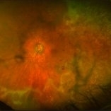
Fluorescein angiogram of an 86-year-old male with progressive outer retinal necrosis and chronic cystoid macular edema. This patient has occlusive vasculitis with non-perfusion, significant retinitis, retinal whitening and intra-retinal hemorrhages. Patient is immunosupressed with a history of kidney transplantation. VA 20/60. Patient was treated with intravitreal foscarnet and admitted to the hospital for an infectious disease and transplant team consultation.
Photographer: Nichole Lewis
Condition/keywords: cystoid macular edema (CME), intraretinal hemorrhage, non-perfusion, occlusive vasculitis, progressive outer retinal necrosis (PORN), retinal whitening, retinitis
-
---thumb.jpg/image-square;max$300,300.ImageHandler) Infectious Retinitis
Infectious Retinitis
Feb 15 2013 by From the Collections of Thomas M. Aaberg, MD and Thomas M. Aaberg Jr., MD
Gross pathologic specimen of an eye with presumed infectious retinitis, showing extensive retinal whitening (necrosis) and peripheral chorioretinal atrophy.
Condition/keywords: occlusive retinitis, pathology, retinal necrosis
 Loading…