Search results (162 results)
-
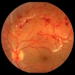 Retinal Vasculitis with Hemorrhages and Cotton Wool Spots
Oct 16 2012 by Jeffrey G. Gross, MD, FASRS
Retinal vasculitis with hemorrhages and cotton wool spots.
Condition/keywords: cotton wool spots, retinal vasculitis
-
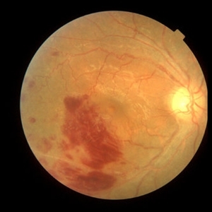
 Acute Idiopathic Occlusive Retinal Vasculitis
May 31 2014 by Hamid Ahmadieh, MD
Color fundus photograph of the right eye of a 28-year-old woman with sudden drop of vision due to acute occlusive retinal vasculitis leading to extensive nerve fiber layer infarction and retinal hemorrhages.
Photographer: Naghmeh Nozhat, Negah Eye Center, Tehran
Condition/keywords: color fundus photograph, cotton wool spots, retinal hemorrhage, retinal ischemia
-
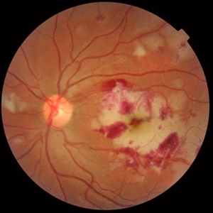
 Behcet's Disease
Mar 13 2013 by Hamid Ahmadieh, MD
Color fundus photograph of the right eye of a 23-year-old man with retinal vasculitis and branch retinal vein occlusion (BRVO) due to Behcet's disease .
Photographer: Solmaz Shahmohammad, Negah Eye Center, Tehran
Imaging device: Heidelberg Spectralis
Condition/keywords: branch retinal vein occlusion (BRVO), retinal vasculitis
-
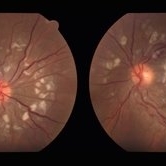
 Behcet's Disease
Nov 25 2012 by Mallika Goyal, MD
Fundus photograph of left eye of a 23-year-old gentleman with Behcet's Disease shows occlusive retinal vasculitis with optic disc pallor and macular ischemia. This eye has no light perception; other eye has similar fundus appearance.
Photographer: Mallika Goyal, MD, Apollo Health City, Hyderabad, India
Condition/keywords: macular ischemia, occlusive vasculitis, optic disc pallor
-
 Acute Idiopathic Occlusive Retinal Vasculitis
May 31 2014 by Hamid Ahmadieh, MD
Color fundus photograph of the left eye of a 28-year-old woman with acute drop of vision due to occlusive retinal vasculitis leading to extensive nerve fiber layer infarction and retinal hemorrhages.
Photographer: Naghmeh Nozhat, Negah Eye Center, Tehran
Condition/keywords: color fundus photograph, cotton wool spots, retinal hemorrhage, retinal ischemia
-
 Idiopathic Retinal Vasculitis, Aneurysms, and Neuroretinitis (IRVAN)
Oct 16 2012 by S. Natarajan, MD, FASRS, FRCS (GLASGOW) , FICO, D.Sc, FELA
Fundus photograph of a young male with IRVAN Syndrome
Photographer: Prof. Dr. S. Natarajan
Condition/keywords: aneurysm, neuroretinitis, retinal vasculitis
-
 Idiopathic Retinal Vasculitis, Aneurysms, and Neuroretinitis (IRVAN)
Oct 16 2012 by S. Natarajan, MD, FASRS, FRCS (GLASGOW) , FICO, D.Sc, FELA
FFA photograph of a 28-year-old male with IRVAN Syndrome.
Photographer: Prof. Dr. S. Natarajan
Condition/keywords: aneurysm, neuroretinitis, retinal vasculitis
-
 SLE Retinopathy
Nov 14 2016 by Mitzy E Torres Soriano, MD
25-year-old female patient with systemic lupus erythematosus. Photographs show cotton wool spots, intraretinal hemorrhages and vascular tortuosity. FA demonstrated retinal vasculitis and OCT revealed cystoid macular edema. In this case diagnosis of SLE was made after ocular manifestation.
Photographer: Grupo Laser Vision, Rosario, Argentina
Condition/keywords: cotton wool spots, occlusive retinal vasculitis, occlusive vasculitis, systemic lupus erythematosus, vasculopathy
-
 Idiopathic Retinal Vasculitis, Aneurysms, and Neuroretinitis (IRVAN)
Oct 16 2012 by S. Natarajan, MD, FASRS, FRCS (GLASGOW) , FICO, D.Sc, FELA
Fundus photograph of a 28-year-old male with IRVAN Syndrome.
Photographer: Prof. Dr. S. Natarajan
Condition/keywords: aneurysm, neuroretinitis, retinal vasculitis
-
---thumb.jpg/image-square;max$300,300.ImageHandler) Idiopathic Retinal Vasculitis, Aneurysms, and Neuroretinitis (IRVAN)
Idiopathic Retinal Vasculitis, Aneurysms, and Neuroretinitis (IRVAN)
Oct 16 2012 by S. Natarajan, MD, FASRS, FRCS (GLASGOW) , FICO, D.Sc, FELA
IRVAN
Condition/keywords: aneurysm, neuroretinitis, retinal vasculitis
-
 Idiopathic Retinal Vasculitis, Aneurysms, and Neuroretinitis (IRVAN)
Oct 16 2012 by S. Natarajan, MD, FASRS, FRCS (GLASGOW) , FICO, D.Sc, FELA
FFA photograph of a 28-year-old male with IRVAN Syndrome showing aneurysms around disc.
Photographer: Prof. Dr. S. Natarajan
Condition/keywords: aneurysm, neuroretinitis, retinal vasculitis
-
---thumb.jpg/image-square;max$300,300.ImageHandler) Idiopathic Retinitis, Vasculitis, Aneurysms, and Neuroretinitis (IRVAN)
Idiopathic Retinitis, Vasculitis, Aneurysms, and Neuroretinitis (IRVAN)
Nov 13 2013 by Hamid Ahmadieh, MD
Color fundus photograph of the right eye of a 35-year-old woman with idiopathic retinitis, vasculitis, aneurysms, and neuroretinitis (IRVAN).
Photographer: Solmaz Shahmohammad , Negah Eye Center , Tehran
Condition/keywords: aneurysm, neuroretinitis, retinal vasculitis, retinitis
-
 Retinal Vasculopathy With Retinal Vasculitis and Ischemia
Jul 23 2014 by John S. King, MD
Purtscher's-like retinopathy.
Photographer: UPMC
Condition/keywords: lupus, systemic lupus erythematosus (SLE) retinopathy, systemic lupus erythematosus (SLE) vasculitis
-
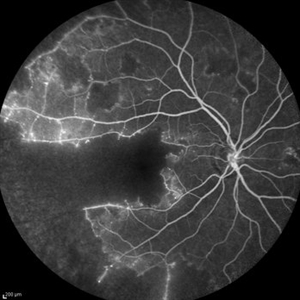 Idiopathic Occlusive Retinal Vasculitis (Late Stage)
May 31 2014 by Hamid Ahmadieh, MD
Late phase FA image of the right eye of a 28-year-old woman with idiopathic occlusive retinal vasculitis 6 months after the onset.
Photographer: Solmaz Shahmohammad, Negah Eye Center, Tehran
Imaging device: Heidelberg Spectralis
Condition/keywords: capillary closure, fluorescein leakage, macular infarction
-
---thumb.JPG/image-square;max$300,300.ImageHandler) Behcet's Disease
Behcet's Disease
Nov 25 2012 by Mallika Goyal, MD
Fundus photograph of right eye of a 23-year-old gentleman with Behcet's Disease shows occlusive retinal vasculitis with optic disc pallor and macular ischaemia. Other eye has similar appearance with no light perception.
Photographer: Mallika Goyal, MD, Apollo Health City, Hyderabad, India
Condition/keywords: macular ischemia, occlusive vasculitis
-
 Idiopathic Retinal Vasculitis, Aneurysms, and Neuroretinitis (IRVAN)
Oct 16 2012 by S. Natarajan, MD, FASRS, FRCS (GLASGOW) , FICO, D.Sc, FELA
FFA photograph of a 28-year-old male with IRVAN Syndrome.
Photographer: Prof. S. Natarajan
Condition/keywords: aneurysm, neuroretinitis, retinal vasculitis
-
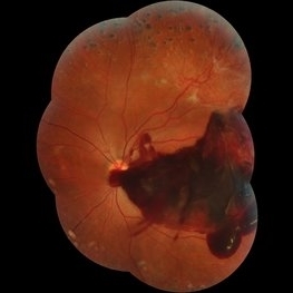 Subhyaloid Hemorrhage
Jul 7 2015 by Hamid Ahmadieh, MD
Color fundus photograph of the left eye of a 25-year-old woman with severe subhyaloid hemorrhage due to an advanced vasoproliferative vitreoretinopathy secondary to a severe idiopathic occlusive retinal vasculitis.
Photographer: Soulmaz Shahmohammad, Negah Eye Center, Tehran, Iran
Condition/keywords: color fundus photograph, occlusive vasculitis, subhyaloid hemorrhage
-
 Retinal Vasculitis
Aug 30 2012 by Abdhish R. Bhavsar, MD, FASRS
Fundus fluorescein angiography in a young female with retinal vasculitis.
Photographer: Abdhish R. Bhavsar, MD Retina Center of Minnesota
Condition/keywords: retinal vasculitis
-
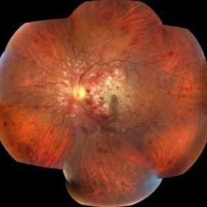 Acute Necrotizing Retinal Vasculitis as Onset of Systemic Lupus Erythematosus.
Sep 3 2016 by ADRIANO FERREIRA
A 28-year-old white man was referred to the rheumatology clinic with gradually and rapid deterioration of the vision (both eyes). In this picture we can observe cotton wool spots in the papillomacular area and extensive hemorrhages in the left eye.
Photographer: Claudio Zett Lobo
Imaging device: TRC50DXi TOPCON
Condition/keywords: systemic lupus erythematosus (SLE) vasculitis, vasculitis
-
 Idiopathic Retinal Vasculitis, Aneurysms, and Neuroretinitis (IRVAN)
Oct 16 2012 by S. Natarajan, MD, FASRS, FRCS (GLASGOW) , FICO, D.Sc, FELA
Red free photograph of a 28-year-old male with IRVAN Syndrome.
Photographer: Prof. Dr. S. Natarajan
Condition/keywords: aneurysm, neuroretinitis, retinal vasculitis
-
 Leptospirosis Neuroretinitis
May 30 2014 by Mitzy E Torres Soriano, MD
50-year-old man, presented with sudden onset of reduced vision in the left eye. Visual acuity (VA) was count fingers. Fundoscopic examination revealed soft exudation adjacent the optic nerve, macular edema with hard exudates in star shape arrangement and retinal vasculitis. OCT confirmed macular edema. There were no systemic symptoms. History of alcoholism and crack cocaine addiction. Systemic work up revealed a positive leptospira. He was treated with oral doxycicline (100mg twice daily) and prednisona (1mg/kg with gradual taper) for two weeks. Follow up at six months showed an improvement of VA to 20/60 with partial resolution of clinical findings at fundoscopic exam. Leptospirosis should be ruled out in every case of neuroretinitis.
Photographer: Mitzy E. Torres Soriano, MD; Centro medico Cagua-Estado Aragua. Venezuela
Imaging device: Retinal Camera TRC-NW8, TOPCON
Condition/keywords: leptospirosis, macular star, neuroretinitis, retinal vasculitis
-
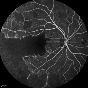 Idiopathic Occlusive Retinal Vasculitis (Late Stage)
May 31 2014 by Hamid Ahmadieh, MD
Late phase FA image of the right eye of a 28-year-old woman with idiopathic occlusive retinal vasculitis 6 months after the onset.
Photographer: Solmaz Shahmohammad, Negah Eye Center, Tehran
Imaging device: Heidelberg Spectralis
Condition/keywords: capillary closure, macular infarction
-
 Idiopathic Retinal Vasculitis, Aneurysms, and Neuroretinitis (IRVAN)
Oct 16 2012 by S. Natarajan, MD, FASRS, FRCS (GLASGOW) , FICO, D.Sc, FELA
Red free photograph of a 28-year-old male with IRVAN Syndrome.
Photographer: Prof. Dr. S. Natarajan
Condition/keywords: aneurysm, neuroretinitis, retinal vasculitis
-
---thumb.jpg/image-square;max$300,300.ImageHandler) Bilateral Retinal Vasculitis
Bilateral Retinal Vasculitis
Oct 29 2013 by Maurice F. Rabb
47 year old white man with bilateral retinal vasculitis.
Condition/keywords: retinal vasculitis
 Loading…