Search results (35 results)
-
 Lattice Degeneration
Nov 9 2012 by Norman Byer
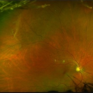
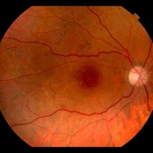
This is a more typical classical example of lattice degeneration in a 42-year-old woman in a photograph taken without scleral indentation. It shows much more marked vascular changes than the previous case. Note the tapering of the blood columns as the vessels approach the lesion and also the white sheathing of the vessel walls. Note also the continuity of the blood vessels on opposite sides of the lesion with the characteristic white lattice lines. More than 45 years ago Vogt pointed this out as a proof that these white lines were actually caused by changed blood vessels. Note also that this lesion shows a combination of several individual features of lattice degeneration. In addition to the white lines, there is a reddish crater-like area beneath the main horizontal white line. There is a prominent horizontal zone below this white line showing a snailtrack appearance. Also, there are two tiny atrophic retinal holes outside the photograph on the right end of this lesion. This eye contained five such retinal holes and they have all remained unchanged for more than 10 years of observation without treatment.
Condition/keywords: atrophic retinal hole, lattice degeneration, moderate snail track, tapering blood columns, white lattice lines, white sheath vessel
-
 Lattice Lesion
Nov 9 2012 by Norman Byer
This is the same lesion as shown in the previous case. Two retinal holes are present, and you can look through the upper hole into the dark subretinal space. This is, therefore, a true subclinical retinal detachment but it has changed only slightly in the past 13 years. About 75% of such holes in lattice lesions show a tiny adjacent zone of subretinal fluid. After the hole forms from gradual progressive thing of the retina, a tiny amount of fluid from the pocket of liquified vitreous over the lesion passes through the hole to the subretinal space
Condition/keywords: lattice degeneration, liquefied vitreous, retinal hole, subretinal fluid
-
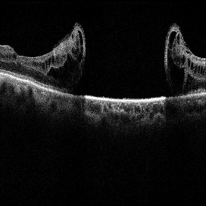
 Multiple Retinal Holes
Sep 10 2017 by JEFFERSON R SOUSA, Tecg.º (Biomedical Systems Technology)
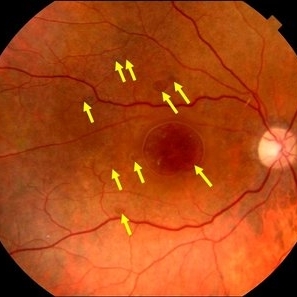
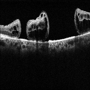
Patient 57-years-old, male, attended the clinic with complaint of low visual acuity and history of already having undergone a surgical procedure in another service. In previous evaluation of retinal mapping and retinography, being confirmed in optical coherence tomography, several retinal holes were observed in the posterior pole. Each arrow represents a hole. Nine retinal holes in the posterior pole
Photographer: JEFFERSON R SOUSA - Study Center and Ophthalmological Research Dr. Andre M V Gomes, Dr. Suel Abujamra Institute São Paulo-Brazil
Imaging device: Retinografo Topcon TRC-50 DX, Imaginet, campo de 50 graus. Flash 75
Condition/keywords: retinal hole
-
 Flat Lattice Lesion
Nov 9 2012 by Norman Byer
This 24-year-old woman had a flat lattice lesion without holes observed with no change for six years. She then developed two tiny retinal holes in this lesion and three years later the clinical retinal detachment shown here. She responded well to surgery. Even though such atrophic holes and lattice lesions may occasionally lead to a clinical detachment, it is important to understand that the mere presence of such holes is not an indication for prophylactic treatment. The reason for this is that we now know statistically that fewer than 1 percent of such cases lead to a retinal detachment.
Condition/keywords: lattice degeneration, retinal hole, scleral depression
-
 Lattice Lesion
Nov 9 2012 by Norman Byer
This is the same lesion as seen in the previous case seen now with scleral indentation. Here you can see directly into the subretinal space through the two retinal holes. The holes appear dark because the shadow of the scleral indentation lies directly beneath them.
Condition/keywords: lattice degeneration, retinal hole, scleral indentation
-
 Myopia with Lattice Degeneration and White Without Pressure in the Setting of Marfan's Syndrome
Aug 31 2020 by Sophia El Hamichi, MD
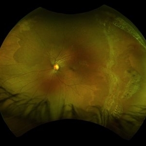
A 1-year-old female with Marfan's syndrome, myopia OU, congenital nystagmus and exotopia OD. Ultra-wide field imaging of her left eye showed lattice degeneration with atrophic retinal holes temporally, in addition to multiple sections of white without pressure.
Imaging device: Optos
Condition/keywords: atrophic retinal hole, lattice degeneration, Marfan's syndrome, myopia, Optos, ultra-wide field imaging
-
 Lattice Degeneration
Nov 9 2012 by Norman Byer
This lesion in a 51-year-old woman is also an example of lattice degeneration but shows only a uniform reddish crater with no other features. This lesion has remained exactly the same for 9 years but such red craters sometimes give rise to punched-out atrophic retinal holes which may lead to subclinical retinal detachment. This sequence of events will be shown in the next two slide pairs.
Condition/keywords: lattice degeneration, lattice lesion, reddish crater
-
 Lattice Lesion
Nov 9 2012 by Norman Byer
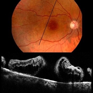
This is a photograph of a lattice lesion in a 23-year-old girl taken without scleral indentation. Just to the left of the center of the slide is a slightly pigmented lesion almost oval in shape with a retinal hole in each end. Ten years earlier at the age of 13 this lesion appeared exactly like the one in the previous case as a pure red crater. Five years later two new round retinal holes were seen, one in each end, with a tiny bit of subretinal fluid within the lattice lesion only. Five years later still the appearance was as shown in this slide pair with the subretinal fluid now extending slightly beyond the lattice lesion as far as the curved row of tiny yellow exudates seen just to the right of the center of the slide. It is now actually a small subclinical retinal detachment. The next slide pair will show this better using scleral indentation.
Condition/keywords: lattice degeneration, lattice lesion, pigmented lesion, reddish crater, retinal hole, subretinal fluid, yellow exudate
-
 Parallel Lattice Lesions
Nov 9 2012 by Norman Byer
This is an example of parallel lattice lesions. The anterior one is faintly seen and not in focus. The posterior lesion shows a prominent whitish meshwork with modeled reddish areas which sometimes may be mistaken for retinal holes.
Condition/keywords: lattice degeneration, lattice lesion, parallel lattice lesions, reddish areas, scleral indentation
-
 Multiple Retinal Holes
Sep 10 2017 by JEFFERSON R SOUSA, Tecg.º (Biomedical Systems Technology)
Patient 57-years-old, male, attended the clinic with complaint of low visual acuity and history of already having undergone a surgical procedure in another service. In previous evaluation of retinal mapping and retinography, being confirmed in optical coherence tomography, several retinal holes were observed in the posterior pole.
Photographer: JEFFERSON R SOUSA - Study Center and Ophthalmological Research Dr. Andre M V Gomes, Dr. Suel Abujamra Institute São Paulo-Brazil
Imaging device: Topcon TRC-50 DX, Imaginet, campo de 50 graus. Flash 75 / Optical coherence tomography system OCT CIRRUS 4000, Protocol Line 125 degrees.
Condition/keywords: retinal hole
-
 Bullous Retinoschisis with Outer Retinal Holes
Jun 15 2020 by Olivia Rainey
Ultra-widefield pseudocolor fundus photograph of a 56-year-old female with bullous retinoschisis with outer retinal holes affecting her right eye. The physician noted superotemporal retinoschisis in her monoculcar functioning eye. There was no demarcation line and no inner or outer layer breaks at her first appointment in February of 2020. On 6/15/20 she had a new onset outer holes and SRF tracking inferiorly. The physician recommended observation, however if this continues to progress we have discussed indications for barrier laser.
Photographer: Olivia Rainey, OCT-C, COA
Imaging device: Optos California
Condition/keywords: bullous retinoschisis, Optos, outer layer breaks, outer layer hole, pseudocolor, subretinal fluid, superior retina, ultra-wide field imaging
-
 Juvenile Retinoschisis
Oct 10 2015 by Hamid Ahmadieh, MD
Merged color fundus photograph of the right eye of a 30-year-old man with juvenile retioschisis. Involvement of the retinal periphery with typical large inner layer retinal holes is visible.
Photographer: Shabnam Pooreh, Negah Eye Center, Tehran, Iran
Condition/keywords: color fundus photograph, inner layer holes, juvenile retinoschisis
-
 Multiple Retinal Holes
Sep 10 2017 by JEFFERSON R SOUSA, Tecg.º (Biomedical Systems Technology)
Patient 57-years-old, male, attended the clinic with complaint of low visual acuity and history of already having undergone a surgical procedure in another service. In previous evaluation of retinal mapping and retinography, being confirmed in optical coherence tomography, several retinal holes were observed in the posterior pole.
Photographer: JEFFERSON R SOUSA - Study Center and Ophthalmological Research Dr. Andre M V Gomes, Dr. Suel Abujamra Institute São Paulo-Brazil
Imaging device: OCT CIRRUS 4000, Protocol Horizontal line.
Condition/keywords: retinal hole
-
 Juvenile Retinoschisis
Oct 10 2015 by Hamid Ahmadieh, MD
Typical changes including large inner layer retinal holes in the retinal periphery of a 30-year-old man with juvenile retinoschisis.
Photographer: Shabnam Pooreh, Negah Eye Center, Tehran, Iran
Condition/keywords: color fundus photograph, inner layer holes, juvenile retinoschisis
-
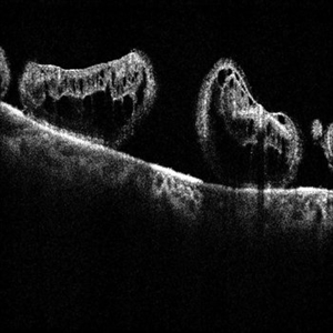
 Multiple Retinal Holes
Sep 10 2017 by JEFFERSON R SOUSA, Tecg.º (Biomedical Systems Technology)
Patient 57-years-old, male, attended the clinic with complaint of low visual acuity and history of already having undergone a surgical procedure in another service. In previous evaluation of retinal mapping and retinography, being confirmed in optical coherence tomography, several retinal holes were observed in the posterior pole.
Photographer: JEFFERSON R SOUSA - Study Center and Ophthalmological Research Dr. Andre M V Gomes, Dr. Suel Abujamra Institute São Paulo-Brazil
Imaging device: Retinografo Topcon TRC-50 DX, Imaginet, campo de 50 graus. Flash 75
Condition/keywords: retinal hole
-
 Multiple Retinal Holes
Sep 10 2017 by JEFFERSON R SOUSA, Tecg.º (Biomedical Systems Technology)
Patient 57-years-old, male, attended the clinic with complaint of low visual acuity and history of already having undergone a surgical procedure in another service. In previous evaluation of retinal mapping and retinography, being confirmed in optical coherence tomography, several retinal holes were observed in the posterior pole.
Photographer: JEFFERSON R SOUSA - Study Center and Ophthalmological Research Dr. Andre M V Gomes, Dr. Suel Abujamra Institute São Paulo-Brazil
Imaging device: OCT CIRRUS 4000, Protocol Horizontal line.
Condition/keywords: retinal hole
-
 Multiple Retinal Holes
Sep 10 2017 by JEFFERSON R SOUSA, Tecg.º (Biomedical Systems Technology)
Patient 57-years-old, male, attended the clinic with complaint of low visual acuity and history of already having undergone a surgical procedure in another service. In previous evaluation of retinal mapping and retinography, being confirmed in optical coherence tomography, several retinal holes were observed in the posterior pole.
Photographer: JEFFERSON R SOUSA - Study Center and Ophthalmological Research Dr. Andre M V Gomes, Dr. Suel Abujamra Institute São Paulo-Brazil
Imaging device: OCT CIRRUS 4000, Protocol Line, transverse 125 degrees.
Condition/keywords: retinal hole
-
 ARN (#2) Five Days Since Initial Visit
May 27 2019 by John S. King, MD
60-year-old African American female who had been treated for iridocyclitis for at least a week sent in for vitritis and a nasal fundus lesion. Complaints included redness, floaters, photophobia, and decreased vision. Husband had recent shingles. Acuity was 20/60-2 with IOP of 12, and small KP in Art's triangel, 1-2+ a/c cell, 2-3+ ant vit cell, diffuse arteriolar sheathing, multiple areas of retinal whitening in periphery and mid-periphery (see Photo #1). PCR of a/c was performed, and intravitreal GCV administered, and VACV 2g qid and ASA started.... PCR positive for HZV, pred taper was started two days after presentation as the infection had begun to stablize..... Five days from presentation the vision was 20/60, inflammation and areas of retinal whitening had improved (see Photo #2).... One week later acuity was 20/30, the a/c was quiet and KP resolved; ant vitreous cell decreased; and there was further improvement in retinal appearance without any signs of retinal holes or detachment; she is now on low dose maint VACV (see photo#3)
Photographer: Maysee Yang
Imaging device: Optos CA
Condition/keywords: acute retinal necrosis, Herpes zoster
-
 ARN (#3) This is comparison between the latest visit (left) and one week prior (which is the right photo, and same one as photo #2)
May 27 2019 by John S. King, MD
60-year-old African American female who had been treated for iridocyclitis for at least a week sent in for vitritis and a nasal fundus lesion. Complaints included redness, floaters, photophobia, and decreased vision. Husband had recent shingles. Acuity was 20/60-2 with IOP of 12, and small KP in Art's triangel, 1-2+ a/c cell, 2-3+ ant vit cell, diffuse arteriolar sheathing, multiple areas of retinal whitening in periphery and mid-periphery (see Photo #1). PCR of a/c was performed, and intravitreal GCV administered, and VACV 2g qid and ASA started.... PCR positive for HZV, pred taper was started two days after presentation as the infection had begun to stablize..... Five days from presentation the vision was 20/60, inflammation and areas of retinal whitening had improved (see Photo #2).... One week later acuity was 20/30, the a/c was quiet and KP resolved; ant vitreous cell decreased; and there was further improvement in retinal appearance without any signs of retinal holes or detachment; she is now on low dose maint VACV (see photo#3)
Photographer: Maysee Yang
Imaging device: Optos CA
Condition/keywords: acute retinal necrosis, Herpes zoster
-
 Multiple Retinal Holes
Sep 10 2017 by JEFFERSON R SOUSA, Tecg.º (Biomedical Systems Technology)
Patient 57-years-old, male, attended the clinic with complaint of low visual acuity and history of already having undergone a surgical procedure in another service. In previous evaluation of retinal mapping and retinography, being confirmed in optical coherence tomography, several retinal holes were observed in the posterior pole.
Photographer: JEFFERSON R SOUSA - Study Center and Ophthalmological Research Dr. Andre M V Gomes, Dr. Suel Abujamra Institute São Paulo-Brazil
Imaging device: OCT CIRRUS 4000, Protocol Horizontal line.
Condition/keywords: retinal hole
-
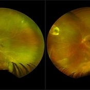
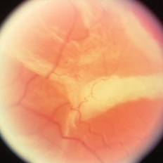
 Total retinal Detachment multiple holes
Sep 26 2022 by Denica Rodriguez
60 year old Male presented with two week old Macula off Retinal detachment with multiple tears.
Photographer: Denica Rodriguez
Imaging device: Optos California
Condition/keywords: color fundus photograph, color photo, macula-off, optos, pseudocolor, Retinal detachment, retinal holes, retinal tear, Retinal tear with detachment, superior arcade, superior field, superior retina, total retinal detachment
-
 Outer Layer Holes with Rolled Edges in Retinoschisis
Apr 2 2019 by Gary R. Cook, MD, FACS
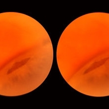
Another view of a 71-year-old white female with rolled edges of outer layer retinal holes in bullous, adult-type retinoschisis OS.
Condition/keywords: bullous retinoschisis, retinoschisis
-
 ARN (#1) Initial Photo
May 27 2019 by John S. King, MD
60-year-old African American female who had been treated for iridocyclitis for at least a week sent in for vitritis and a nasal fundus lesion. Complaints included redness, floaters, photophobia, and decreased vision. Husband had recent shingles. Acuity was 20/60-2 with IOP of 12, and small KP in Art's triangel, 1-2+ a/c cell, 2-3+ ant vit cell, diffuse arteriolar sheathing, multiple areas of retinal whitening in periphery and mid-periphery (see Photo #1). PCR of a/c was performed, and intravitreal GCV administered, and VACV 2g qid and ASA started.... PCR positive for HZV, pred taper was started two days after presentation as the infection had begun to stablize..... Five days from presentation the vision was 20/60, inflammation and areas of retinal whitening had improved (see Photo #2).... One week later acuity was 20/30, the a/c was quiet and KP resolved; ant vitreous cell decreased; and there was further improvement in retinal appearance without any signs of retinal holes or detachment; she is now on low dose maint VACV (see photo#3)
Photographer: Maysee Yang
Imaging device: Optos CA
Condition/keywords: acute retinal necrosis, Herpes zoster
-
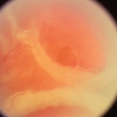
 Peripheral retinal degenerations
Jan 29 2024 by Anupama Kiran Kumar
Fundus photo of a young man who underwent barrage laser after he presented to the clinic with floaters and was diagnosed to have lattices with horse shoe tears and retinal holes.
Photographer: Dr Anupama Kiran Kumar DNB FVR , Narayana Nethralaya Bangalore
Imaging device: Mirante SLO/OCT (Nidek Co., Gamagori, Japan)
Condition/keywords: lattice degeneration, peripheral retinal degeneration
-
 Outer Layer Holes with Rolled Edges in Retinoschisis
Apr 2 2019 by Gary R. Cook, MD, FACS
71-year-old white female with outer layer retinal holes with rolled edges in an area of bullous, adult-type retinoschisis OS
Condition/keywords: bullous retinoschisis, retinoschisis
 Loading…