Search results (100 results)
-
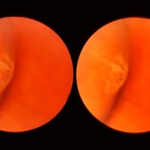 Meridional Fold
Nov 9 2012 by Norman Byer
This is the same lesion as in the previous photograph. With the scleral indentation placed more posterior, we now can see that the fold ends over a small collection of subretinal fluid and that there is a very tiny retinal hole just below the posterior end of the retinal fold.
Condition/keywords: peripheral cystoid degeneration, retinal fold, retinal hole, scleral indentation, subretinal fluid
-
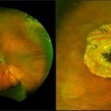
 Operculated Retinal Hole
Oct 12 2012 by Jeffrey G. Gross, MD, FASRS
Operculated retinal hole with RD.
Condition/keywords: operculated retinal hole, retinal degeneration
-
 Lattice Degeneration
Nov 9 2012 by Norman Byer
This is a more typical classical example of lattice degeneration in a 42-year-old woman in a photograph taken without scleral indentation. It shows much more marked vascular changes than the previous case. Note the tapering of the blood columns as the vessels approach the lesion and also the white sheathing of the vessel walls. Note also the continuity of the blood vessels on opposite sides of the lesion with the characteristic white lattice lines. More than 45 years ago Vogt pointed this out as a proof that these white lines were actually caused by changed blood vessels. Note also that this lesion shows a combination of several individual features of lattice degeneration. In addition to the white lines, there is a reddish crater-like area beneath the main horizontal white line. There is a prominent horizontal zone below this white line showing a snailtrack appearance. Also, there are two tiny atrophic retinal holes outside the photograph on the right end of this lesion. This eye contained five such retinal holes and they have all remained unchanged for more than 10 years of observation without treatment.
Condition/keywords: atrophic retinal hole, lattice degeneration, moderate snail track, tapering blood columns, white lattice lines, white sheath vessel
-
 White Retinal Tuft
Nov 9 2012 by Norman Byer
After six years, the previous lesion looked like this. The former flap has been completely avulsed and is now a free operculum. The white zone around the tear represents the small area of detachment and subretinal fluid. It is still asymptomatic and does not require treatment.
Condition/keywords: does not require treatment, free operculum, operculated retinal hole, subretinal fluid, white retinal tuft
-
 Lattice Degeneration
Nov 9 2012 by Norman Byer
This 16-year-old girl has lattice degeneration and also this large oval retinal hole with a surrounding narrow zone of subretinal fluid. This lesion illustrates how large the atrophic holes of lattice degeneration may be. Occasionally the hole can be as large as the initial lattice lesion and can therefore obliterate all other evidence of its true identity. This was almost true in this case, but there does remain a small whitish remnant of the original lattice lesion at the lower end of the oval hole.
Condition/keywords: lattice degeneration, retinal hole, subretinal fluid, white lattice lines
-
 Atrophic Holes in Lattice Lesion
Nov 9 2012 by Norman Byer
In this 26-year-old woman, these two atrophic holes in a lattice lesion led to a clinical retinal detachment which was operated on successfully. In retinal detachments of this type resulting from non tractional atrophic holes, it has been found that 50% occur before the age of 30 years.
Condition/keywords: atrophic retinal hole, lattice lesion
-
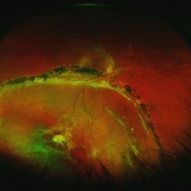
 Operculated Retinal Hole in Retinal Detachment
Oct 12 2012 by Jeffrey G. Gross, MD, FASRS
Operculated retinal hole in retinal detachment.
Condition/keywords: operculated retinal hole, retinal degeneration
-
 Operculated Hole and CHRPE
Jan 16 2018 by Carolyn Daley
58-year-old woman with an operculated hole and CHRPE in the right eye. Patient is asymptomatic so no treatment was recommended at this time.
Photographer: Carolyn Daley
Imaging device: Optos ultra wide field image
Condition/keywords: congenital hypertrophy of the retinal pigment epithelium (CHRPE), operculated retinal hole, Optos, ultra-wide field imaging
-
 Lattice Degeneration
Nov 9 2012 by Norman Byer
Lattice degeneration in a 42-year-old man which has produced four atrophic holes in a linear arrangement surrounded by a subclinical retinal detachment of unknown duration. By age 63, 21 years later, a posterior vitreous detachment was diagnosed in this eye, which was not present four years earlier. Nevertheless, the appearance seen here has remained exactly the same for 30 years, more than eight years with a concurrent PVD.
Condition/keywords: atrophic retinal hole, lattice degeneration, posterior vitreous detachment
-
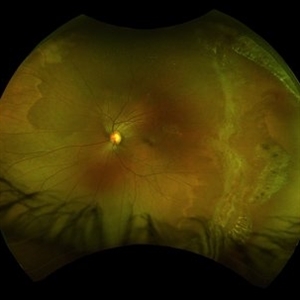
 Chronic Inferior Retinal Detachment
Mar 1 2017 by Philip J. Polkinghorne, MD
Color photograph of chronic retinal detachment with pigment demarcation line and atrophic holes visible. The vision was recorded at 20/20, and follow up is 3 years.
Photographer: Alex Fraser
Condition/keywords: atrophic retinal hole, demarcation line
-
 Lattice Lesion
Nov 9 2012 by Norman Byer
This is the same lesion as shown in the previous case. Two retinal holes are present, and you can look through the upper hole into the dark subretinal space. This is, therefore, a true subclinical retinal detachment but it has changed only slightly in the past 13 years. About 75% of such holes in lattice lesions show a tiny adjacent zone of subretinal fluid. After the hole forms from gradual progressive thing of the retina, a tiny amount of fluid from the pocket of liquified vitreous over the lesion passes through the hole to the subretinal space
Condition/keywords: lattice degeneration, liquefied vitreous, retinal hole, subretinal fluid
-
 Asymptomatic Lesion
Nov 9 2012 by Norman Byer
This is the same lesion as seen in the previous slide pair. Here the scleral indentation is carried more posterior revealing a tiny, round, full thickness retinal hole. This is not a tear produced by traction even though vitreous is always attached to these flaps. You will note that the hole is round and is separated by a slight distance from the flap itself. It is probably the result of long continued atrophy and devitalization of the retina. A posterior vitreous was not detached. This lesion has not changed its appearance for more than a year of observation, but the age of the hole is actually unknown.
Condition/keywords: asymptomatic, atrophy, full thickness retinal hole, posterior scleral indentation, retinal hole, round hole
-
 Full-thickness Macular Hole
Aug 28 2012 by Sharon Fekrat, MD FACS FASRS
65 year old woman with a recurrent full thickness macular hole following previous 20 g pars plana vitrectomy in the right eye as well as an iatrogenic retinal hole in the papillomacular bundle. Both retinal defects are captured here in this Zeiss Stratus OCT image.
Photographer: Michael P. Kelly, FOPS Director, Duke Eye Labs, Duke University Eye Center, Durham, NC
Imaging device: Zeiss Cirrus
Condition/keywords: retinal break
-
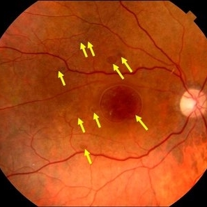 Multiple Retinal Holes
Sep 10 2017 by JEFFERSON R SOUSA, Tecg.º (Biomedical Systems Technology)
Patient 57-years-old, male, attended the clinic with complaint of low visual acuity and history of already having undergone a surgical procedure in another service. In previous evaluation of retinal mapping and retinography, being confirmed in optical coherence tomography, several retinal holes were observed in the posterior pole. Each arrow represents a hole. Nine retinal holes in the posterior pole
Photographer: JEFFERSON R SOUSA - Study Center and Ophthalmological Research Dr. Andre M V Gomes, Dr. Suel Abujamra Institute São Paulo-Brazil
Imaging device: Retinografo Topcon TRC-50 DX, Imaginet, campo de 50 graus. Flash 75
Condition/keywords: retinal hole
-
 Flat Lattice Lesion
Nov 9 2012 by Norman Byer
This 24-year-old woman had a flat lattice lesion without holes observed with no change for six years. She then developed two tiny retinal holes in this lesion and three years later the clinical retinal detachment shown here. She responded well to surgery. Even though such atrophic holes and lattice lesions may occasionally lead to a clinical detachment, it is important to understand that the mere presence of such holes is not an indication for prophylactic treatment. The reason for this is that we now know statistically that fewer than 1 percent of such cases lead to a retinal detachment.
Condition/keywords: lattice degeneration, retinal hole, scleral depression
-
 Lattice Degeneration With Atrophic Hole
Feb 19 2015 by H. Michael Lambert, MD
Color photo of Lattice degeneration with atrophic hole.
Condition/keywords: atrophic retinal hole, lattice degeneration
-
 Lattice Lesion
Nov 9 2012 by Norman Byer
This is the same lesion as seen in the previous case seen now with scleral indentation. Here you can see directly into the subretinal space through the two retinal holes. The holes appear dark because the shadow of the scleral indentation lies directly beneath them.
Condition/keywords: lattice degeneration, retinal hole, scleral indentation
-
 Lattice Lesion
Nov 9 2012 by Norman Byer
When this boy was first examined at the age of six years, he had only the red crater form of lattice at this location. This photograph shows the same lesion at age 11 and there is now a small round atrophic hole with a tiny round zone of detachment around it. It has not changed for four years.
Condition/keywords: atrophic retinal hole, lattice degeneration, lattice lesion, reddish crater, round hole
-
 Lattice Lesion
Nov 9 2012 by Norman Byer
This lattice lesion in a 44-year-old man shows an atrophic retinal hole surrounded by discrete yellowish and pigmented areas. These have been caused by secondary pigment migration and proliferation in the retinal pigment epithelium. There is a small doughnut like elevation of the retina between the edge of the hole and the line of pigment. The lesion and the hole have remained exactly the same for seven years.
Condition/keywords: atrophic retinal hole, elevated retina, lattice degeneration, lattice lesion, proliferation of retinal pigment epithelium, scleral indentation
-
 Myopia with Lattice Degeneration and White Without Pressure in the Setting of Marfan's Syndrome
Aug 31 2020 by Sophia El Hamichi, MD
A 1-year-old female with Marfan's syndrome, myopia OU, congenital nystagmus and exotopia OD. Ultra-wide field imaging of her left eye showed lattice degeneration with atrophic retinal holes temporally, in addition to multiple sections of white without pressure.
Imaging device: Optos
Condition/keywords: atrophic retinal hole, lattice degeneration, Marfan's syndrome, myopia, Optos, ultra-wide field imaging
-
 Lattice Degeneration
Nov 9 2012 by Norman Byer
This lesion in a 51-year-old woman is also an example of lattice degeneration but shows only a uniform reddish crater with no other features. This lesion has remained exactly the same for 9 years but such red craters sometimes give rise to punched-out atrophic retinal holes which may lead to subclinical retinal detachment. This sequence of events will be shown in the next two slide pairs.
Condition/keywords: lattice degeneration, lattice lesion, reddish crater
-
 Lattice Lesion
Nov 9 2012 by Norman Byer
This lattice lesion in a 36-year-old woman shows a snail track feature on the left combined with a reddish crater and retinal hole to the right. The hole has caused a small subclinical detachment. The next slide pair will show more of this lesion.
Condition/keywords: lattice lesion, reddish crater, retinal hole, snail track, subclinical detachment
-
 Lattice Lesion
Nov 9 2012 by Norman Byer
In this 47-year-old woman, this lattice lesion with a small hole in the right end has led to a subclinical retinal detachment which extends to the margin of the subtle yellowish zone almost at the upper edge of the photograph. This patient did not desire surgery, and the area of detachment has changed only a small amount in the past eight years. The risk of a clinical retinal detachment developing from lattice degeneration is less than 1 percent. In those cases where it does though, about 3 quarters are caused by a tractional tear and about one quarter are caused by an atrophic hole as in this case.
Condition/keywords: atrophic retinal hole, lattice degeneration, lattice lesion, retinal hole, yellowish zone
-
 Lattice Lesion
Nov 9 2012 by Norman Byer
This is a photograph of a lattice lesion in a 23-year-old girl taken without scleral indentation. Just to the left of the center of the slide is a slightly pigmented lesion almost oval in shape with a retinal hole in each end. Ten years earlier at the age of 13 this lesion appeared exactly like the one in the previous case as a pure red crater. Five years later two new round retinal holes were seen, one in each end, with a tiny bit of subretinal fluid within the lattice lesion only. Five years later still the appearance was as shown in this slide pair with the subretinal fluid now extending slightly beyond the lattice lesion as far as the curved row of tiny yellow exudates seen just to the right of the center of the slide. It is now actually a small subclinical retinal detachment. The next slide pair will show this better using scleral indentation.
Condition/keywords: lattice degeneration, lattice lesion, pigmented lesion, reddish crater, retinal hole, subretinal fluid, yellow exudate
-
 Parallel Lattice Lesions
Nov 9 2012 by Norman Byer
This is an example of parallel lattice lesions. The anterior one is faintly seen and not in focus. The posterior lesion shows a prominent whitish meshwork with modeled reddish areas which sometimes may be mistaken for retinal holes.
Condition/keywords: lattice degeneration, lattice lesion, parallel lattice lesions, reddish areas, scleral indentation
 Loading…