Search results (32 results)
-
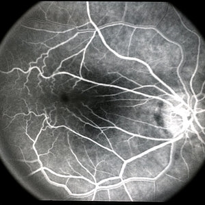
 Cilioretinal Artery Occlusion
Sep 2 2012 by Hyung-Woo Kwak, MD
Cloudiness localized to the area of papillomacular bundle normally perfused by retinal vessel.
Imaging device: Zeiss F450 plus
Condition/keywords: cilioretinal artery occlusion
-
---thumb.jpg/image-square;max$300,300.ImageHandler) Retinoblastoma To Chemothermotherapy
Retinoblastoma To Chemothermotherapy
Oct 4 2013 by Maurice F. Rabb
A 7 week old girl with a family history of retinoblastoma was found to have a small retinoblastoma in each eye. In the right eye the tumor was adjacent to the optic disc in the papillomacular bundle and measured 2 X 2 X 2 mm. Its temporal margin was 1.0 mm from the foveola and it overhung 20% of the optic disc surface. There was not clinical or ultrasonographic evidence of vitreous seeking or optic nerve invation. In the left eye there was a solitary tumor 1mm superonasal to the optic disc. The tumor measured 1 X 1 X 1 mm. The foveal reflex was normal in both eyes. Both tumors showed a fluorescein angiographic pattern compatible with retinoblastoma with rapid filling and late hyperfluorescence.
Condition/keywords: retina
-
 Full-thickness Macular Hole
Aug 28 2012 by Sharon Fekrat, MD FACS FASRS
65 year old woman with a recurrent full thickness macular hole following previous 20 g pars plana vitrectomy in the right eye as well as an iatrogenic retinal hole in the papillomacular bundle. Both retinal defects are captured here in this Zeiss Stratus OCT image.
Photographer: Michael P. Kelly, FOPS Director, Duke Eye Labs, Duke University Eye Center, Durham, NC
Imaging device: Zeiss Cirrus
Condition/keywords: retinal break
-
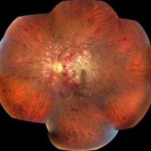 Acute Necrotizing Retinal Vasculitis as Onset of Systemic Lupus Erythematosus.
Sep 3 2016 by ADRIANO FERREIRA
A 28-year-old white man was referred to the rheumatology clinic with gradually and rapid deterioration of the vision (both eyes). In this picture we can observe cotton wool spots in the papillomacular area and extensive hemorrhages in the left eye.
Photographer: Claudio Zett Lobo
Imaging device: TRC50DXi TOPCON
Condition/keywords: systemic lupus erythematosus (SLE) vasculitis, vasculitis
-
---thumb.jpg/image-square;max$300,300.ImageHandler) Retinoblastoma To Chemothermotherapy
Retinoblastoma To Chemothermotherapy
Oct 4 2013 by Maurice F. Rabb
A 7 week old girl with a family history of retinoblastoma was found to have a small retinoblastoma in each eye. In the right eye the tumor was adjacent to the optic disc in the papillomacular bundle and measured 2 X 2 X 2 mm. Its temporal margin was 1.0 mm from the foveola and it overhung 20% of the optic disc surface. There was not clinical or ultrasonographic evidence of vitreous seeking or optic nerve invation. In the left eye there was a solitary tumor 1mm superonasal to the optic disc. The tumor measured 1 X 1 X 1 mm. The foveal reflex was normal in both eyes. Both tumors showed a fluorescein angiographic pattern compatible with retinoblastoma with rapid filling and late hyperfluorescence.
Condition/keywords: retina
-
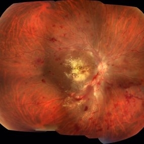 Acute Necrotizing Retinal Vasculitis as Onset of Systemic Lupus Erythematosus.
Sep 3 2016 by ADRIANO FERREIRA
A 28-year-old white man was referred to the rheumatology clinic with gradually and rapid deterioration of the vision (both eyes). In this picture, we can observe cotton wool spots in the papillomacular area and extensive hemorrhages in posterior polo and in the middle periphery. Hard exudates are present in macular area (macular edema)
Photographer: Claudio Zett Lobo
Imaging device: TRC50DXi TOPCON
Condition/keywords: systemic lupus erythematosus (SLE) vasculitis, vasculitis
-
 OCT of Papillomacular Fold in Posterior Microphthalmos
Jul 30 2014 by Jordan L. Heffez, MD
OCT from a healthy 47-year-old gentleman with BCVA 20/50 OU and no known past ocular history. Note the lack of epiretinal tissue as a cause of retina malformation.
Condition/keywords: macular fold
-
 Papilloma Conjunctiva
Sep 2 2016 by JEFFERSON R SOUSA, Tecg.º (Biomedical Systems Technology)
Female patient, 26-years-old without complaint of ocular symptoms, only the of spot avermelha in the right eye. Absence of changes in positions of the look. Intraocular pressure visual acuity and fundoscopy without changes.
Photographer: JEFFERSON R SOUSA - Study Center and Ophthalmological Research Dr. Andre M V Gomes, Institute Dr. Suel Abujamra São Paulo-Brazil
Imaging device: Topcon TRC-50 Dx - Angulation of field photo of 35 Degrees, flash 36, Digital system Imaginet
Condition/keywords: conjunctival cysts, papilloma, papilloma conjunctiva, tumor
-
 Papillomacular Fold in Posterior Microphthalmos
Jul 30 2014 by Jordan L. Heffez, MD
Fundus photo of a healthy 47-year-old gentleman with BCVA 20/50 OU and no known past ocular history.
Condition/keywords: macular fold
-
 Papillomacular Fold in Posterior Microphthalmos
Jul 30 2014 by Jordan L. Heffez, MD
Fundus photo of a healthy 47-year-old gentleman with BCVA 20/50 OU and no known past ocular history.
Condition/keywords: macular fold
-
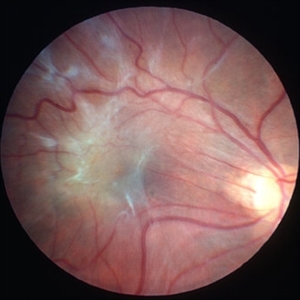 Macular Pucker
Mar 29 2013 by Henry J. Kaplan, MD
Large epiretinal membrane with straightened vessels in the papillomacular bundle and distorsion of retinal vessels.
Condition/keywords: epiretinal membrane (ERM), macular pucker
-
 Posterior Microphthalmos
Nov 22 2015 by Mallika Goyal, MD
Bilateral posterior microphthalmos, high hypermetropia, crowded disc, papillomacular fold, retinitis punctata albescens (white retinal flecks over the retinal midperiphery and periphery) in a 22-year-old male patient. B-scan revealed sclera-choroidal thickening. Best corrected VA was 20/80 each eye.
Photographer: Mallika Goyal, MD, Apollo Health City, Jubilee Hills, Hyderabad, India
Condition/keywords: posterior microphthalmos
-
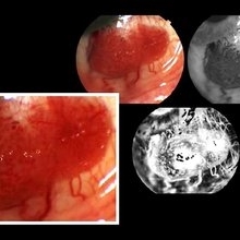
 Unilateral Subretinal Toxocara Granuloma
Apr 10 2018 by Albert Lin, MD
9-year-old male with chronic poor vision, band keratopathy, and posterior synechia in the right eye with a unilateral subretinal toxocara granuloma in the papillomacular bundle.
Photographer: Morgan Ladner, University of Mississippi Medical Center, Department of Ophthalmology
Condition/keywords: toxocara granuloma
-
 Combined Hamartoma
Oct 5 2016 by Guruprasad S. Ayachit, MBBS,MS
Fundus photograph of a 9-year-old boy with an ill-defined lesion extending from nasal to the disc going on to include the papillomacular bundle; 14X10 mm in greatest dimensions. There is a thick epiretinal membrane causing distortion and straightening of temporal vascular arcade.
Photographer: Shravan Masurkar, M M Joshi Eye Institute, Hubli
Imaging device: Topcon TRC50DX
Condition/keywords: combined hamartoma
-
 Posterior Microphthalmos
Nov 16 2015 by Mallika Goyal, MD
Bilateral posterior microphthalmos, high hypermetropia, crowded disc, papillomacular fold, retinitis punctata albescens. B-scan revealed sclera-choroidal thickening.
Photographer: Mallika Goyal, MD
Condition/keywords: posterior microphthalmos
-
 Unilateral Subretinal Toxocara Granuloma
Apr 10 2018 by Albert Lin, MD
9-year-old male with chronic poor vision, band keratopathy, and posterior synechia in the right eye with a unilateral subretinal toxocara granuloma in the papillomacular bundle.
Photographer: Jody Watkins, University of Mississippi Medical Center, Department of Ophthalmology
Imaging device: Optos ultrawide field photo
Condition/keywords: toxocariasis
-
 Posterior Microphthalmos
Nov 22 2015 by Mallika Goyal, MD
OCT of the left eye of a male patient with bilateral posterior microphthalmos showing elevated papillomacular fold. Best corrected VA was 20/80 each eye.
Photographer: Mallika Goyal, MD, Apollo Health City, Jubilee Hills, Hyderabad, India
Condition/keywords: posterior microphthalmos
-
 Posterior Microphthalmos
Nov 22 2015 by Mallika Goyal, MD
Bilateral posterior microphthalmos, high hypermetropia, crowded disc, papillomacular fold, retinitis punctata albescens (white retinal flecks over the retinal midperiphery and periphery) in a 22-year-old male patient. B-scan revealed sclera-choroidal thickening. Best corrected VA was 20/80 each eye.
Photographer: Mallika Goyal, MD, Apollo Health City, Jubilee Hills, Hyderabad, India
Condition/keywords: posterior microphthalmos
-
 Macular Pucker
Mar 29 2013 by Henry J. Kaplan, MD
Angiogram of a macular pucker better demonstrates vessel straightening in papillomacular bundleand tortousity in the area of membrane.
Condition/keywords: epiretinal membrane (ERM), macular pucker
-
 Posterior Microphthalmos
Nov 22 2015 by Mallika Goyal, MD
Bilateral posterior microphthalmos, high hypermetropia, crowded disc, papillomacular fold, retinitis punctata albescens (white retinal flecks over the retinal midperiphery and periphery) in a 22-year-old male patient. B-scan revealed sclera-choroidal thickening. Best corrected VA was 20/80 each eye.
Photographer: Mallika Goyal, MD, Apollo Health City, Jubilee Hills, Hyderabad, India
Condition/keywords: posterior microphthalmos
-
 Posterior Microphthalmos
Nov 22 2015 by Mallika Goyal, MD
OCT of the right eye of a male patient with bilateral posterior microphthalmos showing elevated papillomacular fold. Best corrected VA was 20/80 each eye.
Photographer: Mallika Goyal, MD, Apollo Health City, Jubilee Hills, Hyderabad, India
Condition/keywords: posterior microphthalmos
-
 Posterior Microphthalmos
Nov 22 2015 by Mallika Goyal, MD
Bilateral posterior microphthalmos, high hypermetropia, crowded disc, papillomacular fold, retinitis punctata albescens (white retinal flecks over the retinal midperiphery and periphery) in a 22-year-old male patient. B-scan revealed sclera-choroidal thickening. Best corrected VA was 20/80 each eye.
Photographer: Mallika Goyal, MD, Apollo Health City, Jubilee Hills, Hyderabad, India
Condition/keywords: posterior microphthalmos
 Loading…