Search results (32 results)
-
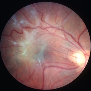
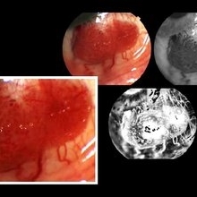 Papilloma Conjunctiva
Sep 2 2016 by JEFFERSON R SOUSA, Tecg.º (Biomedical Systems Technology)
Female patient, 26-years-old without complaint of ocular symptoms, only the of spot avermelha in the right eye. Absence of changes in positions of the look. Intraocular pressure visual acuity and fundoscopy without changes.
Photographer: JEFFERSON R SOUSA - Study Center and Ophthalmological Research Dr. Andre M V Gomes, Institute Dr. Suel Abujamra São Paulo-Brazil
Imaging device: Topcon TRC-50 Dx - Angulation of field photo of 35 Degrees, flash 36, Digital system Imaginet
Condition/keywords: conjunctival cysts, papilloma, papilloma conjunctiva, tumor
-
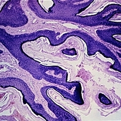 Slide 5-21
Feb 20 2019 by Lancaster Course in Ophthalmology
Hyperkeratotic squamous papilloma consisting of thin, keratin-covered fronds with dermal cores.
Condition/keywords: dermal cores, papilloma
-
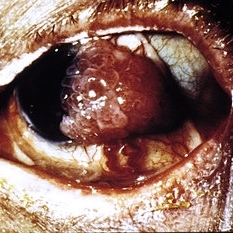 Slide 7-16
Feb 25 2019 by Lancaster Course in Ophthalmology
Papilloma of the conjunctiva.
Condition/keywords: conjunctiva, papilloma
-
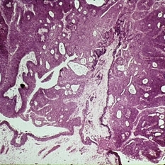 Slide 7-17
Feb 25 2019 by Lancaster Course in Ophthalmology
Papilloma of the conjunctiva showing fronds of acanthotic epithelium with central fibrovascular cores.
Condition/keywords: acanthosis, conjunctiva, papilloma
-
 Papillomacular Fold in Posterior Microphthalmos
Jul 30 2014 by Jordan L. Heffez, MD
Fundus photo of a healthy 47-year-old gentleman with BCVA 20/50 OU and no known past ocular history.
Condition/keywords: macular fold
-
 Papillomacular Fold in Posterior Microphthalmos
Jul 30 2014 by Jordan L. Heffez, MD
Fundus photo of a healthy 47-year-old gentleman with BCVA 20/50 OU and no known past ocular history.
Condition/keywords: macular fold
-
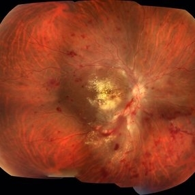 Acute Necrotizing Retinal Vasculitis as Onset of Systemic Lupus Erythematosus.
Sep 3 2016 by ADRIANO FERREIRA
A 28-year-old white man was referred to the rheumatology clinic with gradually and rapid deterioration of the vision (both eyes). In this picture, we can observe cotton wool spots in the papillomacular area and extensive hemorrhages in posterior polo and in the middle periphery. Hard exudates are present in macular area (macular edema)
Photographer: Claudio Zett Lobo
Imaging device: TRC50DXi TOPCON
Condition/keywords: systemic lupus erythematosus (SLE) vasculitis, vasculitis
-
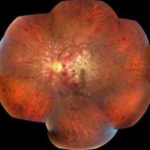 Acute Necrotizing Retinal Vasculitis as Onset of Systemic Lupus Erythematosus.
Sep 3 2016 by ADRIANO FERREIRA
A 28-year-old white man was referred to the rheumatology clinic with gradually and rapid deterioration of the vision (both eyes). In this picture we can observe cotton wool spots in the papillomacular area and extensive hemorrhages in the left eye.
Photographer: Claudio Zett Lobo
Imaging device: TRC50DXi TOPCON
Condition/keywords: systemic lupus erythematosus (SLE) vasculitis, vasculitis
-
 AMG on Papillomacular Bundle
Mar 16 2025 by PUJA NEGI
Patient came to our OPD with history of AMG done for macular hole . On examination we found that the AMG had displaced over the papillomacular bundle from the hole.
Photographer: DR Nuzhat
Condition/keywords: amniotic membrane graft, macular hole
-
 Central Retinal Artery Occlusion Secondary to Endophthalmitis
Oct 24 2022 by Kelli Nyenhuis
Ultra-widefield fluorescein angiogram of a 64 year old female who developed a Central Retinal Artery Occlusion following acute endophthalmitis. The physician commented that there is no vascular filling with the exception of the papillomacular bundle. The patient's vision was scHM at the time the image was taken.
Photographer: Kelli Nyenhuis
Imaging device: Optos California
Condition/keywords: central retinal artery occlusion (CRAO), endophthalmitis, fluorescein angiogram (FA), left eye, non-perfusion, Optos, ultra-wide field imaging
-
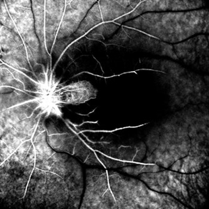
 Cilioretinal Artery Occlusion
Sep 2 2012 by Hyung-Woo Kwak, MD
Cloudiness localized to the area of papillomacular bundle normally perfused by retinal vessel.
Imaging device: Zeiss F450 plus
Condition/keywords: cilioretinal artery occlusion
-
 Cilioretinal Artery Sparing CRAO
May 1 2025 by Tejaswita Verma
Fundus photo of a middle aged male with CRAO partially sparing cilioretinal artery and papillomacular bundle. Vision 6/60.
Photographer: Dr. Tejaswita Verma
Imaging device: MIRANTE
Condition/keywords: CRAO with cilioretinal sparing
-
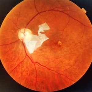
 Combined Hamartoma
Oct 5 2016 by Guruprasad S. Ayachit, MBBS,MS
Fundus photograph of a 9-year-old boy with an ill-defined lesion extending from nasal to the disc going on to include the papillomacular bundle; 14X10 mm in greatest dimensions. There is a thick epiretinal membrane causing distortion and straightening of temporal vascular arcade.
Photographer: Shravan Masurkar, M M Joshi Eye Institute, Hubli
Imaging device: Topcon TRC50DX
Condition/keywords: combined hamartoma
-
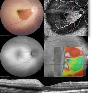 CRAO with cilioretinal sparing - Multimodal imaging
Jun 28 2023 by Maneesh M Bapaye, MD, MBA
A 34 year old male patient presented with sudden onset vision loss of 1 week duration. Visual acuity at presentation was 20/200. Fundus examination revealed diffuse retinal whitening with sparing of papillomacular bundle and fovea due to patent cilioretinal artery. Autofluorescence shows peripheral hypoAF, patent capillaries can be seen only in area of cilioretinal supply, OCT shows thickening of inner retinal layers temporal to fovea Systemic examination revealed that patient had valvular heart disease with multiple valves involved.
Photographer: Maneesh Bapaye
Condition/keywords: cilioretinal sparing, CRAO, multimodal imaging
-
 Full-thickness Macular Hole
Aug 28 2012 by Sharon Fekrat, MD FACS FASRS
65 year old woman with a recurrent full thickness macular hole following previous 20 g pars plana vitrectomy in the right eye as well as an iatrogenic retinal hole in the papillomacular bundle. Both retinal defects are captured here in this Zeiss Stratus OCT image.
Photographer: Michael P. Kelly, FOPS Director, Duke Eye Labs, Duke University Eye Center, Durham, NC
Imaging device: Zeiss Cirrus
Condition/keywords: retinal break
-
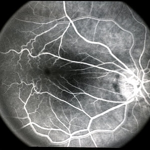 Macular Pucker
Mar 29 2013 by Henry J. Kaplan, MD
Angiogram of a macular pucker better demonstrates vessel straightening in papillomacular bundleand tortousity in the area of membrane.
Condition/keywords: epiretinal membrane (ERM), macular pucker
-
 Macular Pucker
Mar 29 2013 by Henry J. Kaplan, MD
Large epiretinal membrane with straightened vessels in the papillomacular bundle and distorsion of retinal vessels.
Condition/keywords: epiretinal membrane (ERM), macular pucker
-
 OCT of Papillomacular Fold in Posterior Microphthalmos
Jul 30 2014 by Jordan L. Heffez, MD
OCT from a healthy 47-year-old gentleman with BCVA 20/50 OU and no known past ocular history. Note the lack of epiretinal tissue as a cause of retina malformation.
Condition/keywords: macular fold
-
 Posterior Microphthalmos
Nov 16 2015 by Mallika Goyal, MD
Bilateral posterior microphthalmos, high hypermetropia, crowded disc, papillomacular fold, retinitis punctata albescens. B-scan revealed sclera-choroidal thickening.
Photographer: Mallika Goyal, MD
Condition/keywords: posterior microphthalmos
-
 Posterior Microphthalmos
Nov 22 2015 by Mallika Goyal, MD
Bilateral posterior microphthalmos, high hypermetropia, crowded disc, papillomacular fold, retinitis punctata albescens (white retinal flecks over the retinal midperiphery and periphery) in a 22-year-old male patient. B-scan revealed sclera-choroidal thickening. Best corrected VA was 20/80 each eye.
Photographer: Mallika Goyal, MD, Apollo Health City, Jubilee Hills, Hyderabad, India
Condition/keywords: posterior microphthalmos
 Loading…