 Initializing download.
-
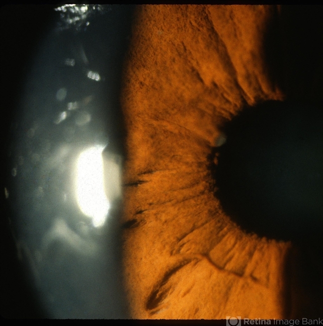
 By Ronald C. Gentile, MD
The New York Eye and Ear Infirmary of Mount Sinai - Uploaded on Oct 22, 2012.
- Last modified by Chayal Patel on Nov 20, 2012.
- Reviewed by Chayal Patel
- Rating
- Appears in
- Sarcoidosis Panuveitis
- Condition/keywords
- sarcoidosis panuveitis
- Photographer
- The New York Eye & Ear Infirmary Department of Medical Imaging
- Imaging device
- Photo slit lamp biomicroscope
- Description
- Anterior segment photo of the iris and pupillary margin shows a Koeppe nodule at the 9:30 position. Koeppe nodules consist of inflammatory cell precipitates.


