Search results (883 results)
-
---thumb.JPG/image-square;max$300,300.ImageHandler) Weiss Ring (Floater)
Weiss Ring (Floater)
Jul 10 2013 by Jason S. Calhoun
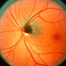
Patient comes in complaining of a floater towards the nasal aspect of her vision. Fundus photograph with anterior shot, shows a weiss ring pulled off from the optic nerve.
Photographer: Jason S. Calhoun, Department of Ophthalmology, Mayo Clinic Jacksonville, Florida
Condition/keywords: floaters, Weiss ring
-
 Toxoplasma Neuroretinitis (Jensen`s Disease)
Feb 25 2013 by Henry J. Kaplan, MD
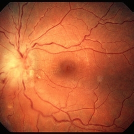
Toxoplasma neuroretinitis in the left eye of a patient with macular star formation, retinitis adjacent to the optic nerve head with disc swelling.
Condition/keywords: Jensen disease, ocular toxoplasmosis, toxoplasmosis
-
---thumb.JPG/image-square;max$300,300.ImageHandler) Weiss Ring (Floater)
Weiss Ring (Floater)
Jul 10 2013 by Jason S. Calhoun
Patient comes in complaining of a floater towards the nasal aspect of her vision. Fundus photograph with anterior shot, shows a weiss ring pulled off from the optic nerve.
Photographer: Jason S. Calhoun, Department of Ophthalmology, Mayo Clinic Jacksonville, Florida
Condition/keywords: floaters, Weiss ring
-
---thumb.jpg/image-square;max$300,300.ImageHandler) Myelinated Nerve Fiber Layer
Myelinated Nerve Fiber Layer
Feb 20 2013 by From the Collections of Thomas M. Aaberg, MD and Thomas M. Aaberg Jr., MD
Myelinated nerve fiber layer Optic nerve Fundus Photo
Condition/keywords: myelinated nerve fibers
-
 Ocular Toxocariasis slide 1
Oct 22 2012 by Ronald C. Gentile, MD
40-year-old man from South America was referred for a peripheral retinal scar in his left eye. He had a history of conjunctivitis as a child with exposure to multiple pets (cats and dogs). Fundus photo revealed a peripheral scarred sub-retinal granuloma located superior nasal with a retinal fold and traction extending to the optic nerve.
Photographer: The New York Eye & Ear Infirmary Department of Medical Imaging
Condition/keywords: toxocariasis
-
 Coloboma, In Stereo
Oct 1 2012 by Michael P. Kelly, FOPS
This is a stereo retinal fundus photograph of a coloboma, with the optic nerve centered, using a Zeiss FF3C retinal fundus camera.
Photographer: Michael P. Kelly, FOPS Director, Duke Eye Labs, Duke University Hospital, Duke Eye Center, Durham, NC
Condition/keywords: coloboma, fundus photograph, stereo pair
-
 Optic Disc Drusen
Jul 31 2016 by Mitzy E Torres Soriano, MD
Optic Disc Drusen (Right eye)
Photographer: Mitzy E. Torres Soriano. Retina Department. Hospital Provincial del Centenario. Rosario, Argentina
Imaging device: TOPCON
Condition/keywords: optic disc drusen, optic nerve drusen
-
---thumb.jpg/image-square;max$300,300.ImageHandler) CMV Retinitis in a Patient with the Diagnosis of AIDS
CMV Retinitis in a Patient with the Diagnosis of AIDS
Feb 27 2013 by Henry J. Kaplan, MD
Color fundus photograph, right eye: CMV neuroretinitis (retinitis which begins from the arcades and is accompanied by hemorrhage and also has involved the optic nerve).
Condition/keywords: AIDS
-
 Traumatic Macular Hole with Retinal Detachment and PVR
Sep 27 2012 by Pauline T Merrill, MD, FASRS
Fundus photo of a 13-year-old boy s/p soccer ball injury 1 month previously. In addition to full-thickness macular hole and total retinal detachment with grade C PVR, note pigment granules visible in vitreous over optic nerve.
Photographer: Karen Parque, Illinois Retina Associates, Chicago, IL
Condition/keywords: proliferative vitreoretinopathy (PVR), traumatic macular hole
-
 Optic Nerve Malformation/Coloboma/ Bergmeister's Papilla
Feb 19 2013 by From the Collections of Thomas M. Aaberg, MD and Thomas M. Aaberg Jr., MD
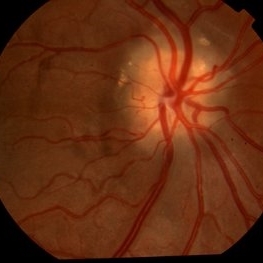
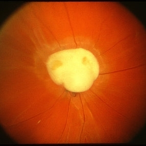
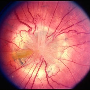
Morning glory appearance; no history.
Condition/keywords: Bergmeister's Papillae, coloboma
-
 Superior Crescent Optic Nerve
Feb 19 2013 by From the Collections of Thomas M. Aaberg, MD and Thomas M. Aaberg Jr., MD
Associated with Bergmeister's Papillae.
Condition/keywords: Bergmeister's Papillae
-
 Optic Nerve Head Drusen With Idiopathic CNV
Feb 17 2017 by Kristen Wagner
22-year-old female fundus photograph of a right eye with Optic Nerve Drusen with Idiopathic CNV.
Photographer: Kristen Wagner, COT, OSC Ophthalmic Photographer, Tennessee Retina, Nashville TN
Condition/keywords: choroidal neovascularization (CNV), drusen of optic disc, optic disc drusen
-
 Optic Nerve Coloboma With 2 Pits, Nasal and Temporal Color
Nov 21 2013 by Alexandre Durao Alves Pereira, MD
Fundus photograph, color, red free, blue lite and FAF of a optic nerve coloboma with 2 pits, one nasal and other temporal.
Photographer: Alexandre Pereira
Imaging device: Visucam 300
Condition/keywords: color photo, optic nerve coloboma
-
 Gyrate Atrophy of Choroid and Retina
Apr 19 2014 by Mallika Goyal, MD
Right eye fundus of a 45-year-old male patient with advanced gyrate atrophy of the choroid and retina with macular sparing. Optic nerve head is healthy.
Photographer: Mallika Goyal, MD, Apollo Health City, Hyderabad, India
Condition/keywords: choroid, gyrate atrophy
-
 Toxocara Granuloma
Feb 25 2013 by Henry J. Kaplan, MD
Toxocara granuloma of the optic nerve head.
Condition/keywords: ocular toxoplasmosis, toxocara granuloma, toxocariasis
-
 Optic Disc Coloboma
Apr 25 2017 by Nimrod Dar
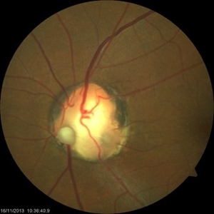
9 year-old patient, noticed a gradual deterioration in her visual acuity at her LE (6/15). On her examination, a double optic disc can be seen. OCT scan revealed an intra retinal fluid and macular schisis.
Photographer: Nimrod Dr, MD
Condition/keywords: coloboma of the optic nerve
-
 Superior Peripapillary Hemorrhage
Jul 13 2013 by Jason S. Calhoun
Patient was seen for acute vision loss in the right eye. Patient has glaucoma. VA was 20/70 in the right eye. Had vitrectomy back in May 2012 for ERM stripping. Also had trabectome with cataract surgery in December of 2012. Fundus photos presents a superior peripapillary Hemorrhage of the optic nerve. Patient will be followed up in one month.
Photographer: Jason S. Calhoun, Department of Ophthalmology, Mayo Clinic Jacksonville, Florida
Imaging device: TOPCON TRC 50-EX
Condition/keywords: peripapillary hemorrhage
-
 Melanocytoma
May 2 2013 by Henry J. Kaplan, MD
Melanocytoma of the optic nerve head.
Condition/keywords: melanocytoma
-
---thumb.jpg/image-square;max$300,300.ImageHandler) Optic nerve melanocytoma 2
Optic nerve melanocytoma 2
Jan 11 2013 by Alex P. Hunyor, MD
Optic nerve melanocytoma, left eye
Condition/keywords: optic disc melanocytoma
-
---thumb.JPG/image-square;max$300,300.ImageHandler) Myelinated Nerve Fiber Layer
Myelinated Nerve Fiber Layer
Jul 12 2013 by Jason S. Calhoun
Myelinated nerve fiber layer surrounding the nasal aspect of the optic nerve in the left eye.
Photographer: Jason S. Calhoun, Department of Ophthalmology, Mayo Clinic Jacksonville, Florida
Condition/keywords: myelinated nerve fibers
-
 Papilledema
Sep 8 2012 by Hamid Ahmadieh, MD
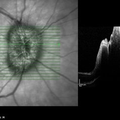
OCT of the optic nerve head of the right eye of a 55-year-old woman with a malignant intracranial tumor.
Photographer: Hamid Ahmadieh, MD, Ophthalmic Research Center, Labbafinejad Medical Center, Shahid Beheshti University of Medical Sciences
Imaging device: Heidelberg Spectralis
Condition/keywords: malignant intracranial tumor, optical coherence tomography (OCT), papilledema
-
 Morning Glory Optic Nerve; Coloboma
Feb 20 2013 by From the Collections of Thomas M. Aaberg, MD and Thomas M. Aaberg Jr., MD
No history.
Condition/keywords: Morning Glory Syndrome, optic nerve coloboma
-
---thumb.JPG/image-square;max$300,300.ImageHandler) SLE retinopathy
SLE retinopathy
Nov 18 2013 by Mallika Goyal, MD
Occlusive retinitis in a lady with SLE; optic nerve head pallor suggestive of prior optic neuritis or ischaemic optic neuropathy.
Photographer: Mallika Goyal, MD, Apollo Health City, Hyderabad
Condition/keywords: systemic lupus erythematosus (SLE) retinopathy
-
 Papilledema
Sep 8 2012 by Hamid Ahmadieh, MD
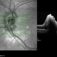
OCT of the optic nerve head of the left eye of a 55-year-old woman with a malignant intracranial tumor.
Photographer: Hamid Ahmadieh, MD, Ophthalmic Research Center, Labbafinejad Medical Center, Shahid Beheshti University of Medical Sciences
Imaging device: Heidelberg Spectralis
Condition/keywords: malignant intracranial tumor, optical coherence tomography (OCT), papilledema
-
 Probable Optic Nerve Coloboma
Feb 19 2013 by From the Collections of Thomas M. Aaberg, MD and Thomas M. Aaberg Jr., MD
No history.
Condition/keywords: color photo, optic nerve coloboma
 Loading…