Search results (48 results)
-
 APMPPE With Serous Macular Detachment
Jun 2 2014 by Rameez N Hussain, MD
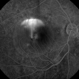
Acute posterior multifocal placoid pigment epitheliopathy (APMPPE) with serous macular detachment.
Photographer: Rameez N Hussain MD, Vitreo Retinal Services, Giridhar Eye Institute, Cochin, India
Imaging device: Zeiss FF4
Condition/keywords: acute posterior multifocal placoid pigment epitheliopathy (APMPPE), serous retinal detachment
-
 APMPPE With Serous Macular Detachment SD-OCT
Jun 2 2014 by Rameez N Hussain, MD
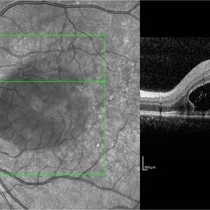
SD OCT image of acute posterior multifocal placoid pigment epitheliopathy (APMPPE) with serous macular detachment.
Photographer: Rameez N Hussain MD, Vitreo Retinal Services, Giridhar Eye Institute, Cochin, India
Imaging device: Heidelberg Spectralis
Condition/keywords: acute posterior multifocal placoid pigment epitheliopathy (APMPPE), serous retinal detachment
-
 "Hang in There"
Apr 20 2021 by Tomas Minelli, MD
Fundus wide field photograph of a 50-year-old man with a macular detachment associated with a big temporal superior tear. The laser is firmly holding the progression of the tear in the 14th day post- laser. BCVA 20/20
Photographer: Livia Conci, Universtity of São Paulo
Imaging device: Optos Daytona
Condition/keywords: giant retinal tear
-
 Pit Macular Syndrome
Mar 21 2013 by Yusuke Oshima, MD, PhD
Fundus photograph of a 38-year-old man with macular detachment associated with an optic disc pit.
Photographer: Yusuke Takada, Osaka University Graduate School of Medicine
Condition/keywords: congenital optic nerve pit
-
 APMPPE With Serous Macular Detachment 3D SD-OCT
Jun 2 2014 by Rameez N Hussain, MD
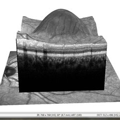
3D SD-OCT of acute posterior multifocal placoid pigment epitheliopathy (APMPPE) with serous macular detachment.
Photographer: Rameez N Hussain MD, Vitreo Retinal Services, Giridhar Eye Institute, Cochin, India
Imaging device: Heidelberg Spectralis
Condition/keywords: acute posterior multifocal placoid pigment epitheliopathy (APMPPE), serous retinal detachment
-
 CSCR Mushroom Cloud
Feb 23 2015 by James J. Bedrick, MD
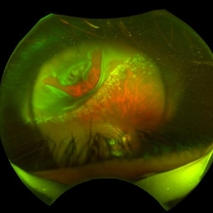
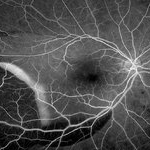
Late transit FA of a large active sub-foveal CSCR leak. You may view this pair in stereo to appreciate the plume of leakage within this large serous RD of the macula. This patient presented with a BCVA of 20/200 and fluorescein and historic evidence of prior episodes of leakage. After discussion of known treatment options including observation, he elected to be treated initially with oral rifampin and BCVA improved to 20/40 with persistent metamorphosis and a shallower persistent macular detachment over several visits. Rifampin was discontinued and he then received sub-threshold micro-pulse laser photocoagulation with an 810 diode which resulted in the patient reporting full restoration of his vision subjectively within a month. He failed to keep his follow-up appointment.
Photographer: Diana Bodnar, COT
Imaging device: Topcon 50X with Merge capture station
Condition/keywords: CSCR subfoveal leak
-
 Myopic Traction Maculopathy
May 31 2014 by Rameez N Hussain, MD
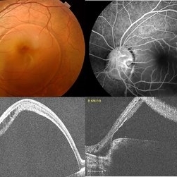
Spectral domain optical coherence tomography of macular detachment in posterior staphyloma - myopic traction maculopathy (MTM).
Photographer: Rameez N Hussain MD, Vitreo Retinal Services, Giridhar Eye Institute, Cochin, India
Imaging device: Heidelberg Spectralis
Condition/keywords: high myopia, macular detachment, myopic traction maculopathy, pathologic myopia, posterior staphyloma
-
 Exudative Macular Detachment After Intensive Laser Photocoagulation
Mar 12 2016 by Sjakon G Tahija, MD
Fundus photograph of 44-year-old man with exudative detachment of the macula after vitrectomy and ILM peeling for proliferative diabetic retinopathy combined with intensive endolaser photocagulation.
Photographer: Avris Siahaan, Klinik Mata Nusantara
Condition/keywords: exudative detachment, pan-retinal photocoagulation (PRP)
-
 Myopic Traction Maculopathy
May 31 2014 by Rameez N Hussain, MD
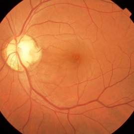
Color photograph of macular detachment in a posterior staphyloma - myopic traction maculopathy (MTM).
Photographer: Rameez N Hussain MD, Vitreo Retinal Services, Giridhar Eye Institute, Cochin, India
Imaging device: Zeiss
Condition/keywords: high myopia, macular detachment, myopic traction maculopathy, pathologic myopia, posterior staphyloma
-
 Choroidal Rupture, Subretinal and Vitreous Hemorrhage Secondary to Blunt Trauma
Dec 29 2012 by Humberto Ruiz-Garcia, MD
SD-OCT obtained at 72 hours which shows neurosensory macular detachment and severe thinning (impending macular hole).
Photographer: Humberto Ruiz-Garcia
Imaging device: Cirrus HD OCT
Condition/keywords: traumatic macular hole
-
 Macular Detachment from Multiple Myeloma
Mar 10 2013 by Edwin H. Ryan, MD
84-year-old white male with blurred vision over 2 months. Has incidental BVO.
Imaging device: stratus
Condition/keywords: macular detachment, myeloma
-
 Macular Detachment from Multiple Myeloma
Mar 10 2013 by Edwin H. Ryan, MD
84-year-old white male with blurred vision over 2 months. Has incidental BVO.
Condition/keywords: macular detachment, myeloma
-
 Macular Detachment from Multiple Myeloma
Mar 10 2013 by Edwin H. Ryan, MD
84 year-old white male with blurred vision over 2 months. Has incidental BVO.
Imaging device: stratus
Condition/keywords: macular detachment, myeloma
-
 Optic Nerve Pit
Feb 19 2013 by From the Collections of Thomas M. Aaberg, MD and Thomas M. Aaberg Jr., MD
Optic nerve pit with serous macular detachment.
Condition/keywords: macular detachment, optic nerve pit
-
 Posterior Scleritis
Dec 8 2013 by Mallika Goyal, MD
Resolving fluid at macula 1 week after pulsed intravenous steroids for posterior scleritis with serous macular detachment in a young lady.
Photographer: Mallika Goyal, MD, Apollo Health City, Hyderabad, India
Condition/keywords: posterior scleritis
-
 Macular Detachment from Multiple Myeloma
Mar 10 2013 by Edwin H. Ryan, MD
84 year-old white male with blurred vision over 2 months. Has incidental BVO.
Imaging device: stratus
Condition/keywords: macular detachment, myeloma
-
 Posterior Scleritis
Dec 8 2013 by Mallika Goyal, MD
Rapid onset serous macular detachment in a young lady with scleritis. Treated with pulsed intravenous steroids with resolution.
Photographer: Mallika Goyal, MD, Apollo Health City, Hyderabad, India
Condition/keywords: posterior scleritis
-
 Macular Detachment from Multiple Myeloma
Mar 10 2013 by Edwin H. Ryan, MD
84-year-old male with blurred vision over 2 months. Has incidental BVO.
Condition/keywords: macular detachment, myeloma
-
 Macular Detachment from Multiple Myeloma
Mar 10 2013 by Edwin H. Ryan, MD
84 year-old white male with blurred vision over 2 months. Has incidental BVO.
Imaging device: stratus
Condition/keywords: macular detachment, myeloma
-
 Macular Detachment from Multiple Myeloma
Mar 10 2013 by Edwin H. Ryan, MD
84-year-old white male with blurred vision over 2 months. Has incidental BVO.
Condition/keywords: macular detachment, myeloma
-
 Posterior Scleritis
Dec 8 2013 by Mallika Goyal, MD
Flat macula 3 weeks after pulsed intravenous steroids for posterior scleritis with serous macular detachment in a young lady.
Photographer: Mallika Goyal, MD, Apollo Health City, Hyderabad, India
Condition/keywords: posterior scleritis
-
 Macular Detachment from Multiple Myeloma
Mar 10 2013 by Edwin H. Ryan, MD
84 year-old white male with blurred vision over 2 months. Has incidental BVO.
Condition/keywords: macular detachment, myeloma
-
 Optic Disc Pit
Nov 8 2021 by Michael Grinton
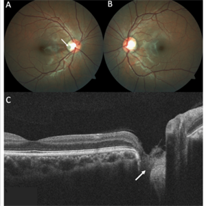
Optic disc pits are rare congenital abnormalities of the optic nerve head. Colour fundus image of an asymptomatic 18-year old male shows an optic disc pit in the right eye (A, white arrow); a small, grey, oval shaped excavation in the temporal segment of the optic disc. These pits are usually unilateral (B shows normal colour fundus of left eye) and asymptomatic. Imaging with optical coherence tomography (C) shows the optic disc pit in cross section (white arrow) and normal macular structure. In some patients with the condition, fluid can accumulate underneath the macular (serous macular detachment).
Condition/keywords: Optic disc pit, Optic nerve pit, Optic pit
-
 FA 40 Seconds - Large Hemorrhage With Macular Detachment Due to AMD
Nov 7 2019 by John S. King, MD
81-year-old white female with three day history of seeing a "dark blob" nasally OD; no blood thinners; vision was 20/100- J16 with 2+NSC OD; OCT (not shown) had large SRF that included the fovea and extended out temporally. Posterior segment showed a large amount of SRF in the macula with some SRH in the inferior portion of the macula, hemorrhagic PEDs temporally with some RPE scarring and SRH in the periphery. On the FA there is blockage by the SRH and SRPE heme; there is staining peripherally; there is a wave of leakage that extends out into the macula and pools into to subretinal space.
Photographer: Brandon Peter
Condition/keywords: retinal pigment epithelium, subretinal hemorrhage, wet age-related macular degeneration (wet AMD)
-
 Optic Disc Pit with Coloboma (Hybrid Anomaly)
Jun 10 2021 by Janani Sreenivasan
Optic disc pit is a rare anomaly of the optic nerve head that can be associated with maculopathy leading to progressive visual deterioration. It belongs to the spectrum of congenital cavitary anomalies of the optic disc which encompasses extrapapillary cavitation, optic disc coloboma, and morning glory. Very rarely, optic disc pits are seen in combination with optic disc colobomas. Histopathologically, disc pit is defined as herniation of dysplastic retinal tissue into an excavation, rich in collagen, which can stretch into the subarachnoid space via a defect in the lamina cribrosa. Interestingly, this structural abnormality leading to a non-physiological communication between the intraocular and extraocular spaces is a common feature among all the congenital cavitary disc anomalies. Optic disc pit maculopathy is characterized by intraretinal and subretinal fluid at the area of macula. The origin of the retinal fluid remains unclear. Possible sources include the vitreous cavity, the subarachnoid space and the orbital space surrounding the dura. It has been estimated that approximately 25% to 75% of patients will develop serous detachment and/or retinoschisis of the central macula at some stage of their life. On fundus examination, ODPs typically appear as single grayish, round or oval depressions at the optic disc. Most commonly, they are detected at the inferotemporal segment of the disc, but may also be observed elsewhere, including the central area.The coexisting macular detachment can be related to lamellar or full-thickness macular holes, cystoid changes, retinal pigment epithelium atrophy and eventually to irreversible loss of vision,especially in longstanding cases. Herewith, we present a 32-years-old male patient presenting with an unusual combination of optic disc pit with maculopathy and optic disc coloboma (hybrid anomaly) in the same eye with corrected visual acuity of 3/60.
Photographer: Dr Janani Sreenivasan
Imaging device: Zeiss Cirrus HD-OCT
Condition/keywords: coloboma of optic disc, hybrid anomaly, macular detachment, optic disc, optic disc pit
 Loading…