Search results (30 results)
-
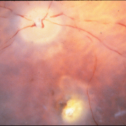
 Orange Pigment Overlying a Lesion Suspicious for a Choroidal Melanoma
Jan 16 2019 by John S. King, MD
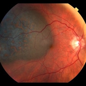
76-year-old white male saw his eye doctor with a three week complaint of photopsias and a shadow in his vision. Found to have a 10.5/12.5/2.5 (medium reflectivity) pigmented, choroidal mass associated with SRF and orange pigment (hyper-autofluorescence of lipofuscin), and without drusen or halo. See photo
Photographer: Stacey Coleman
Imaging device: Topcon 50
Condition/keywords: lipofuscin, orange pigment
-
 Juxtapapillary Choroidal Melanoma With Lipofuscin
Oct 25 2015 by Dwain G. Fuller, MD, JD
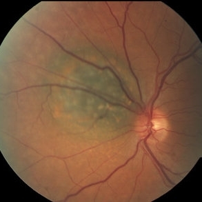
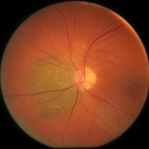
Fundus photograph of a juxtapapillary choroidal melanoma with lipofuscin.
Condition/keywords: lipofuscin
-
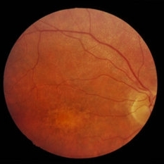
 RP Variant
Dec 22 2014 by H. Michael Lambert, MD
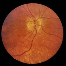
Fundus photo with lipofuscin accumulation in the central macula.
Condition/keywords: RP variant
-
 RP Variant
Dec 22 2014 by H. Michael Lambert, MD
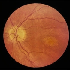
Fundus photo with lipofuscin accumulation in the central macula.
Condition/keywords: RP variant
-
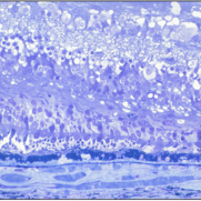
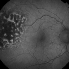 Hyper-autofluorescence of Orange Pigment Overlying a Lesion Suspicious for a Choroidal Melanoma
Jan 16 2019 by John S. King, MD
76-year-old white male saw his eye doctor with a three week complaint of photopsias and a shadow in his vision. Found to have a 10.5/12.5/2.5 (medium reflectivity) pigmented, choroidal mass associated with SRF and orange pigment (hyper-autofluorescence of lipofuscin, see image), and without drusen or halo.
Photographer: Stacey Coleman
Imaging device: Topcon 50
Condition/keywords: lipofuscin, orange pigment
-
 RP Variant
Dec 22 2014 by H. Michael Lambert, MD
Fundus photo with lipofuscin accumulation in the central macula.
Condition/keywords: RP variant
-
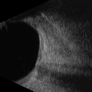
 Large, Dome-Shaped Peripheral Choroidal Melanoma - Widefield Color
Feb 13 2020 by Michael Seider, MD
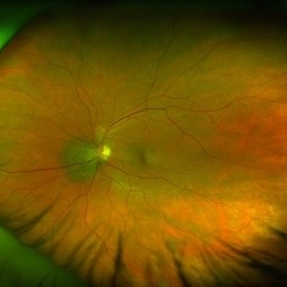
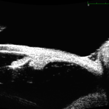
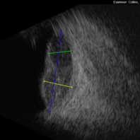
Large, dome-shaped peripheral choroidal melanoma of the left eye with inferior exudative retinal detachment. Note the lack of obvious orange pigment over the tumor and apparent drusen anteriorly. A lack of ophthalmoscopically obvious lipofuscin is not uncommon among larger choroidal melanomas. B-Scan ultrasonography (transverse, 10 o’clock) confirms a low-moderate internally reflective dome-shaped choroidal lesion with a small adjacent retinal detachment. Ultrasound biomicroscopy (radial, 10 o’clock) confirms no ciliary body involvement of the tumor.
-
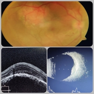 Circumscribed Choroidal Hemangioma
Jul 3 2020 by Dhaivat Shah
A 30-year-old young male presented with drop in vision in right eye since 1 year (6/60). Fundus examination revealed choroidal hemangioma superotemporal to macula. Choroidal hemangioma is an unusual benign vascular tumor of the choroid. It can be circumscribed solitary or diffuse tumor with the later having other systemic associations. Circumscribed choroidal hemangiomas (CCHs) are usually unilateral, unifocal hamartomatous vascular tumor affecting people in second to fourth decade. It appers as round to oval, orangish-red mass in posterior pole with smooth homogenous surface mostly present in macular and peripapillary area. Hyperopic shift is seen in sub-foveal tumors in contrast to para-foveal ones which are usually asymptomatic or present with metamorphopsia or photopsia and diminished vision secondary to exudative retinal detachment. B-scan shows highly reflective tumor without any shadowing or acoustic solidity with high anterior A scan spike. EDI-OCT here depicts a smooth gently sloping choridal mass with compressed choriocapillaries and enlarged medium and large choroidal vessels. Over a period of time structural abnormalities of the outer retina can be visualised. Ancillary testing using Fluorescein Angiography shows lacy hyper-fluorescence during early arterial phase followed by increased hyper-fluorescence due to progressive profuse leakage from pin point foci during arterial and venous phase. Indocyanine green angiography shows lacy diffuse fluorescent tumor in early phase followed by hypo-fluorescent tumor due to dye wash out in late phase. Intrinsic auto-fluorescence is also seen in CCHs from lipofuscin and fresh sub-retinal fluid. Tumor is relatively hyper-intense with respect to vitreous in T1-weighted images in iso-intense in T2-weighted images of MRI. Asymptomatic cases need no treatment, while patients showing vision loss with presence or absence of exudative retinal detachment can be treated with photodynamic therapy which is preferred treatment due to site specific action. Selective occlusion of choroidal neovascularization can be achieved while the neurosensory retinal layers and Bruch membrane are almost unaffected, leaving retinal function intact. Green or rarely red wavelength laser photocoagulation is used to create a chorioretinal adhesion and resolve the SRF. Other treatment modalities include Transpupilary thermotherapy, external beam irradiation, proton beam therapy, brachytherapy and gamma knife.
Photographer: Miss Deepika Nagle
Imaging device: Zeiss
Condition/keywords: B scan ultrasound, choroidal hemangioma, fundus photograph, optical coherence tomography (OCT), photodynamic therapy
-
 Large, Irregularly Shaped Choroidal Melanoma - B Scan (Transverse)
Feb 13 2020 by Michael Seider, MD
Large, irregularly shaped choroidal melanoma with overlying subretinal fluid and inferior exudative retinal detachment in the right eye of a 93-year-old woman. Note the extensive overlying orange pigment (lipofuscin) which is hyper-autofluorescent. B-Scan ultrasonography confirms low tumor internal reflectivity, adjacent retinal detachment and multi-lobulated shape. Especially because of the poor baseline vision and the severe vision loss expected from radiotherapy (because of the larger tumor size and proximity to the optic nerve), this older woman elected primary enucleation.
-
 Small, Peripapillary Choroidal Melanoma of the Left Eye - OCT
Feb 13 2020 by Michael Seider, MD
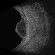
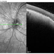
Small, peripapillary choroidal melanoma of the left eye. Note the diffuse borders, clumped overlying orange pigment (lipofuscin) and lack of drusen. The standard fundus photograph reveals the true color of the lesion. The wide-field Optos photograph shows the lesion as being more green than the true color, although the lipofuscin remains prominently orange. Wide-field fundus autofluoresence shows a bright signal corresponding to the orange pigment and also superior and inferior to the optic nerve, likely from previous exposure to subretinal fluid. Optical coherence tomography confirms the subretinal fluid seen on examination. B-Scan ultrasonography shows a very low-lying choroidal lesion adjacent to the optic nerve.
-
 Large, Dome-Shaped Peripheral Choroidal Melanoma - B Scan
Feb 13 2020 by Michael Seider, MD
Large, dome-shaped peripheral choroidal melanoma of the left eye with inferior exudative retinal detachment. Note the lack of obvious orange pigment over the tumor and apparent drusen anteriorly. A lack of ophthalmoscopically obvious lipofuscin is not uncommon among larger choroidal melanomas. B-Scan ultrasonography (transverse, 10 o’clock) confirms a low-moderate internally reflective dome-shaped choroidal lesion with a small adjacent retinal detachment. Ultrasound biomicroscopy (radial, 10 o’clock) confirms no ciliary body involvement of the tumor.
-
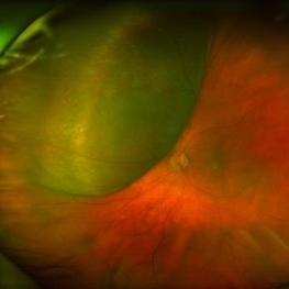
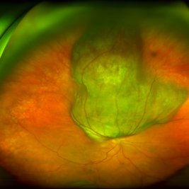
 Small, Peripapillary Choroidal Melanoma of the Left Eye - Widefield Color
Feb 13 2020 by Michael Seider, MD
Small, peripapillary choroidal melanoma of the left eye. Note the diffuse borders, clumped overlying orange pigment (lipofuscin) and lack of drusen. The standard fundus photograph reveals the true color of the lesion. The wide-field Optos photograph shows the lesion as being more green than the true color, although the lipofuscin remains prominently orange. Wide-field fundus autofluoresence shows a bright signal corresponding to the orange pigment and also superior and inferior to the optic nerve, likely from previous exposure to subretinal fluid. Optical coherence tomography confirms the subretinal fluid seen on examination. B-Scan ultrasonography shows a very low-lying choroidal lesion adjacent to the optic nerve.
-
 Autosomal Recessive Bestrophinopathy
Apr 7 2022 by Nassim Alejandro Abreu Arbaje, MD
Fundus autofluorescence photo of a 22 year old boy with macular hypoautofluorescence due to the pigmentary changes in the posterior pole and some hyperautofluorescence due to the pooling of lipofuscin
Photographer: Nassim Abreu
Imaging device: Topcon Triton Plus
Condition/keywords: Autosomal recessive bestrophinopathy
-
 Small, Peripapillary Choroidal Melanoma of the Left Eye - Standard Color
Feb 13 2020 by Michael Seider, MD
Small, peripapillary choroidal melanoma of the left eye. Note the diffuse borders, clumped overlying orange pigment (lipofuscin) and lack of drusen. The standard fundus photograph reveals the true color of the lesion. The wide-field Optos photograph shows the lesion as being more green than the true color, although the lipofuscin remains prominently orange. Wide-field fundus autofluoresence shows a bright signal corresponding to the orange pigment and also superior and inferior to the optic nerve, likely from previous exposure to subretinal fluid. Optical coherence tomography confirms the subretinal fluid seen on examination. B-Scan ultrasonography shows a very low-lying choroidal lesion adjacent to the optic nerve.
-
 Best Vitelliform Macular Dystrophy
Dec 10 2020 by McGill University Health Centre
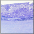
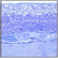
Postmortem eyes from 101-year-old female. Past clinical history includes a poor vision for many years due to macular degeneration. The last Visual acuity test recorded 6/15 OD and 6/6 OS. IOP 14 and 18 torr OS. Histopathology: Disclosed and yellow 2x2mm macular lesion. Microscopic examination: elevated placoid macular lesion with overlying pigment granules. Electron microscopy examination: pigment granules with abundant lipofuscin and melanolysosomes, photoreceptor cells markedly attenuated (less degenerated at the periphery) Numerous calcified drusen throughout the retina particularly in the posterior pole. RPE lipofuscin content is elevated in Best’s dystrophy. The extractability of the PRE lipofuscin fluorophores is reduced (it is normal during senescence). The defect in Best’s dystrophy accelerates this age related change in lipofuscin.
Condition/keywords: Best vitelliform macular dystrophy (BVMD), histopathology, pathology
-
 Large, Dome-Shaped Peripheral Choroidal Melanoma - UBM
Feb 13 2020 by Michael Seider, MD
Large, dome-shaped peripheral choroidal melanoma of the left eye with inferior exudative retinal detachment. Note the lack of obvious orange pigment over the tumor and apparent drusen anteriorly. A lack of ophthalmoscopically obvious lipofuscin is not uncommon among larger choroidal melanomas. B-Scan ultrasonography (transverse, 10 o’clock) confirms a low-moderate internally reflective dome-shaped choroidal lesion with a small adjacent retinal detachment. Ultrasound biomicroscopy (radial, 10 o’clock) confirms no ciliary body involvement of the tumor.
-
 Large, Irregularly Shaped Choroidal Melanoma - B Scan (Radial)
Feb 13 2020 by Michael Seider, MD
Large, irregularly shaped choroidal melanoma with overlying subretinal fluid and inferior exudative retinal detachment in the right eye of a 93-year-old woman. Note the extensive overlying orange pigment (lipofuscin) which is hyper-autofluorescent. B-Scan ultrasonography confirms low tumor internal reflectivity, adjacent retinal detachment and multi-lobulated shape. Especially because of the poor baseline vision and the severe vision loss expected from radiotherapy (because of the larger tumor size and proximity to the optic nerve), this older woman elected primary enucleation.
-
 Best Vitelliform Macular Dystrophy
Dec 10 2020 by McGill University Health Centre
Postmortem eyes from 101-year-old female. Past clinical history includes a poor vision for many years due to macular degeneration. The last visual acuity test recorded 6/15 OD and 6/6 OS. IOP 14 and 18 torr OS. Histopathology: Disclosed and yellow 2x2mm macular lesion. Microscopic examination: elevated placoid macular lesion with overlying pigment granules. Electron microscopy examination: pigment granules with abundant lipofuscin and melanolysosomes, photoreceptor cells markedly attenuated (less degenerated at the periphery) Numerous calcified drusen throughout the retina particularly in the posterior pole. RPE lipofuscin content is elevated in Best’s dystrophy. The extractability of the PRE lipofuscin fluorophores is reduced (it is normal during senescence). The defect in Best’s dystrophy accelerates this age related change in lipofuscin.
Condition/keywords: Best vitelliform macular dystrophy (BVMD), histopathology, pathology
-
 Autosomal Recessive Bestrophinopathy
Apr 7 2022 by Nassim Alejandro Abreu Arbaje, MD
Fundus autofluorescence photo of a 22 year old boy with macular hypoautofluorescence due to the pigmentary changes inside the temporal arcades and some hyperautofluorescence due to the pooling of lipofuscin
Photographer: Nassim Abreu
Imaging device: Topcon Triton Plus
Condition/keywords: Autosomal recessive bestrophinopathy
-
 Amelanotic Choroidal Melanoma
May 18 2020 by McGill University Health Centre
The enucleation image shows a large amelanotic tumor with large areas of hemorrhage and necrosis. Note the several dilated blood vessels and an adjacent retinal detachment with lipofuscin pigment on its surface (arrow).
Condition/keywords: amelanotic melanoma, enucleation, mushroom-shaped
-
 Best Vitelliform Macular Dystrophy
Dec 10 2020 by McGill University Health Centre
Postmortem eyes from 101-year-old female. Past clinical history includes a poor vision for many years due to macular degeneration. The last visual acuity test recorded 6/15 OD and 6/6 OS. IOP 14 and 18 torr OS. Histopathology: Disclosed and yellow 2x2mm macular lesion. Microscopic examination: elevated placoid macular lesion with overlying pigment granules. Electron microscopy examination: pigment granules with abundant lipofuscin and melanolysosomes, photoreceptor cells markedly attenuated (less degenerated at the periphery) Numerous calcified drusen throughout the retina particularly in the posterior pole. RPE lipofuscin content is elevated in Best’s dystrophy. The extractability of the PRE lipofuscin fluorophores is reduced (it is normal during senescence). The defect in Best’s dystrophy accelerates this age related change in lipofuscin.
Condition/keywords: Best vitelliform macular dystrophy (BVMD), fundus photograph
-
 Small, Peripapillary Choroidal Melanoma of the Left Eye - B-Scan
Feb 13 2020 by Michael Seider, MD
Small, peripapillary choroidal melanoma of the left eye. Note the diffuse borders, clumped overlying orange pigment (lipofuscin) and lack of drusen. The standard fundus photograph reveals the true color of the lesion. The wide-field Optos photograph shows the lesion as being more green than the true color, although the lipofuscin remains prominently orange. Wide-field fundus autofluoresence shows a bright signal corresponding to the orange pigment and also superior and inferior to the optic nerve, likely from previous exposure to subretinal fluid. Optical coherence tomography confirms the subretinal fluid seen on examination. B-Scan ultrasonography shows a very low-lying choroidal lesion adjacent to the optic nerve.
-
 Large, Irregularly Shaped Choroidal Melanoma - Widefield Color
Feb 13 2020 by Michael Seider, MD
Large, irregularly shaped choroidal melanoma with overlying subretinal fluid and inferior exudative retinal detachment in the right eye of a 93-year-old woman. Note the extensive overlying orange pigment (lipofuscin) which is hyper-autofluorescent. B-Scan ultrasonography confirms low tumor internal reflectivity, adjacent retinal detachment and multi-lobulated shape. Especially because of the poor baseline vision and the severe vision loss expected from radiotherapy (because of the larger tumor size and proximity to the optic nerve), this older woman elected primary enucleation.
-
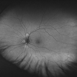 Small, Peripapillary Choroidal Melanoma of the Left Eye - Widefield FAF
Feb 13 2020 by Michael Seider, MD
Small, peripapillary choroidal melanoma of the left eye. Note the diffuse borders, clumped overlying orange pigment (lipofuscin) and lack of drusen. The standard fundus photograph reveals the true color of the lesion. The wide-field Optos photograph shows the lesion as being more green than the true color, although the lipofuscin remains prominently orange. Wide-field fundus autofluoresence shows a bright signal corresponding to the orange pigment and also superior and inferior to the optic nerve, likely from previous exposure to subretinal fluid. Optical coherence tomography confirms the subretinal fluid seen on examination. B-Scan ultrasonography shows a very low-lying choroidal lesion adjacent to the optic nerve.
-
 Best Vitelliform Macular Dystrophy
Dec 10 2020 by McGill University Health Centre
Postmortem eyes from 101-year-old female. Past clinical history includes a poor vision for many years due to macular degeneration. The last visual acuity test recorded 6/15 OD and 6/6 OS. IOP 14 and 18 torr OS. Histopathology: Disclosed and yellow 2x2mm macular lesion. Microscopic examination: elevated placoid macular lesion with overlying pigment granules. Electron microscopy examination: pigment granules with abundant lipofuscin and melanolysosomes, photoreceptor cells markedly attenuated (less degenerated at the periphery) Numerous calcified drusen throughout the retina particularly in the posterior pole. RPE lipofuscin content is elevated in Best’s dystrophy. The extractability of the PRE lipofuscin fluorophores is reduced (it is normal during senescence). The defect in Best’s dystrophy accelerates this age related change in lipofuscin.
Condition/keywords: Best vitelliform macular dystrophy (BVMD), histopathology, pathology
 Loading…