 Initializing download.
-
 By Michael Seider, MD
The Southern California Permanente Medical Group - Uploaded on Feb 13, 2020.
- Last modified by Caroline Bozell on Feb 14, 2020.
- Rating
- Appears in
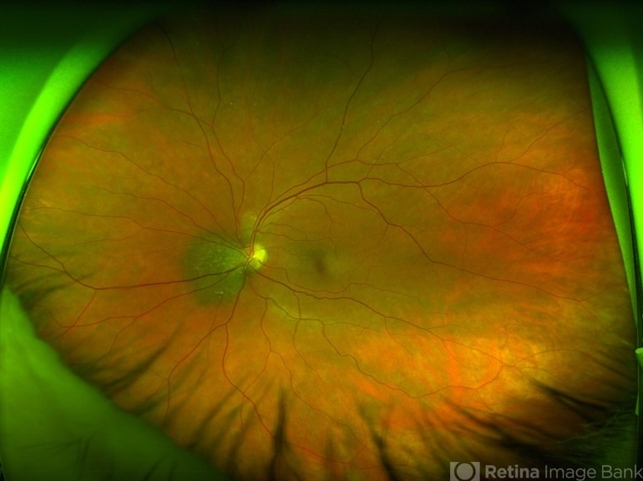
- Small peripapillary choroidal melanoma of the left eye
- Description
- Small, peripapillary choroidal melanoma of the left eye. Note the diffuse borders, clumped overlying orange pigment (lipofuscin) and lack of drusen. The standard fundus photograph reveals the true color of the lesion. The wide-field Optos photograph shows the lesion as being more green than the true color, although the lipofuscin remains prominently orange. Wide-field fundus autofluoresence shows a bright signal corresponding to the orange pigment and also superior and inferior to the optic nerve, likely from previous exposure to subretinal fluid. Optical coherence tomography confirms the subretinal fluid seen on examination. B-Scan ultrasonography shows a very low-lying choroidal lesion adjacent to the optic nerve.