Search results (153 results)
-
 Lattice Degeneration
Nov 9 2012 by Norman Byer
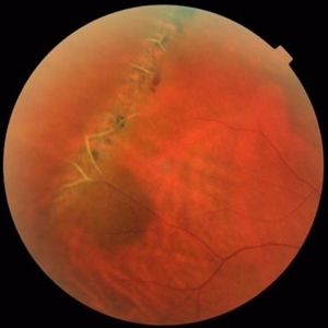
This is a more typical classical example of lattice degeneration in a 42-year-old woman in a photograph taken without scleral indentation. It shows much more marked vascular changes than the previous case. Note the tapering of the blood columns as the vessels approach the lesion and also the white sheathing of the vessel walls. Note also the continuity of the blood vessels on opposite sides of the lesion with the characteristic white lattice lines. More than 45 years ago Vogt pointed this out as a proof that these white lines were actually caused by changed blood vessels. Note also that this lesion shows a combination of several individual features of lattice degeneration. In addition to the white lines, there is a reddish crater-like area beneath the main horizontal white line. There is a prominent horizontal zone below this white line showing a snailtrack appearance. Also, there are two tiny atrophic retinal holes outside the photograph on the right end of this lesion. This eye contained five such retinal holes and they have all remained unchanged for more than 10 years of observation without treatment.
Condition/keywords: atrophic retinal hole, lattice degeneration, moderate snail track, tapering blood columns, white lattice lines, white sheath vessel
-
---thumb.jpg/image-square;max$300,300.ImageHandler) Lattice Degeneration
Lattice Degeneration
-
 Lattice Degeneration
Nov 9 2012 by Norman Byer
This 16-year-old girl has lattice degeneration and also this large oval retinal hole with a surrounding narrow zone of subretinal fluid. This lesion illustrates how large the atrophic holes of lattice degeneration may be. Occasionally the hole can be as large as the initial lattice lesion and can therefore obliterate all other evidence of its true identity. This was almost true in this case, but there does remain a small whitish remnant of the original lattice lesion at the lower end of the oval hole.
Condition/keywords: lattice degeneration, retinal hole, subretinal fluid, white lattice lines
-
 Lattice Degeneration
Nov 9 2012 by Norman Byer
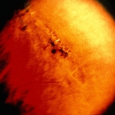
This is lattice degeneration in a 10-year-old boy showing an almost pure snailtrack feature with only a hint of a reddish crater in the center. It has not changed over 10 years. The photograph was taken with scleral indentation.
Condition/keywords: lattice degeneration, reddish crater, scleral indentation, snail track
-
 Lattice Degeneration and Choroidal Nevus
Oct 10 2015 by Hamid Ahmadieh, MD
Color fundus photograph of the right eye of a 46-year-old woman with a typical lattice degeneration and an adjacent choroidal nevus.
Photographer: Solmaz Shahmohammad, Negah Eye Center, Tehran, Iran
Condition/keywords: choroidal nevus, color fundus photograph, lattice degeneration
-
 Retinal Break at Site of Lattice Degeneration with Scleral Indentation
Nov 9 2012 by Norman Byer
This is the same case as the previous photograph. With scleral indentation slightly more posterior, the flap is seen to be associated with a large retinal tear. This is a tractional tear and it is possible that in this case the cryotherapy itself may have increased the vitreoretinal traction at this site and in this way led to this new tear. The age of the tear is unknown because it was asymptomatic, and even though the eye is aphakic the tear has not caused a clinical retinal detachment.
Condition/keywords: retinal flap, scleral indentation, tractional retinal tear, vitreoretinal traction
-
 Lattice Degeneration
Nov 9 2012 by Norman Byer
Lattice degeneration in a 42-year-old man which has produced four atrophic holes in a linear arrangement surrounded by a subclinical retinal detachment of unknown duration. By age 63, 21 years later, a posterior vitreous detachment was diagnosed in this eye, which was not present four years earlier. Nevertheless, the appearance seen here has remained exactly the same for 30 years, more than eight years with a concurrent PVD.
Condition/keywords: atrophic retinal hole, lattice degeneration, posterior vitreous detachment
-
 Lattice Degeneration
Nov 9 2012 by Norman Byer
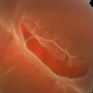
In this 54-year-old woman, lattice degeneration has led to a large horseshoe tractional tear around the posterior side on one end of the lesion resulting in a clinical retinal detachment. Note the very attenuated blood column passing through the white sheath vessel that crosses the tear. This demonstrates that the white blood vessels and a fragment of attached tissue are the only structures which have escaped the tearing effect of the strong vitreoretinal traction which occurred. This usually is true, although, in some cases this bridging vessel may bleed.
Condition/keywords: bridging vessel, lattice degeneration, tractional retinal tear, white sheath vessel
-
 Retinal Tear at the Posterior Edge of Lattice Degeneration
Mar 1 2014 by Homayoun Tabandeh, MD, FASRS
Retinal tear at the posterior edge of lattice degeneration.
Condition/keywords: lattice degeneration, retinal tear
-
 Lattice Degeneration
Nov 9 2012 by Norman Byer
Lesion immediately adjacent to the ora serrata in an 18-year-old boy probably represents lattice degeneration characterized primarily by a reddish crater. It has remained unchanged for more than three years.
Condition/keywords: lattice degeneration, ora serrata, reddish crater
-
 Acute Posterior Vitreous Detachment
Nov 9 2012 by Norman Byer
This large and complicated retinal tear in a 51-year-old man resulted from an acute posterior vitreous detachment which concentrated its tractional forces around this area of lattice degeneration. Because of the powerful traction, there is an additional central tear splitting the large retinal flap and almost severing one of its arms. The traction was strong enough to completely rupture the blood vessel just to the left of the flap. Marking the ruptured peripheral end of the blood vessel is a yellow depigmented thrombus.
Condition/keywords: acute posterior vitreous detachment, depigmented thrombus, lattice degeneration, retinal tear, tractional retinal detachment
-
 Lattice Degeneration
May 2 2013 by Henry J. Kaplan, MD
Pigmented lattice degeneration with lattice "wicker" caused by sclerotic blood vessels.
Condition/keywords: lattice degeneration, peripheral retinal degeneration
-
 Wagner Disease
May 2 2013 by Henry J. Kaplan, MD
Pigmented lattice degeneration in Wagner disease.
Condition/keywords: lattice degeneration, Wagner disease
-
 Lattice Lesion
Nov 9 2012 by Norman Byer
This lattice lesion in a 30- year-old woman also shows combined features with a reddish crater above and a parallel snailtrack appearance just below it. Please note especially another interesting feature. From the left end of the lesion, there is a faint thin yellow line slanting down toward the right just below the shadow of the scleral indentation. This line identifies the dome of the pocket of liquified vitreous which is present over every lesion of lattice degeneration.
Condition/keywords: lattice degeneration, lattice lesion, liquefied vitreous, reddish crater, scleral indentation, snail track
-
 Lattice Degeneration
Nov 9 2012 by Norman Byer
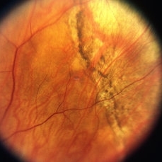
This is a very subtle example of lattice degeneration showing the mildest possible changes in a 27-year-old man. In the upper left there is a vein directed toward the center of the slide. Just above and to the right of the pigment spot it veers to the right and then abruptly disappears as it passes through the lattice lesion. As it leaves the lesion, it resumes its normal appearance going down to the right. In a similar manner, the arteriole in the lower left enters the lesion just to the right of the pigment spot, then disappears as it passes through the lesion and reappears later as it emerges. The only change in this lesion in 12 years was the appearance of the pigment spot.
Condition/keywords: lattice degeneration
-
 Acute Retinal Detachment
Nov 9 2012 by Norman Byer
This 54-year-old man was referred because of sudden symptoms in his opposite eye in which he had suffered an acute retinal detachment secondary to a horseshoe tear around lattice degeneration. During the examination, the fellow eye shown here was also found to have this large horseshoe tear about 1 o’clock hour (4 disc diameters) in size. A tear occurred around a lattice lesion which is present on the flap but is out of focus. This tear had been asymptomatic even though it was caused by a posterior vitreous detachment and illustrates that even very large tears may produce no symptoms or mild symptoms that are easily overlooked.
Condition/keywords: lattice degeneration, posterior vitreous detachment
-
 Lattice Lesion
Nov 9 2012 by Norman Byer
This is the same lesion as shown in the previous case. Two retinal holes are present, and you can look through the upper hole into the dark subretinal space. This is, therefore, a true subclinical retinal detachment but it has changed only slightly in the past 13 years. About 75% of such holes in lattice lesions show a tiny adjacent zone of subretinal fluid. After the hole forms from gradual progressive thing of the retina, a tiny amount of fluid from the pocket of liquified vitreous over the lesion passes through the hole to the subretinal space
Condition/keywords: lattice degeneration, liquefied vitreous, retinal hole, subretinal fluid
-
 Lattice Lesion
Nov 9 2012 by Norman Byer
This lattice lesion in a 36-year-old woman has remained unchanged over a period of 13 years. It shows a moderate snailtrack feature with discrete yellow dots visible on the surface of the lesion and especially along the posterior border. One of these can be well seen just below the lesion superimposed over the dark shadow of the scleral indentation. The exact nature of these yellow dots is still not entirely clear.
Condition/keywords: lattice degeneration, moderate snail track, scleral indentation, yellow dots
-
 Flat Lattice Lesion
Nov 9 2012 by Norman Byer
This 24-year-old woman had a flat lattice lesion without holes observed with no change for six years. She then developed two tiny retinal holes in this lesion and three years later the clinical retinal detachment shown here. She responded well to surgery. Even though such atrophic holes and lattice lesions may occasionally lead to a clinical detachment, it is important to understand that the mere presence of such holes is not an indication for prophylactic treatment. The reason for this is that we now know statistically that fewer than 1 percent of such cases lead to a retinal detachment.
Condition/keywords: lattice degeneration, retinal hole, scleral depression
-
 Lattice Lesion
Nov 9 2012 by Norman Byer
This is the same lesion as seen in the previous case seen now with scleral indentation. Here you can see directly into the subretinal space through the two retinal holes. The holes appear dark because the shadow of the scleral indentation lies directly beneath them.
Condition/keywords: lattice degeneration, retinal hole, scleral indentation
-
 Lattice Combined with Tiny Round Hole
Nov 9 2012 by Norman Byer
This 45 year-old man shows the snail track form of lattice combined with a tiny round hole. There is a tiny subclinical retinal detachment confined to the lesion itself.
Condition/keywords: glial vitreous tuft, lattice degeneration, round hole, snail track
-
 Lattice Lesion
Nov 9 2012 by Norman Byer
When this boy was first examined at the age of six years, he had only the red crater form of lattice at this location. This photograph shows the same lesion at age 11 and there is now a small round atrophic hole with a tiny round zone of detachment around it. It has not changed for four years.
Condition/keywords: atrophic retinal hole, lattice degeneration, lattice lesion, reddish crater, round hole
-
 Lattice Lesion
Nov 9 2012 by Norman Byer
This lattice lesion in an 18-year-old girl shows the combination of a reddish crater, several prominent pigment clumps and white lines. Please note that the white vessel changes involve both arterioles and venules.
Condition/keywords: lattice degeneration, lattice lesion, pigment clumps, reddish crater, white lattice lines
-
 Lattice Degeneration With Atrophic Hole
Feb 19 2015 by H. Michael Lambert, MD
Color photo of Lattice degeneration with atrophic hole.
Condition/keywords: atrophic retinal hole, lattice degeneration
-
 Lattice Lesion
Nov 9 2012 by Norman Byer
This lattice lesion in a 44-year-old man shows an atrophic retinal hole surrounded by discrete yellowish and pigmented areas. These have been caused by secondary pigment migration and proliferation in the retinal pigment epithelium. There is a small doughnut like elevation of the retina between the edge of the hole and the line of pigment. The lesion and the hole have remained exactly the same for seven years.
Condition/keywords: atrophic retinal hole, elevated retina, lattice degeneration, lattice lesion, proliferation of retinal pigment epithelium, scleral indentation
 Loading…