Search results (241 results)
-
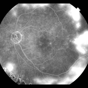
 Central Retinal Artery Occlusion
Aug 23 2012 by Gerardo Garcia-Aguirre, MD
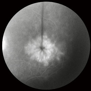
Fluorescein angiogram, late phase, of a central retinal artery occlusion, showing very delayed filling and wide areas of capillary nonperfusion.
Photographer: Noemí Hernández, Asociación para Evitar la Ceguera en México
Condition/keywords: capillary nonperfusion, central retinal artery occlusion (CRAO), vessel sheathing
-
 Central Serous Chorioretinopathy, Fluorescein Angiogram
Aug 23 2012 by Gerardo Garcia-Aguirre, MD
Fluorescein angiogram, late phase, showing hyperfluorescent spot, larger than earlier phases.
Photographer: Noemí Hernández, Asociación para Evitar la Ceguera en México
Imaging device: Zeiss FF4
Condition/keywords: central serous chorioretinopathy (CSCR)
-
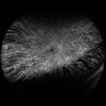
 Angioid Streaks
Sep 29 2012 by Hamid Ahmadieh, MD
Late phase ICG angiography image of the left eye of a 59-year-old man with angioid streaks.
Photographer: Hamid Ahmadieh, MD; Ophthalmic Research Center, Labbafinejad Medical Center, Shahid Beheshti University of Medical Sciences
Imaging device: Heidelberg Spectralis
Condition/keywords: angioid streaks, indocyanine green (ICG) angiography
-
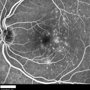
 Proliferative Diabetic Retinopathy - Neovascularization on the Disc
Aug 23 2012 by Gerardo Garcia-Aguirre, MD
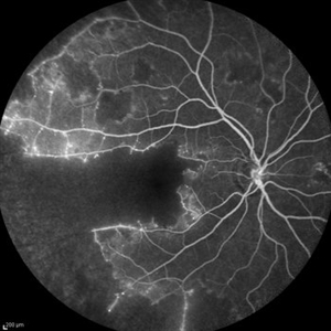
Fluorescein angiogram, late phase, showing microaneurysms, wide areas of capillary non-perfusion, and leakage secondary to neovascularization on the disc.
Photographer: Noemí Hernández, Asociación para Evitar la Ceguera en México
Condition/keywords: microaneurysms, neovascularization of the disc (NVD)
-
 Acute Posterior Multifocal Placoid Pigment Epitheliopathy
Sep 15 2012 by Roy D. Brod, MD
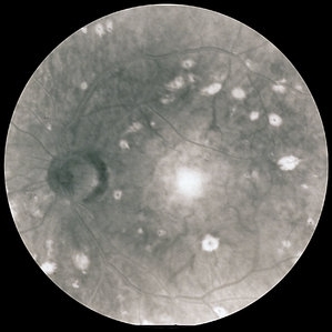
Late phase fluorescein angiogram demonstrating staining of placoid lesions in patient with APMPPE.
Photographer: Julia Walker
Condition/keywords: acute posterior multifocal placoid pigment epitheliopathy (APMPPE)
-
 Central Serous Chorioretinopathy, Fluorescein Angiogram
Aug 23 2012 by Gerardo Garcia-Aguirre, MD
Fluorescein angiogram, late phase, showing a small, hyperfluorescent spot.
Photographer: Noemí Hernández, Asociación para Evitar la Ceguera en México
Imaging device: Zeiss FF4
Condition/keywords: central serous chorioretinopathy (CSCR)
-
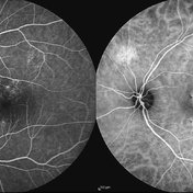
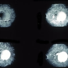
 Fundus Flavimaculatus and CNV
Nov 14 2013 by Hamid Ahmadieh, MD
Late phase FA and ICG angiography images of the right eye of a 35-year-old woman with subfoveal CNV secondary to fundus flavimaculatus .
Photographer: Nayereh Hadipour, Negah Eye Center, Tehran
Condition/keywords: choroidal neovascularization (CNV), fundus flavimaculatus, indocyanine green (ICG) angiography, retinal flecks
-
 Chronic Active Central Serous Chorioretinopathy (CSCR)
Sep 11 2012 by Hamid Ahmadieh, MD
Late phase FA & ICG angiography images of a 30-year-old man with chronic active CSCR.
Photographer: Hamid Ahmadieh, MD, Ophthalmic Research Center, Labbafinejad Medical Center, Shahid Beheshti University of Medical Sciences
Imaging device: Heidelberg Spectralis
Condition/keywords: central serous chorioretinopathy (CSCR), indocyanine green (ICG) angiography
-
 Central Serous Choroidopathy, CSR, with Foci of Leakage
Oct 9 2012 by James B. Soque, CRA, OCT-C, COA, FOPS
50 y/o WM with Central Serous Choroidopathy Left eye. VA OS cc 20/80. Topcon 3D 1000 SD OCT composite image reveals Sub RPE detachment in several locations, and subretinal fluid blister. Pin Point Registration shows leakage along horizontal line axis, SR leakage, and RPE detachments OS. Color, Early, and Late phase FA photos enclosed above. FA shows obvious ‘smoke stack’ appearance of leakage in superonasal fovea, and 3 other foci of leakage. 3D 1000 SD OCT with pin point registration image shown.
Photographer: James Soque CRA COA
Imaging device: Topcon 3D OCT 1000 System
Condition/keywords: central serous retinopathy (CSR)
-
 Cystoid Macular Edema (CME)
Sep 11 2012 by Hamid Ahmadieh, MD
Late phase FA & ICG angiography imagings of the left eye a 17-year-old boy with CME & retinal periphlebitis secondary to chronic intermediate uveitis.
Photographer: Hamid Ahmadieh, MD, Ophthalmic Research Center, Labbafinejad Medical Center, Shahid Beheshti University of Medical Sciences
Imaging device: Heidelberg Spectralis
Condition/keywords: cystoid macular edema (CME), indocyanine green (ICG) angiography, intermediate uveitis
-
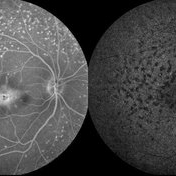
 "Mud-Splatter" of Posterior Pole and Peripheral Radial Streaks in a Carrier of Ocular Albinism
Jan 22 2019 by John S. King, MD
14-year-old healthy white female with family history of ocular albinism was seen by Dr. Hruby for a second opinion. Father and some of his brothers were positive for a history of ocular albinism. Va cc 20/30 J1+ OU; no nystagmus; no TIDs; no foveal hypoplasia. A "mud-spatter" appearance to the posterior pole was present, along with peripheral alternating streaks that are very prominent on this late phase FA OS. Dr. Hruby agreed that this was most likely a carrier of Ocular Albinism Type-1 (XR; GPR143 mutation), and possible genetic testing/counselling was discussed.
Photographer: Gretchen Harper
Imaging device: Optos California
Condition/keywords: Nettleship-Falls ocular albinism, ocular albinism
-
 Sickle Cell Retinopathy with Sea Fans (angiogram)
Aug 24 2012 by Geoffrey G. Emerson, MD, PhD, FASRS
Fluorescein angiography (late phase) of a 40-year-old man with African heritage and sickle SC disease. Sea fans are present around the macula (profusely leaking fluorescein dye).
Photographer: Geoffrey Emerson, MD, PhD, Retina Center, Minneapolis
Condition/keywords: sea fan
-
 Bruch’s membrane rupture
Jan 11 2013 by Hyung-Woo Kwak, MD
An area of Bruch’s membrane rupture involving the fovea is seen on indocyanine green angiography: late phase (right).
Photographer: Misook Lee, Kyung Hee Univsersity Hospital, Seoul
Imaging device: Zeiss f 450 plus
Condition/keywords: Bruch's membrane, myopic choroidal neovascularization (CNV)
-
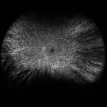
 "Mud-Splatter" of Posterior Pole and Peripheral Radial Streaks in a Carrier of Ocular Albinism
Jan 22 2019 by John S. King, MD
14-year-old healthy white female with family history of ocular albinism was seen by Dr. Hruby for a second opinion. Father and some of his brothers were positive for a history of ocular albinism. Va cc 20/30 J1+ OU; no nystagmus; no TIDs; no foveal hypoplasia. A "mud-spatter" appearance to the posterior pole was present, along with peripheral alternating streaks, which are very prominent in this late phase FA of the right eye. Dr. Hruby agreed that this was most likely a carrier of Ocular Albinism Type-1 (XR; GPR143 mutation), and possible genetic testing/counselling was discussed.
Photographer: Gretchen Harper
Imaging device: Optos California
Condition/keywords: Nettleship-Falls ocular albinism, ocular albinism
-
 Fibrovascular PED
May 2 2013 by Henry J. Kaplan, MD
Fluorescein angiogram of the fibrovascular PED in the same patient; early homogeneous hyperfluorescence in the PED area which is increased in fluorescence to the late phase with a granular hyperfluorescence adjacent to PED in the foveal side which starts in the mid-phase of the F/A and is increased later; #2.
Condition/keywords: exudative age-related macular degeneration, fibrovascular pigment epithelial detachment (PED)
-
 Suspected Multiple Evanescent White Dot Syndrome
Mar 3 2015 by Stuart Alfred, CRA, OCT-C
30 degree, late phase angiogram image of left fundus of a 28-year-old Caucasian female.
Photographer: Stuart Alfred, CRA, OCT-C, Midwest Eye Institute, Greenwood, Indiana
Imaging device: cSLO by Heidelberg engineering (Spectralis)
Condition/keywords: dry age-related macular degeneration (dry AMD), multiple evanescent white dot syndrome (MEWDS), punctate inner choroidopathy (PIC), uveitis
-
 Cystoid Macular Edema
Oct 8 2012 by Jeffrey G. Gross, MD, FASRS
CME, s/p AC-IOL, FA late phase.
Condition/keywords: cystoid macular edema (CME), late phase
-
 Central Serous Choroidopathy, CSR, with Foci of Leakage
Oct 9 2012 by James B. Soque, CRA, OCT-C, COA, FOPS
50 y/o WM with Central Serous Choroidopathy Left eye. VA OS cc 20/80. Topcon 3D 1000 SD OCT image reveals Sub RPE detachment in several locations, and subretinal fluid blister. Color, Early, and Late phase FA photos enclosed. FA shows obvious ‘smoke stack’ appearance of leakage in superonasal fovea, and 3 other foci of leakage. FC Photo shown.
Photographer: James Soque CRA COA
Imaging device: Topcon TRC 50 EX, with OIS V 10.5.74 Software. 5 MP Camera
Condition/keywords: central serous retinopathy (CSR)
-
 Color Fundus Photograph of Macular Infarction Secondary to Subonjunctival Gentamicin Injection
May 16 2014 by Arwa Azmeh, MD, PhD
A 20-year-old male suffered from diplopia since age one. He was diagnosed to have acquired fourth nerve palsy in his left eye. VA at time of diagnosis was 20/20 in OU and Fundus exam was WNL in OU. His history revealed no other complaints. 3 days ago he underwent left superior oblique tucking for relief of his diplopia.The surgery was uneventful and at the end of surgery subconjunctival gentamicin was injected. Immediately following surgery his VA in OS decreased from 20/20 to complete loss of central vision and sensation of HM from the periphery. He was referred to us 3 days after surgery. At time of referral fundus exam of his left eye revealed macular infarction with cherry red spot appearance with few retinal hemorrhages, mild optic disc edema and CWS surrounding optic disc. Peripheral retina had normal color and appearance. The vitreous was clear. Anterior segment was quiet. IOP was WNL. Macular OCT was consistent with macular infarction. FA revealed delay in central retinal artery filling as fluorescein started to appear in the arteries at the level of the optic disc at 28 sec, and in the retinal veins at 38 sec. Macular area remained to be non-perfused throughout the whole FA. In late phases staining of blood vessels walls was noticed. The "wipe out" of large vessels and capillaries persisted in the central area. OCT through foveal area showed diffuse thickening of the retina with severe elevation in the fovea, reduced backscattering from the outer layers of the retina and enhanced reflectivity from the inner retina, due to ischemia. Complete blood count and cardiovascular study were WNL. The final diagnosis was macular infarction secondary to subconjunctival gentamicin injection.
Imaging device: OCT
Condition/keywords: macular infarction, subconjunctival gentamicin
-
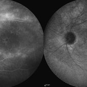
 Central Serous Chorioretinopathy
Nov 13 2012 by Mallika Goyal, MD
Late phase fluorescein angiogram of right eye of a 33-year-old gentleman with bilateral CSC.
Photographer: Mallika Goyal, MD
Condition/keywords: bilateral chronic central serous retinopathy, central serous chorioretinopathy (CSCR)
-
 Sickle SC Sea Fan
Oct 8 2012 by Jeffrey G. Gross, MD, FASRS
Sickle SC sea fan, partial regression, FA late phase leakage.
Condition/keywords: FA late phase leakage, partial regression, sea fan, sickle cell
-
 Angioid Streaks
Sep 29 2012 by Hamid Ahmadieh, MD
Late phase ICG angiography image of the right eye of a 59-year-old man with angioid streaks.
Photographer: Hamid Ahmadieh, MD; Ophthalmic Research Center, Labbafinejad Medical Center, Shahid Beheshti University of Medical Sciences
Imaging device: Heidelberg Spectralis
Condition/keywords: angioid streaks, indocyanine green (ICG) angiography
-
 Idiopathic Occlusive Retinal Vasculitis (Late Stage)
May 31 2014 by Hamid Ahmadieh, MD
Late phase FA image of the right eye of a 28-year-old woman with idiopathic occlusive retinal vasculitis 6 months after the onset.
Photographer: Solmaz Shahmohammad, Negah Eye Center, Tehran
Imaging device: Heidelberg Spectralis
Condition/keywords: capillary closure, fluorescein leakage, macular infarction
-
 Ocular ischaemic syndrome FA 2
Jan 11 2013 by Alex P. Hunyor, MD
Ocular ischaemic syndrome, left eye - fluorescein angiogram, late phase.
Condition/keywords: ocular ischemic syndrome
-
 Central Serous Choroidopathy, CSR, with Foci of Leakage
Oct 9 2012 by James B. Soque, CRA, OCT-C, COA, FOPS
50 y/o WM with Central Serous Choroidopathy Left eye. VA OS cc 20/80. Topcon 3D 1000 SD OCT image reveals Sub RPE detachment in several locations, and subretinal fluid blister. Color, Early, and Late phase FA photos enclosed. FA shows obvious ‘smoke stack’ appearance of leakage in superonasal fovea, and 3 other foci of leakage. Late FA Photo shown.
Photographer: James Soque CRA COA
Imaging device: Topcon TRC 50 EX, with OIS V 10.5.74 Software. 5 MP Camera
Condition/keywords: central serous chorioretinopathy (CSCR), central serous retinopathy (CSR)
 Loading…